c cc
2017 The Korean Academy of Prosthodontics
This is an Open Access article distributed under the terms of the Creative Commons Attribution Non-Commercial License (http://creativecommons.org/licens- es/by-nc/3.0) which permits unrestricted non-commercial use, distribution, and reproduction in any medium, provided the original work is properly cited.
*Corresponding Author: Hyeong-Seob Kim
Department of Prosthodontics, School of Dentistry, Kyung Hee University 26, Kyungheedae-ro, Dongdaemun-gu, Seoul 02447, Republic of Korea +82 (0)958 9340: e-mail, [email protected]
Article history: Received May 31, 2017 / Last Revision July 28, 2017 / Accepted July 31, 2017
서론
완전 무치악 환자를 임플란트를 이용하여 치료하기 위해서 는 수술 전에 식립될 임플란트의 개수, 위치, 종류 등이 결정되 어야 한다. 이를 위해서는 먼저 최종 보철물의 형태가 결정되 어야 하며 그에 따른 구체적인 치료계획이 세워져야 한다.1만 일, 최종 보철물의 형태를 고려하지 않고, 잔존골이 풍부한 곳 에 임플란트를 먼저 식립하고 보철 치료를 진행한다면, 적절 한 보철물의 제작이 어려울 수 있기 때문이다.
최종 보철물의 형태를 고려한 치료계획 수립 시 비용이 1차 적인 결정요인이 될 수 있으므로 의료진은 환자의 요구사항을
잘 경청할 필요가 있다. 비용 및 환자의 요구에 따라 고정성 혹 은 가철성 보철물을 결정한 후 이상적인 최종 보철물에 대한 치료계획을 수립해야 한다. 2
본 증례는 8년 전 상악 우측 측절치를 제외하고 전악 임플란 트 식립된 무치악 환자가 고정성 보철수복을 주소로 내원하여 먼저, 고정성 보철 수복이 가능한지 평가한 후, 전치부는 지르 코니아 하부구조(substructure) 위에 단일 구조(monolithic) 지르 코니아 전부 도재관을, 구치부는 맞춤형 지대주(customized abut- ment)를 제작한 후 단일 구조(monolithic) 지르코니아 전부도재관 을 이용하여 최종 수복한 증례로 심미 및 기능적으로 만족할 만한 결과를 얻었기에 이를 보고하고자 한다.
최종 보철물에 대한 고려 없이 전악 임플란트 식립된 환자의 고정성 보철 수복 증례
천영훈∙배아란∙권긍록∙김형섭*
경희대학교 치과대학 치과보철학교실
Fixed prosthesis restoration in edentulous patient fully implanted without considering definitive prosthesis: A case report
Young-Hoon Chun, Ahran Pae, Kung-Rock Kwon, Hyeong-Seob Kim*
Department of Prosthodontics, School of Dentistry, Kyunghee University, Seoul, Republic of Korea
The most important factor in the treatment of fully edentulous patients using implants is the shape of the definitive prosthesis. After the shape of the definitive prosthesis is deter- mined, residual bone analysis and selection of the implant type, number and position should be followed. In this case, for restoration of an edentulous patient fully implanted (except the maxillary right lateral incisor) without considering definitive prosthesis, facial esthetics and possibility of fixed type prosthesis were evaluated using complete den- ture. It was determined that the fixed type prosthesis was possible. Implants that could not be used for the definitive prosthesis were excluded from the treatment plan and fixed type provisional restorations were fabricated. After four months of provisional restorations, the patient showed stable occlusion and esthetic satisfaction. Definitive prosthesis was made of zirconia using CAD/CAM (computer aided design and computer aided manufacturing). The results were satisfactory during the 3 months of follow-up period after termination of treatment. (J Korean Acad Prosthodont 2017;55:427-35)
Keywords: Edentulous; Full mouth rehabilitation; Implant; Prosthesis design; Computer aided design and computer aided manufacturing
증례
진단 및 준비
환자는 63세의 남성으로“동네 치과에서 식립한 임플란트에 대해 고정성 보철수복을 원한다”는 주소로 내원하였다. 8년 전 상악 우측 측절치를 제외한 전악에 다양한 종류의 임플란트를 식립하였으며 (Table 1), 당시 제작된 임시 총의치를 사용하고 있 었다.
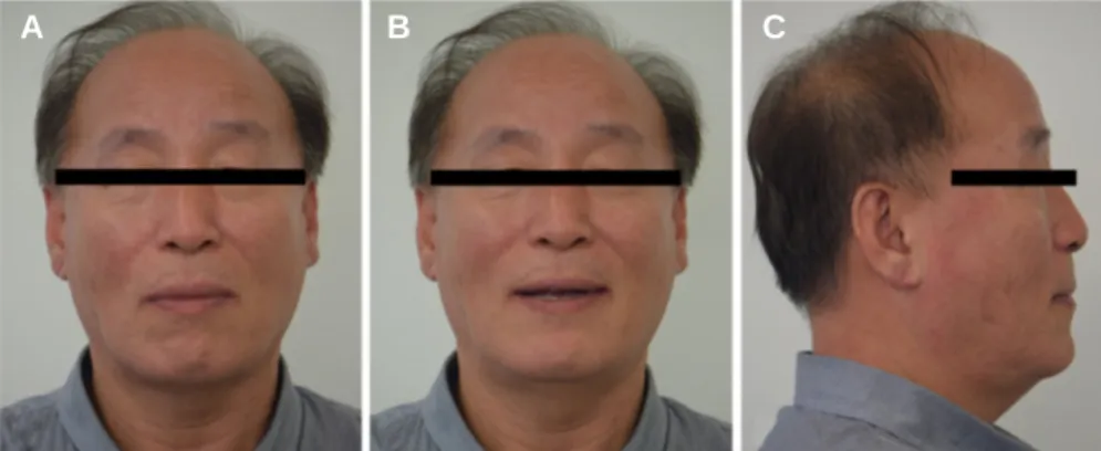
환자는 긴 안모로, 입을 다물 경우 구강주변 근육이 과긴장 되었으며, 상악 구순 지지도는 부족한 편이었다 (Fig. 1). 구내 관
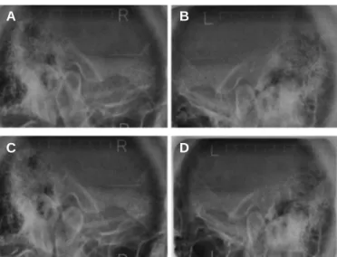
찰 시 일부 치유 지대주가 탈락하였고, 불연속적인 치조정 및 불량한 구강위생 상태가 관찰되었다 (Fig. 2). 방사선 영상에서 는 상악 우측 측절치를 제외하고 모든 부위에 식립된 임플란 트와 일부 소실된 치유 지대주를 확인할 수 있다 (Fig. 3). 경두개 방사선 영상에서는 우측 과두의 전방변위를 확인할 수 있다 (Fig. 4).
안모의 심미성을 회복하고, 악관절 및 교합의 안정화를 위해 총의치를 제작하기로 하였다. 수직고경 분석에 있어서 Turner와 Missirlian3이 제안한 항목들의 사용 및 과거 사진과의 비교를 통 해 현재 수직고경을 유지하기로 결정하였으며, 제작된 총의치 를 이용해 악관절과 교합의 안정성을 재평가하였다.
Fig. 2. Initial intra-oral view. (A) Occlusal view of maxilla, (B) Lateral view (right side), (C) Frontal view, (D) Lateral view (left side), (E) Occlusal view of mandible.
A
C
B D
E
Fig. 1. Initial extra-oral view (old denture wearing state); long face and lack of maxillary lip support. (A) Frontal view in centric occlusion, (B) Frontal view at rest posi- tion, (C) Lateral view.
A B C
고정성 보철 가능성 평가 및 치료계획
임플란트를 이용한 완전 무치악 환자의 치료 시, 환자가 부 족한 안모지지, 골격성 3급 악간관계, 교차교합, 순측으로 과도 하게 경사진 치조제 등을 보일 경우에는 고정성 보철수복보다 는 가철성 보철수복을 추천한다.4본 증례에서는 최종 보철물
의 형태를 결정하기 위해 사용 중인 총의치를 복제하여 의치 변연을 제거한 후 구내 시적 하였다.5,6구내 및 구외 분석을 통 해 의치변연이 없어도 심미적인 안모가 유지됨을 확인하였으 며, 임플란트가 전악에 골고루 분포하고 있으므로 고정성 수 복이 가능하다고 판단하였다. 동시에 전치부의 경우 치은색조 를 이용한 수복이 필요함을 확인하였다 (Fig. 5).
Table 1. Distribution of implant type and abutment connection type Implant
Implant type Abutment
distribution connection type
#16, 17 Snucon (Screw fixture SF, Locking SNUCONE KOREA, Daejeon, Korea)
#23, 31, 41 Superline (Dentium, Seoul, Korea) Screw Rest of all Oneplant (Warantec, Seoul, Korea) Screw
Fig. 4. Initial trans-cranial view. (A) Right TMJ at rest position, (B) Left TMJ at rest position, (C) Right TMJ in MIP (maximal intercuspal position); condylar ante- rior displacement, (D) Left TMJ in MIP.
Fig. 3. Initial panoramic view; various types of implant fixture fully implanted except right maxillary lateral incisor.
A B
C D
Fig. 5. Evaluation of fixed type prostheses availability. (A) Duplication of complete denture with labial flange removed, (B) Try-in of duplicated denture with labial flange removed, (C) Evaluation of incisor exposure, (D) Evaluation of smile line, (E) Evaluation of facial support.
A
C
B
D E
다양한 종류의 임플란트가 식립되었기 때문에 개인트레이 (individual tray)를 제작한 후 pick up 및 transfer type coping을 모두 이 용하여 fixture level 인상채득(Imprint II Garant Regular body, 3M ESPE, St. Paul, MN, USA)하였다. 작업모델(GC Fujirock EP, GC Europe N.V., Leuven, Belgium) 제작한 후, 고정성 record base 및 gothic arch trac- ing (Gothic Arch Tracer & Gothic par, Chansdental, Seoul, Korea) 사용 하여 안정적인 악간 관계 채득 후 교차부착(cross mounting)하였 다 (Fig. 6).7
작업모델 및 CAD (exocad DentalCAD, exocad, Darmstadt, Germany) 를 이용하여 새롭게 제작한 총의치 치아 위치를 기준으로 치 아의 크기 및 위치를 수정하면서 임플란트 위치를 3차원적으 로 평가하였다. 지나치게 구개 측으로 위치한 상악 우측 제1대 구치와 후방 임플란트와 지나치게 근접하여 임플란트 주위염 을 유발할 가능성이 있는8하악 우측 제2소구치 위치의 임플란 트는 치료계획에서 제외하였다 (Fig. 7).
Fig. 6. (A) Impression of maxillary implants, (B) Impression of mandibular implants, (C) Gothic arch tracing and implant supported record base, (D) Maxillary working model, (E) Mandibular working model, (F) Interocclusal record taking.
Fig. 7. (A) Diagnostic model and wax denture, (B) Maxillary implants location analysis using CAD, (C) Mandibular implants location analysis using CAD, (D) Working model and wax denture, (E) Maxillary implants path analysis using silicone index, (F) Mandibular implants path analysis using silicone index, (G) Implant of the right max- illary first molar invasive of lingual space, (H) Implants of the right mandibular second premolar and first molar were too closely located mesio-distally, (I) Implants of the right maxillary first molar and right mandibular second premolar are excluded from treatment plan.
A
D
B
E
C
F
A
D
B
E
C
F
G H I
고정성 임시 보철물 수복
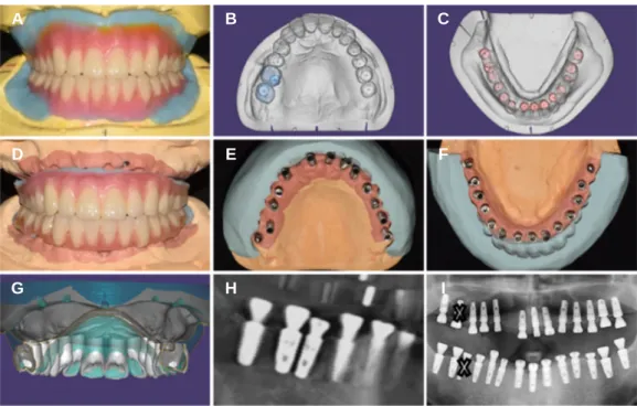
고정성 임시 보철물 제작을 위해 총의치 치아배열을 참고하 여 납형 형성하였으며, 임플란트의 응력 분산을 고려하여 전 방 및 측방운동 시 군기능 교합양식을9부여하였다. 실리콘 인 덱스(EXAFINE putty type, GC Co., Tokyo, Japan)를 이용해 시멘트 유지형의 보철물(cement-retained prosthesis) 제작이 가능함을 확인 한 후, 맞춤형 지대주(titanium customized abutment, EZEN Dental Laboratory, Daegu, Korea)를 제작하였으며, 시멘트 유지형의 임시 보철물(provisional prosthesis)을 장착하였다 (Fig. 8).10
지속적인 평가 및 조정을 통해 교합은 안정화되었으나, 전치
부에서 잉여 시멘트로 인한 염증반응이 나타났으며, 구치부 치아가 너무 길어 보인다는 환자의 의견을 고려하여 두 번째 임시 보철물 제작하였다. 두 번째 임시 보철물에서는 전치부 를 나사 유지형(screw retained provisional prosthesis)으로 변경하였 으며, 구치부에도 치은색조를 적용하였다 (Fig. 9).
4개월 동안 고정성 임시 보철물을 사용하면서 안정적인 중 심교합과 전방유도 및 군기능 교합양식을 확인하였다. 측두하 악관절에서의 불편감은 없었으며, 구강 내 연조직은 임시 보 철물에 잘 적응한 것으로 판단하였다. 전치부와 교합면은 입 술과 조화로운 관계를 보였다.
Fig. 8. 1stfixed type provisional prostheses. (A) Wax pattern for provisional prostheses, (B) Implants path analysis using silicon index (maxilla), (C) Implants path analy- sis using silicon index (mandible), (D) Computer aided design of customized abutment (maxilla), (E) Customized abutment setting (maxilla), (F) Fixed type provision- al prostheses (maxilla), (G) Computer aided design of customized abutment (mandible), (H) Customized abutment setting (mandible), (I) Fixed type provisional prostheses (mandible).
A
D
B
E
C
F
G H I
Fig. 9. 2ndfixed type provisional prostheses. (A) Intra oral view, (B) Extra oral view.
A B
최종 보철물
최종 보철물은 구치부의 경우 탈착 용이성을 고려하여 나 사-시멘트 유지형태(screw-cement retained prosthesis, SCRP)의 단일 구조(monolithic) 지르코니아 도재관(rainbow Trans, Genoss, Suwon, Korea)을 사용하고, 전치부의 경우 치은색조(Noritake super porce- lain Ex-3 tissue, Kuraray Noritake Dental Inc., Myoshi(Aichi), Japan)가 적용된 나사유지형태의 지르코니아 하부구조(zirconia substruc- ture)에 단일구조 지르코니아 도재관(rainbowTMTrans)을 합착하기 로 결정하였다 (Fig. 10).11,12
부가중합형 실리콘 인상재로 최종 인상 채득하고, 주모형 (GC Fujirock EP, GC Europe N.V.) 제작하였다. 상악 임시 보철물 상
태에서 안궁 이전하였으며, 전치부 임시 보철물 사이에 pattern resin (GC Corporation, Tokyo, Japan) 적용과 동시에 하악 양측의 제 2대구치 임시 보철물로 교합을 안정시킨 상태에서 바이트(O- bite, DMG, Hamburg, Germany)를 채득하였다. 그리고 상악 양측의 제2대구치와 전치부의 임시 보철물로 안정적인 교합을 확인 한 후 다시 바이트 채득하여 교차부착(cross mounting)하였다.
비가역성 하이드로콜로이드 알지네이트로(AROMA FINE PLUS, GC Corporation, Tokyo, Japan) 인상을 채득한 임시 보철물의 석고 모형과 최종 인상을 통해 얻은 석고 모형을 디지털 스캐 너(Rainbow Scanner, Dentium, Yongin, Korea)와 소프트웨어(exocad DentalCAD, exocad)를 이용하여 중첩 후 최종 보철물을 제작하였 다 (Fig. 11).
Fig. 10. (A) SCRP type monolithic zirconia crown, (B) Screw type zirconia substructure and cement type monolithic zirconia crown.
A B
Fig. 11. (A) Final impression of maxilla, (B) Final impression of mandible, (C) Model of provisional prostheses, (D) Working model of maxilla, (E) Working model of mandible, (F) Cross mounting, (G) Computer aided design (CAD) of Definitive prostheses (maxilla), (H) CAD of definitive prostheses (mandible), (I) Definitive pros- theses fabrication using computer aided manufacturing (CAM).
A
D
B
E
C
F
G H I
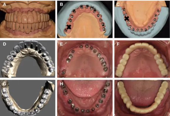
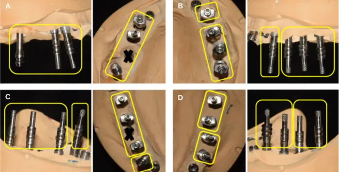
구치부는 식립된 임플란트의 종류, 식립 방향, 탈착 용이성 을 고려하여 연결 고정(splint)하였으며 (Fig. 12), 레진시멘트 (RelyX, 3M ESPE, Neuss, Germany)로 최종 합착 후 구강 외에서 잉 여 시멘트를 제거하였다. 전치부는 나사유지형태의 지르코니 아 하부구조에 지르코니아 단일 도재관을 임시 합착재(Temp- Bond, Kerr, Romulus, MI, USA)를 이용하여 합착하였다.
최종 보철물은 임시 보철물의 안정적인 교합과 형태를 적절 히 재현하였으며, 적절한 전방유도 및 군기능 교합양식을 보 였다 (Fig. 13).
장기적인 유지와 안정성을 위해 야간 유지 장치를 제작하고 사용법에 대해 교육하였으며, 주기적인 follow-up 기간 동안 안 정적인 교합양상을 보였다 (Fig. 14).
Fig. 12. Segmentaion for retrievability of posterior prostheses. (A) Right maxillary posterior implants, (B) Left maxillary posterior implants, (C) Right mandibular pos- terior implants, (D) Left mandibular posterior implants.
A
D B
C
Fig. 14. Evaluation of definitive prostheses. (A) Panoramic view, (B) Right trans-cranial view, (C) Left trans-cranial view.
Fig. 13. Definitive prostheses. (A) Intraoral view, (B) Extraoral view in centric occlusion, (C) Extraoral view on smile.
A B C
A B C
고찰
Misch13는 완전 무치악 환자의 임플란트 고정성 보철물이 충 분한 지지를 얻기 위해서는 상악에는 8개, 하악에는 6개 이상 의 임플란트 식립이 필요하다고 하였다. 본 증례에서 이미 식 립된 27개 임플란트의 골 유착 상태는 매우 양호하였다. 최종 보철물의 안정적인 지지 및 유지를 위해서는 모두 사용하는 것이 추천되나, 상악 우측 제1대구치 위치의 임플란트는 지나 치게 구개측으로 식립되었으며, 지대주 연결 형태는 friction fit connection 형태였기 때문에 맞춤형 지대주 제작이 불가능하여 지대치에서 제외하였다. 또한 Tarnow 등8은 임플란트 사이 거리 가 3 mm 보다 가까울 경우, 치조정의 골소실이 유의하게 증가 한다고 하였는데, 하악 우측 제2소구치와 제1대구치 위치의 임 플란트는 근원심 방향으로 매우 근접해 추후 과도한 골소실 및 임플란트 주위염을 일으킬 가능성이 있었기에 제2소구치 를 지대치에서 제외하였다. 따라서 본 증례에서는 2개 지대치 를 치료계획에서 제외하고 가공치(pontic) 처리하기로 하였다.
임플란트 최종 보철물의 유지형태는 시멘트 유지형과 나사 유지형 보철물로 구분된다. 시멘트 유지형은 기공과정이 간단 하고, 수동적 적합(passive fit), 심미 등의 이점이 있으며, 나사 유 지형은 탈착 및 관리가 용이한 장점이 있다.10이러한 두 가지 유지형태의 장점을 모두 가지는 나사-시멘트 유지형 보철물은 임플란트 지대주와 상부 보철물을 합착한 상태에서 구강 외로 제거가 가능해 잉여 시멘트를 제거할 수 있으며, 지대주와 보 철물의 수동적 적합을 통해 나사 풀림과 임플란트 구성요소의 파절을 줄일 수 있다.11원래 본 증례에서는 비용과 수동적 적합 을 고려해 최종 보철 수복에 시멘트 유지형태를 사용하려 했 으나, 전치부의 경우 임시 보철물 제거 시 잉여 시멘트가 지속 적으로 발견되어 불량한 예후가 의심되었기 때문에 나사유지 형 하부구조(substructure)에 지르코니아 단일 금관을 합착하였으 며, 구치부에는 관리 용이성을 위해 나사-시멘트 유지형의 지 르코니아 단일 금관을 사용하였다.14,15그리고 최종 보철물 제작 을 위해 4개월 동안 사용한 고정성 임시 보철물을 CAD를 이용 하여 주 모형에 중첩하였다. 이를 통해 구체적인 최종 보철물 제작이 가능했으며, 제작된 최종 보철물에서도 안정된 교합과 적절한 구개형태가 재현되었다.
결국 본 증례에서는 최종 보철물의 형태를 고려하지 않은 채 식립한 27개의 임플란트 중 2개의 임플란트를 치료계획에서 제외하였고, 전치부의 경우 나사-시멘트 유지형 보철 수복을 위해 추가적인 재료를 사용하였다. 만약, 최종 보철물 형태를 고려한 정확한 치료계획 하에 임플란트 식립 및 치료가 진행 되었다면 이러한 문제점들을 고려할 필요가 없었을 것이다.1 결론
완전 무치악 환자의 잔존골이 풍부할 경우, 많은 수의 임플 란트 식립를 통해 우수한 지지와 안정을 얻을 수는 있지만 최
종 보철물의 형태를 고려하지 않은 임플란트 식립은 최종 보 철 수복 시 여러 문제점을 일으킬 수 있으며, 과다한 치료비용 을 초래할 수 있다. 따라서 완전 무치악 환자를 임플란트를 이 용하여 치료하기 위해서는 최종 보철물의 형태와 그에 따른 치료계획 설정이 반드시 선행되어야 한다.
ORCID
Ahran Pae http://orcid.org/0000-0001-8758-0754
Hyeong-Seob Kim https://orcid.org/0000-0002-0964-0288
References
1. Almog DM, Sanchez R. Correlation between planned pros- thetic and residual bone trajectories in dental implants. J Prosthet Dent 1999;81:562-7.
2. Misch CE. Dental implant prosthetics. 2nd ed. St. Louis, Mosby;
Elsevier; 2015. p. 187.
3. Turner KA, Missirlian DM. Restoration of the extremely worn dentition. J Prosthet Dent 1984;52:467-74.
4. Mericske-Stern RD, Taylor TD, Belser U. Management of the edentulous patient. Clin Oral Implants Res 2000;11:108-25.
5. Sadowsky SJ. The role of complete denture principles in implant prosthodontics. J Calif Dent Assoc 2003;31:905-9.
6. Neves FD, Mendonça G, Fernandes Neto AJ. Analysis of influence of lip line and lip support in esthetics and selection of maxillary implant-supported prosthesis design. J Prosthet Dent 2004;91:286- 8.
7. Savabi O, Nejatidanesh F. Interocclusal record for fixed implant- supported prosthesis. J Prosthet Dent 2004;92:602-3.
8. Tarnow DP, Cho SC, Wallace SS. The effect of inter-implant dis- tance on the height of inter-implant bone crest. J Periodontol 2000;71:546-9.
9. Carlsson GE. Dental occlusion: modern concepts and their application in implant prosthodontics. Odontology 2009;97:8-17.
10. Wittneben JG, Millen C, Brägger U. Clinical performance of screw- versus cement-retained fixed implant-supported reconstruc- tions-a systematic review. Int J Oral Maxillofac Implants 2014;
29:84-98.
11. Rajan M, Gunaseelan R. Fabrication of a cement- and screw-re- tained implant prosthesis. J Prosthet Dent 2004;92:578-80.
12. Abdulmajeed AA, Lim KG, Närhi TO, Cooper LF. Complete- arch implant-supported monolithic zirconia fixed dental prostheses:
A systematic review. J Prosthet Dent 2016;115:672-7.
13. Misch CE. Dental Implant Prosthetics. 2nd ed. St. Louis, Mosby; Elsevier; 2015. p. 277.
14. Linkevicius T, Puisys A, Vindasiute E, Linkeviciene L, Apse P.
Does residual cement around implant-supported restorations cause peri-implant disease? A retrospective case analysis. Clin Oral Implants Res 2013;24:1179-84.
15. Chee WW, Duncan J, Afshar M, Moshaverinia A. Evaluation of the amount of excess cement around the margins of cement-re- tained dental implant restorations: the effect of the cement application method. J Prosthet Dent 2013;109:216-21.
최종 보철물에 대한 고려 없이 전악 임플란트 식립된 환자의 고정성 보철 수복 증례
천영훈∙배아란∙권긍록∙김형섭*
경희대학교 치과대학 치과보철학교실
임플란트를 이용한 완전 무치악 환자의 치료에 있어 가장 중요한 것은 최종 보철물의 형태이며, 최종 보철물의 형태가 결정된 후 잔존골 분석 및 식 립할 임플란트의 종류, 개수, 위치 등에 대한 선택이 이루어져야 한다. 본 증례는 상악 우측 측절치를 제외하고 전악에 임플란트가 식립된 채로 내 원한 환자에 대해 고정성 보철 수복한 증례이다. 최종 보철물에 대한 고려 없이 임플란트가 식립되었기 때문에 먼저 총의치를 제작하여 안모 분석 및 고정성 보철 수복 가능성을 평가하였다. 고정성 보철 수복이 가능하다고 판단되어 진단 납형, 방사선 영상 및 디지털 분석을 통해 최종 보철물에 사용할 수 없는 임플란트를 치료계획에서 제외한 후 고정성 임시 보철물을 제작하였다. 4개월 동안 고정성 임시 보철물을 사용하면서 안정적인 교 합과 심미적인 만족을 보였기에 CAD/CAM (Computer aided design and computer aided manufacturing)을 이용하여 지르코니아 최종 보철물로 이행하였 고, 치료 종료 후 3개월 동안의 평가에서 만족스러운 결과를 얻었다. (대한치과보철학회지 2017;55:427-35)
주요단어: 완전 무치악; 전악수복; 임플란트; 치료계획; 캐드캠
*교신저자: 김형섭
02447 서울 동대문구 경희대로 26 경희대학교 치과대학 치과보철학교실 02 958 9340: e-mail, [email protected]
원고접수일: 2017년 5월 31일 / 원고최종수정일: 2017년 7월 28일 / 원고채택일: 2017년 7월 31일
2017 대한치과보철학회
이 글은 크리에이티브 커먼즈 코리아 저작자표시-비영리 3.0 대한민국 라이선스에 따라 이용하실 수 있습니다.
c cc