Percutaneous catheter drainage (PCD) is one of the most commonly performed procedures worldwide. To
guide the puncture needle and position the catheter, a variety of imaging tools can be used, such as fluo- roscopy, computed tomography and ultrasonography (US). Among these different procedures, ultrasonogra- phy has gained in popularity for several reasons, includ- ing its easy availability, low cost, lack of radiation haz- ard, good manageability and mobility. The greatest ad- vantage of ultrasound is its real-time capability. The tar- get, the advancing needle and the intervening structures can be visualized at all times, allowing the operator to plan the route, confirm precise needle placement, and
Percutaneous Drainage with Ultrasound Guidance in the Intensive Care Unit
1Doo Kyung Kang, M.D., Je Hwan Won, M.D., Jai Keun Kim, M.D., Kwang Hun Lee, M.D.2, Ji Hyung Kim, M.D.3
Purpose:To determine the efficacy and safety of bedside percutaneous drainage pro- cedures with ultrasound guidance in critically ill patients in the intensive care unit (ICU).
Materials and Methods:Sixty five percutaneous drainage procedures performed at the bedside, in 39 ICU patients, were evaluated. All of the procedures were performed with ultrasound guidance alone. The procedures consisted of percutaneous drainage of abdominal (n=35) and pleural (n=27) fluids, percutaneous cholecystostomy (n=2) and percutaneous nephrostomy (n=1). The clinical responses were classified as ‘com- plete response’, ‘partial response’, ‘failure’ or ‘undetermined’. The medical records were reviewed retrospectively to evaluate the clinical response.
Results:Technical success was achieved in 64 of the 65 procedures (98.5%). The com- plication rate was 13.8% (9 cases). There was no immediate procedure-related death or worsening of the clinical condition of the patients. The clinical responses after drainage were ‘complete response’ in 39 cases (60.9%), ‘partial response’ in 14 (21.9%), ‘failure’ in 3 (4.7%), and ‘undetermined’ in 8 (12.5%).
Conclusion:Bedside drainage procedures with ultrasound guidance are effective and safe to perform when patients are too critically ill to be moved from the ICU to the an- giography room.
Index words :Percutaneous drainage Ultrasound guidance Abscess
Thorax
Interventional procedures
1Department of Diagnostic Radiology, Ajou University, College of Medicine
2Department of Diagnostic Radiology, Yonsei University, College of Medicine
3Department of Diagnostic Radiology, Keon Yang University, College of Medicine
Received September 29, 2003 ; Accepted January 5, 2004
Address reprint requests to : Doo Kyung Kang, M.D., Department of Diagnostic Radiology, Ajou University College of Medicine, San 5, Woncheon-dong, Paldal-gu, Suwon, Kyongi-Do 442-749, Korea.
Tel. 82-31-219-5863 Fax. 82-31-219-5862 E-mail: [email protected]
avoid vital structures (1, 2).
Due to the severity of their illness, and the large amount of life-support equipment involved, such as monitoring devices, ventilators, hemodialysis machines, and infusion pumps, patients in the intensive care unit (ICU) are rarely candidates for routine percutaneous catheter drainage in the interventional suite. When an ICU patient requires a percutaneous drainage proce- dure, it therefore has to be performed at the bedside with ultrasound guidance alone (3).
We retrospectively evaluated percutaneous drainage procedures performed on ICU patients at the bedside using ultrasound guidance alone; these included PCD, percutaneous cholecystostomy, and percutaneous nephrostomy. The technical success rate, procedure-re- lated complications, and clinical efficiency of the proce- dure were analyzed.
Materials and Methods
Patient population
Between May 1998 and May 2003, 65 US-guided per- cutaneous drainage procedures were performed at the bedside of 39 ICU patients in our hospital. The patients were 25 men and 14 women, ranging from 3 to 85 years of age (mean 49.8 years). All of the patients were critical- ly ill, with sepsis or unstable vital signs, and required a large amount of life-support equipment (Table 1).
Therefore, the drainage procedures could not be per- formed in the interventional suite under fluoroscopic guidance. Since our patients were critically ill or in the terminal stage of their disease, the absolute contraindi- cations were limited to bleeding diathesis (platelet count<50,000/mm3, partial thromboplastin time>15
sec) and no safe access route under ultrasonography.
The nature of the drainage procedures is summarized in Table 2.
The percutaneous drainage procedures performed were PCD of abdominal (n=35) or pleural (n=27) fluid, percutaneous cholecystostomy (n=2) and percutaneous nephrostomy (PCN, n=1). Multiple drainage procedures were performed in 17 patients: consisting of 2, 3 and 4 procedures in 9, 7 and 1 patients, respectively. Multiple procedures were commonly performed in patients with bilateral or multiple fluid collections, and in patients with simultaneous abdominal and pleural fluid collec- tions. One patient with panperitonitis after surgery for rectal lymphoma required three drainage catheters for bilateral pleural effusions and an abdominal abscess.
One patient with a traumatic liver laceration and hemo- thorax had a total of four drainage catheters for left-side hemothorax, right-side pleural effusion, right-side he- moperitoneum and left-side abscess cavity.
Drainage procedure
Ultrasound (VST MASTER; Diasonics Ultrasound Corp., U.S.A.) was performed just before the drainage procedure with a 3.5- or 7-MHz convex probe and the skin entry site was determined in this way. The skin en- try site was prepared and draped in a sterile manner and the US probe was covered with a sterile covering. Then, 5 to 10 ml of 0.1 or 0.2% lidocaine were administered for local anesthesia, and a small incision (2-3 mm) was made at the skin entry site with a surgical blade. US- guided puncture was done using a needle-guidance sys- tem with a 22-gauge micro-puncture introducer needle (Cook, Bloomington, U.S.A.) or an 18-gauge Chiba nee- dle (Cook) (Fig. 1). The drainage catheter was inserted
Table 1. Primary Diagnosis in Patients Undergoing US-guided PCD
Primary diagnosis No. of patients
Trauma 10
Malignancy 07
Infection 07
Post-op complication 05
Bowel perforation 04
Liver transplantation 02
Abdominal aortic aneurysm 01
Postpartum bleeding 01
Neuroganglioma 01
Drug toxicity 01
Total 39
Table 2. The Nature of the Percutaneous Drainage Procedures Performed (Numbers in parentheses are the numbers of patients)
Cause of procedure No. of cases
Intraabdominal fluid Abdominal abscess 18
collection (35) hemoperitoneum 06
Ascites 03
Liver abscess 03
Abdominal fluid collection 03
Biloma 02
Pleural fluid Pleural effusion 20
collection (27) Hemothorax 04
empyema 03
Gallbladder (2) Acute cholecystitis 02
Kidney (1) Pyonephrosis 01
Total 65
using the Seldinger technique. When using a 22-gauge needle, first the wire (Cook) was advanced and then the yellow sheath (Cook) was inserted. Then, the 0.035-inch guide-wire (Terumo, Tokyo, Japan) was advanced suffi- ciently and echogenic looping of the guide-wire within the lumen was confirmed. Then, the 18-gauge puncture needle or yellow sheath was removed and a 7 to 10 French pigtail catheter (Boston Scientific, Spencer, U.S.A.) with an inner stylet was introduced along the guide-wire. As soon as the tip of the catheter entered the target lumen, the inner stiffening stylet of the catheter was withdrawn slightly, in order to allow the pigtail loop to reform. With this blunt loop leading the way, the catheter was advanced further into the target lumen (Fig. 2A, B). After performing the PCD of pleural fluid, we routinely checked for the development of pneu- mothorax, by taking a portable chest radiograph in the antero-posterior projection.
In performing PCN, the patient was prepared and draped in the prone position. After administering local anesthesia at the needle entry site, an 18-gauge Chiba needle with an inner stylet was introduced into the pos- terior calyx of the lower pole of the kidney under con- tinuous ultrasound guidance, and the puncture of the collecting system was confirmed by noting the efflux of urine. A 0.035 guide-wire (Terumo) was passed into the renal pelvis and the pigtail catheter was inserted under real-time US guidance. The presence of the complete looping curve of the pigtail catheter in the renal pelvis on the sonogram confirmed the proper position of the
catheter in the urinary system.
Evaluation of technical and clinical results
We retrospectively evaluated the technical success rate, procedure-related complication rate and clinical re- sponse. Technical success was defined as proper posi- tioning of the catheter in the target space with proper drainage. The clinical response was evaluated retrospec- tively by reviewing the medical records, using a modi- fied version of the criteria of Civardi et al. (4). The result
Fig. 1. This US picture shows an 18-gauge Chiba needle insert- ed along a US biopsy guide (curved arrow) into an abdominal abscess cavity. The tip of the needle (straight arrow) is within the abscess cavity.
A B
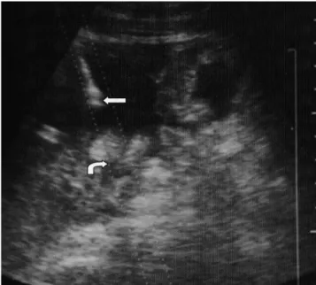
Fig. 2. Percutaneous cholecystostomy in a patient with acute cholecystitis.
A. This US picture shows the curled guide-wire (arrow) within the gall bladder lumen.
B. This US picture shows a typical echogenic double linear image (arrow) indicating the catheter tip within the gall bladder lumen.
was defined as a ‘complete response’ when the lesion was drained completely and the catheter removed (Fig.
3A-C). When PCD improved the clinical status, but an- other procedure (e.g., surgery) was required to cure the condition, or the patient died with a well-functioning catheter, the result was defined as a ‘partial response’.
When the patient obtained no benefit from the proce- dure, the result was defined as a ‘failure’. Finally, when the patient died or was discharged before the clinical re- sponse could be evaluated, the response was defined as
‘undetermined’.
Results
Technical success rate
US-guided PCD was technically successful in 64 of the 65 procedures (98.5%). In one patient, because the pleural effusion was located at a considerable depth, ex- act targeting of the anechoic fluid failed due to poor ul- trasound guidance, resulting from a thick subcutaneous fat layer. In this case, no pleural effusion was drained through the puncture needle, although a blood clot was aspirated.
The procedure time was less than 1 hour in all cases.
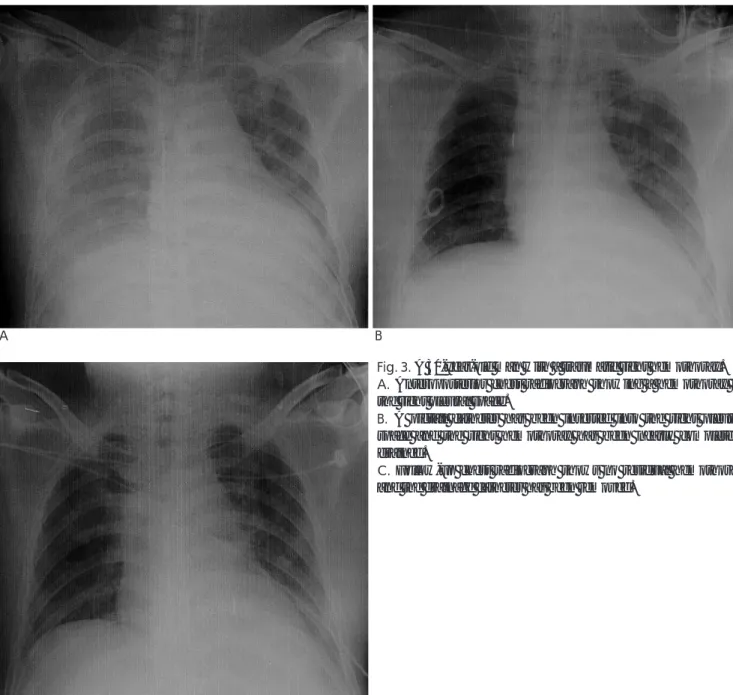
A
C
B
Fig. 3. A 30-year-old man with a traumatic right hemothorax.
A. Anteroposterior chest radiograph showing a hemothorax in the right pleural space.
B. A pigtail catheter has been inserted into the right pleural space and the right hemothorax has been nearly completely drained.
C. Follow-up chest radiograph shows no residual hemothorax and the drainage catheter has been removed.
Complications (six immediate complications and three delayed complications) arose in nine cases (13.8%). No bleeding from any inserted catheter persisted for more than 6 hours after the procedure in any of the patients.
Immediate bleeding occurred in three cases and was sta- bilized within 1 hour by capping the drainage tube.
Other immediate complications were three cases of pneumothorax and these resolved spontaneously. There were no cases of immediate procedure-related death or worsening of the clinical or pathological condition in our series. Delayed complications consisted of two cases of tract infection and one catheter fracture.
Clinical responses
The clinical response was ‘complete’ in 39 cases (60.9%), ‘partial’ in 14 (21.9%), ‘failure’ in 3 (4.7%) and
‘undetermined’ in 8 (12.5%) (Table 3).
Of the ‘partial responses (n=14)’, six patients (n=11) died and one underwent a hemi-nephrectomy after PCN, and two with percutaneous cholecystostomy un- derwent cholecystectomy. Ineffective fluid drainage was the most common cause of failure (n=3). In the three cases of ‘failure’ (three patients), one patient had a chest tube inserted as an additional drainage procedure, and two patients with intraabdominal abscess required surgical therapy. The eight ‘undetermined’ cases (five patients) consisted of patients who either died or were discharged within 5 days of the procedure.
Discussion
Percutaneous drainage is a well-established therapeu- tic technique and is one of the most commonly per- formed procedures worldwide. PCD can be guided us- ing fluoroscopy, US or computed tomography.
Ultrasound has the advantage of being both widely available and portable (1). However, radiologists find US-guided PCD at the bedside inconvenient, because of the additional difficulty caused by the lack of fluoro- scopic guidance. Nevertheless, in some situations the
patient’s clinical condition prevents transport to the in- terventional suite in the radiology department. In such situations, US-guided PCD at the bedside constitutes a safe alternative.
Ultrasound guidance is optimal for targets located su- perficially or at moderate depth in thin to average-sized patients. The use of US as a guidance system may be precluded by the inability to visualize the target because of its depth, because it is obscured by overlying bowel gas or bone, or because of poor penetration by the sound waves in the case of an obese patient. Our one in- stance of failure resulted from targeting an anechoic flu- id with poor ultrasound guidance, due to a thick subcu- taneous fat layer.
Problems can be encountered when performing PCD using US guidance alone, because of the difficulties in- volved in visualizing the needle itself, manipulating the catheter in the target area or obtaining the final catheter position. The improvements made in the resolution of US transducers combined with the development of echogenic polymer-coated needles have improved both needle shaft and tip visibility in clinical practice, com- pared with previous methods involving standard un- coated needles (5). The polymer film has a porous mi- crostructure that entraps microbubbles of air. As the coated needle is advanced into the tissue, the air bub- bles trapped in the polymeric coating create multiple specular reflectors on the surface of the needle (6).
To document the proper position of the catheter in the target and to avoid inaccurate positioning of the side- holes, it is important to visualize the complete loop of the pigtail catheter in the target lumen. Some investiga- tors have proposed several signs that are useful for con- firming proper target puncture. In US-guided percuta- neous transhepatic biliary drainage, hyperechoic lines at the tip of the needle can often be recognized when the duct has successfully been penetrated. These lines widen as the needle is advanced more deeply relative to the probe. During percutaneous transhepatic biliary drainage, this widening occurs at the moment of duct
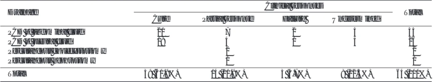
Table 3. Clinical Response in the US-guided Drainage Procedures
Drainage Clinical responses
Cure Partial response Failure Undetermined Total
PCD of abdominal fluid 21 7 2 5 35
PCD of pleural fluid 18 4 1 3 26
Percutaneous cholecystostomy 2 02
Percutaneous nephrostomy 1 01
Total 39 (60.9%) 14 (21.9%) 3 (4.7%) 8 (12.5%) 64 (100%)
penetration, thus providing a reliable indication of suc- cessful puncture. The absence of hyperechoic lines in US images suggests that the needle has shifted away from the center of the probe. Therefore, hyperechoic lines at the tip of the needle are a useful sign, providing confirmation of successful bile duct puncture (7). Using these methods, we can perform US-guided PCD at the bedside more safely and effectively.
A few reports have described bedside drainage proce- dures using US alone. Crass and Karl drained abdominal abscesses at the bedside in three patients who had un- stable septic shock and were undergoing resuscitation, prohibiting their transfer to the radiology department (8). McGahan performed ten aspirations and six drainage procedures with US guidance in an ICU (9). US guidance was successful in all of the aspiration proce- dures, while catheter malpositioning occurred in one pa- tient and required repositioning under fluoroscopic guidance. In another case, a chest tube was inserted by a surgeon. McGahan et al. performed 32 percutaneous drainage procedures with portable US guidance, mostly for thoracentesis or abdominal paracentesis (10). The failure rate was less than 7%. We also had a high techni- cal success rate with US guidance (98.5%), and only one case resulted in failure.
Civardi et al. (11) performed US-guided PCD proce- dures for abdominal abscesses and fluid collection in 50 patients. These procedures resulted in complete recov- ery in 70% of cases, partial success in 20%, and failure in 10%. Lambiase et al. (12) retrospectively reviewed the percutaneous drainage of 335 abscesses in 323 con- secutive patients. The overall cure rate was 62.4%, with a failure rate of 8.95%. The overall complication rate was 9.8%. The recent report by Civardi et al. (4) was the largest study of US-guided PCD of abdominal abscesses.
They reported 886 cases of US-guided PCD from eight clinical institutions and observed an overall cure rate of 90.4%, temporization in 6.1% of cases, and failure in 3.6%. The frequency of complications was 6.6% and there were no drainage-related deaths. Our drainage procedure resulted in complete response in 60.9% of cases, partial response in 21.9%, and failure in 4.7%
(Table 3). The overall complication rate in our study was 13.8%, while the final cure rate was relatively low, be- cause many of our patients were critically ill (Table 1).
Eleven patients (28.2%) in the study population died, or were discharged because they were deemed irremedia- ble, over the course of the study period.
Image-guided percutaneous catheter drainage of
pleural fluid collection is considered the mainstay treat- ment for patients with a diagnostic thoracentesis demonstrating a frank empyema or a complicated para- pneumonic effusion (13). Success is measured by com- plete evacuation of the pleural fluid, so that pulmonary symptoms resolve and sepsis is avoided. Complications are uncommon and can be avoided by meticulous imag- ing and planning, approaching the fluid over the top of a rib, and avoiding the intra-mammary vessels and deep mediastinal structures. Possible complications include bleeding secondary to injury of the intercostal vessels and pneumothorax (13).
The management of acute cholecystitis remains con- troversial, especially in terms of the ability of critically ill hospitalized patients to undergo surgery and the tim- ing of the surgical intervention. Some believe surgical cholecystectomy should be delayed until medical thera- py is instituted. Percutaneous cholecystostomy is a low- risk interventional procedure (14) and is useful in long- term conservative management, either as a cure for very poor surgical candidates or as a temporizing treat- ment until surgical cholecystectomy can be performed (15). In our series, two percutaneous cholecystostomies were performed without complication, such as bile peri- tonitis or bile leakage. The catheters were removed 5 and 8 days following the initial drainage, respectively, at which point an additional surgical procedure was used to cure the condition.
Catheter placement can be performed using two dif- ferent techniques: the trocar or Seldinger techniques (2).
The trocar technique uses a catheter mounted over a stiffening cannula that has a sharp inner stylet, which are inserted into the collected fluid as a unit. The stylet is removed to allow fluid aspiration and confirm the lo- cation of the catheter tip, and then the cannula is re- moved, allowing the distal loop of the catheter to form, securing the catheter in the collected fluid. This tech- nique is best suited for large or superficial fluid collec- tion.
In the Seldinger or guide-wire exchange technique, a puncture needle is inserted into the fluid and a guide- wire is advanced into the fluid through the needle. The needle is then removed, and the guide-wire is used as an anchor and guide, as progressively larger dilators are passed over the guide-wire to prepare for the passage of a catheter and cannula assembly (or simply a catheter).
A catheter is then passed over the guide-wire, and the guide-wire is removed, allowing the distal loop of the catheter to form, securing it in the fluid collection.
Some investigators have suggested that the Seldinger technique is more suitable for large abscess cavities and that the trocar technique is more suitable for small cavi- ties (9, 16). In this study, all of the catheters were placed using the Seldinger technique, regardless of the size of the cavity.
In conclusion, percutaneous drainage procedures per- formed at the bedside with only US guidance are safe and effective in unstable ICU patients. Our relatively low clinical success rate was probably due to the morbid clinical state of the patients, rather than to any limitation in the efficiency of the procedure.
References
1. McGahan JP. Invasive ultrasound principles (biopsy, aspiration, and drainage). In McGahan JP, Goldberg BB, eds. Diagnostic Ultrasound: A Logical Approach. Philadelphia, U.S.A.: Lippincott- Raven, 1998;39-75
2. Caspers JM, Reading CC, McGahan JP, et al: Ultrasound-guided biopsy and drainage of the abdomen and pelvis. In Rumack CM, Wilson SR, Charboneau JW, eds. Diagnostic ultrasound, 2nd. St Louis, U.S.A.: Mosby, 1997;599-628
3. Beagle GL. Bedside diagnostic ultrasound and therapeutic ultra- sound-guided procedures in the intensive care setting. Crit Care Clin 2000;16:59-81
4. Civardi G, Di Candio G, Giorgio A, et al. Ultrasound guided percu- taneous drainage of abdominal abscesses in the hands of the clini- cian: a multicenter Italian study. Eur J Ultrasound 1998;8:91-99 5. Bergin D, Pappas JN, Hwang JJ, Sheafor DH, Paulson EK.
Echogenic polymer coating: does it improve needle visualization in sonographically guided biopsy? AJR Am J Roentgenol 2002;178:
1188-1190
6. Gottlieb RH, Robinette WB, Rubens DJ, Hartley DF, Fultz PJ, Violante MR. Coating agent permits improved visualization of biopsy needles during sonography. AJR Am J Roentgenol 1998;171:
1301-1302
7. Tamada K, Tomiyama T, Wada S, et al. Hyperechoic lines as a sonographic confirmatory sign during percutaneous transhepatic biliary drainage. Abdom Imaging 2001;26:39-42
8. Crass JR, Karl R. Bedside drainage of abscesses with sonographic guidance in the desperately ill. AJR Am J Roentgenol 1982;139:183- 185
9. McGahan JP. Aspiration and drainage procedures in the intensive care unit: percutaneous sonographic guidance. Radiology 1985;
154:531-532
10. McGahan JP, Anderson MW, Walter JP. Portable real-time sono- graphic and needle guidance systems for aspiration and drainage.
AJR Am J Roentgenol 1986;147:1241-1246
11. Civardi G, Fornari F, Cavanna L, Sbolli G, Di Stasi M, Buscarini L.
Ultrasonically guided percutaneous drainage of abdominal fluid collections: a long-term study of its therapeutic efficacy.
Gastrointest Radiol 1990;15:245-250
12. Lambiase RE, Deyoe L, Cronan JJ, Dorfman GS. Percutaneous drainage of 335 consecutive abscesses: results of primary drainage with 1 year follow up. Radiology 1992;184:167-179
13. Klein JS, Schultz S, Heffner JE: Interventional radiology of the chest: image-guided percutaneous drainage of pleural effusions, lung abscess, and pneumothorax. AJR Am J Roentgenol 1995;164:
581-588
14. Lee MJ, Saini S, Brink JA, et al. Treatment of critically ill patients with sepsis of unknown causes: value of percutaneous cholecys- tostomy. AJR Am J Roentgenol 1991;156:1163-1166
15. Werbel GB, Nahrwold DL, Joehl RJ, Vogelzang RL, Rege RV.
Percutaneous cholecystostomy in the diagnosis and treatment of acute cholecystitis in the high-risk patient. Arch Surg 1989;124:782- 785
16. Browning PD, McGahan JP, Gerscovich EO. Percutaneous chole- cystostomy for suspected acute cholecystitis in the hospitalized pa- tient. J Vasc Interv Radiol 1993;4:531-538
대한방사선의학회지 2004;50:167-174
집중치료실에서 시행한 초음파 유도하 경피적 배액술의 유용성1
1아주대학교 의과대학 진단방사선과학교실
2연세대학교 의과대학 진단방사선과학교실
3건양대학교 의과대학 진단방사선과학교실
강두경・원제환・김재근・이광훈2・김지형3
목적:집중치료실에서 치료를 요하는 위중한 환자에서 초음파 유도하에 시행한 경피적배액술의 효과와 안전성에 대하 여 알아보고자 한다.
대상과 방법:집중치료실에 입원한 39명의 환자에서 총 65예의 초음파 유도하 경피적배액술을 시행하였으며 복강내 액
체 저류의 경피적배액술이 35예, 흉수의 경피적배액술이 27예, 경피적 담낭조루술이 2예, 경피적 신루설치술이 1예 였 다. 임상적 반응은‘완전(complete response)’, ‘부분적(partial response)’, ‘실패(failure)’, ‘미결 (undetermined)’
로 분류하였다.
결과:기술적인 성공율은 98.5 %로 65예중 단지 1예에서 실패하였다. 합병증은 9예(13.8%)에서 있었으나 시술과 연 관된 임상적 상태의 악화나 사망은 없었다. 임상적 반응은‘완전’이 39예(60.9%), ‘부분적’이 14예(21.9%), ‘실패’
가 3예(4.7 %), ‘미결’이 8예(12.5%) 이었다.
결론:집중치료실에서 초음파 유도하에서 시행한 경피적배액술은 중재시술실로 이동하기 힘든 위중한 환자에게 안전 하면서도 효과적인 시술 방법이다.