59
New Virulence Factors of Enterohemorrhagic Escherichia coli (EHEC) O157:H7 in Dairy Food Processing
Yong-Il Moon1, Sangnam Oh2, Mi Ri Park2, Seok Jun Son2, Gwang-woong Go3, Minho Song4, Sejong Oh5, Sae Hun Kim6 and Younghoon Kim2
1Dept. of Animal Source Foods, Woosuk University, Wanju 565-701, Korea
2BK21 Plus Graduate Program, Department of Animal Science and Institute of Agricultural Science & Technology, Chonbuk National University, Jeonju 561-756, Korea
3Dept. of Food and Nutrition, Kookmin University, Seoul 136-702, Korea
4Dept. of Animal Science and Biotechnology, Chungnam National University, Daejeon 305-764, Korea
5Division of Animal Science, Chonnam National University, Gwangju 500-757, Korea
6Division of Food Bioscience and Technology, College of Life Sciences and Biotechnology, Korea University, Seoul 136-701, Korea
Abstract
Enterohemorrhagic Escherichia coli (EHEC) O157:H7 is well-characterized as an important food-borne pathogen worldwide and causes human diseases such as diarrhea, hemorrhagic colitis, and hemolytic uremic syndrome (HUS) by producing shiga-like toxin (Stx). It has been reported that a number of dairy foods, including cheese, can act as the source of EHEC O157:H7 infections. In addition to the toxicity of Stx, recently it has been indicated that EHEC O157:H7 possesses virulence factors related to attachment, quorum sensing, and biofilms. Moreover, these novel virulence factors might become critical points to be considered in the future production of food derived from animals. Here, we review the evidences that support these insights on new virulence factors and discuss the potential mechanisms mediating the pathogenesis of EHEC O157:H7 in the dairy food industry.
Keywords: dairy foods, EHEC O157:H7, attachment, quorum sensing, biofilms, virulence
* Corresponding author: Younghoon Kim, Dept. of Animal Science, Chonbuk National University, Jeonju 561-756, Korea.
Tel: +82-63-219-5265, Fax: +82-62-270-2612, E-mail: ykeys2584
@jbnu.ac.kr
Enterohemorrhagic Escherichia coli (EHEC) O157:H7 is a high-virulence microorganism found in ground beef and milk products. Therefore, ruminants serve as critical reservoirs for the transfer of this pathogen to humans (Griffin et al., 1991). Through these routes, EHEC O157:H7 becomes a causative agent of hemorrhagic colitis (HC), hemolytic uremic syndrome (HUS), and thrombocytopenic purpura (TTP). Gene- rally, symptoms for HC include bloody diarrhea and stomach cramps. Similarly, HUS causes bloody diarrhea, acute renal failure, and kidney failure (Ruggenenti et al., 1998). Further- more, TTP has been shown to result in neurological disorders
and kidney failure (Ruggenenti et al., 1998).
Notably, EHEC O157:H7 has been associated with a number of food-borne outbreaks (Hudson et al., 1997). EHEC O157:H7 leads to a characteristic pathology in large intestinal epithelial cells known as attaching and effacing (A/E) lesion.
The Locus of Enterocyte Effacement (LEE) region is a pathogenicity island that encodes specific proteins involved in the formation of A/E lesions (Sperandio et al., 2002).
Since 1982, food-borne outbreaks of EHEC O157:H7 have occurred in several areas in Japan, Europe, and the United States (Bean et al., 1996).
A variety of other foods, such as ready-to-eat fruit, salad, and apple cider have also been involved in outbreaks of EHEC O157:H7 as well as animal recourses foods from
bovine (Mead et al., 1999). Typically, human diseases due to EHEC O157:H7 are linked with the consumption of food or water that has been contaminated with virulent EHEC O157:H7-containing bovine feces. The ability of EHEC O157:H7 to survive in a low pH environment, as well as in refrigerated products, makes it an organism of concern especially because the infective dose for EHEC O157:H7 is very low (less 100 cells; Spika et al., 1986). In 2003, 6,584 samples of ground beef products were analyzed and 20 samples (0.3%) were found to be positive for EHEC O157: H7 (FSIS, 2004).
Fortunately, human diseases resulting from EHEC O157:H7 significantly declined in 2003, which otherwise was steady from 1996 to 2002 (MMWR, 2004). This was probably the result of an FSIS notice issued in October 2002 for the manufacturers of raw ground beef products that they must reassess their Hazard Analysis Critical Control Points (HACCP) plans regarding this pathogen (MMWR, 2004). Presently, beef processing plants do not distribute lots of raw ground beef unless microbiological tests performed at the plant give negative results for EHEC O157:H7 (MMWR, 2004).
Milk and dairy foods are equally important and potential sources for human EHEC infections. In 1999, over 11% of the total number of reported cases of infection caused by EHEC O157:H7 in England were due to dairy products (Vernozy- Rozand et al., 2005). Moreover, recent outbreaks of EHEC O157:H7 occurred among consumers of five Southwestern states in the United States in 2010 with a total of 41 patients. Notably, a majority of patients reported consuming complimentary samples of aged Gouda type cheese produced with raw-milk (McCollum et al., 2012). Therefore, given that many regional or local cheese specialties throughout Europe are manufactured from unpasteurized milk, there is growing concern that these products may pose a threat for transmitting pathogens such as EHEC O157:H7 to the con- sumer. Here, we will review the novel virulence factors of EHEC O157:H7 in regards to dairy food safety, focusing on their specific characteristics and interactions with the host as well as target genes clusters.
1. Shiga-like toxin and EHEC virulence
It was well-established that shiga-like toxin (Stx) produced by EHEC O157:H7 is the main virulence factor for human EHEC infections. Interestingly, EHEC O157:H7 strains have acquired bacteriophages encoding two component AB-5 toxins.
Moreover, there are several variants of shiga toxins with Stx1 being virtually identical to the enterotoxin produced by Shigella dysenteriae type 1 (Smith et al., 2002). On the other hand, multiple variants of Stx2 have been characterized in- cluding Stx2c, d, e, f, and g. Among them, Stx2 and Stx2c have significantly strong correlation with the human infection (Bidet et al., 2005). Structural studies of Stx1 and Stx2 have identified key points to distinguish the differences in their structure (Fraser et al., 2004).
Importantly, the five B-subunits direct cell-binding specificity and allow translocation of the A-subunit into the cell. In the case of Stx1 and Stx2/2c, the B-subunit binds to the gly- colipid receptor Gb3/CD77 (Lingwood, 1996), driving the entry of the A-subunit, which is then cleaved to produce an A1 peptide with N-glycosidase activity that represses protein synthesis via cleavage or degradation of the 28S ribosomal RNA. One possibility why cattle colonized with EHEC O157:H7 do not exhibit clinical symptoms compared to the potentially devastating impact due to colonization in humans is the difference between the Gb3 receptor distribution in humans and cattle (Pruimboom-Brees et al., 2000). Gb3/CD77 exists on human endothelial cells (which line blood vessels), but this is not the case in cattle. Thus, any toxin reaching these cells does not have the same hemorrhagic consequences in both hosts. Significantly, Gb3/CD77 is present on human kidney glomerular cells, thus, causing severe damage to this organ as a critical outcome of human EHEC infection, such as HUS (Adler and Bollu, 1998). In humans, trafficking of Stx from the site of infection in the gastrointestinal (GI) tract occurs, at least in part, by associating with granulocytes that infiltrate in response to the infection (te Loo et al., 2001).
Conversely, cattle do have some Gb3/CD77-positive cells that are present in intestinal crypts and this possibly prevents the entry of Stx (Hoey et al., 2002). Therefore, while both humans and cattle are colonized by EHEC O157, the diffe- rence in Gb3 receptor distribution in the hosts acts as the key determining factor for severe outbreaks in human.
2. Attachment and EHEC virulence
As stated, EHEC O157:H7 colonizes the GI tract and produces a potent Stx, resulting in HC and HUS in a sig- nificant proportion of infected people (Nataro and Kaper, 1998). Interestingly, the EHEC O157:H7 causes A/E lesion, a histopathological and characteristic formation on the intestinal
epithelial cells. This lesion is characterized by the destruction of microvilli and the rearrangement of the cytoskeleton to form pedestal-like structures that cradle the bacteria individually.
The gene cluster involved in the formation of the A/E lesion exists on a pathogenicity island termed the Locus of Enterocyte Effacement (LEE) region (Elliot et al., 1998). The LEE region contains (1) sep and esc genes encoding a type III secretion system (Jarvis et al., 1995); (2) the eae gene encodes an adhesion protein, named intimin, that is responsible for the initial bacterial attachment to the surface of epithelial cells (Jerse and Kaper, 1991); (3) the espABD genes that encode extracellular proteins involved in the type III secretion system. This includes EspABD, which is recognized as an important component that facilitates pore formation on the epithelia cell surface and thus completes the delivery system of proteins from the EHEC O157:H7 into the cytoplasm of the host cell (Knutton et al., 1998); (4) the tir gene encoding the translocated intimin receptor, a partner for intimin (Kenny et al., 1997); and (5) the ler gene (LEE-encoded regulator) that encodes a positive regulator of LEE genes (Mellies et al., 1999). Based on the sequence analysis results of the conserved sequence of LEE for EHEC O157:H7, 41 open reading frames (ORFs) were identified that are highly con- served at the levels of DNA and protein for the type III secretion genes, but were more complex compositions for the esp, eae, and tir genes partially. In addition, the majority of the LEE genes were described to be in five primary polycistronic operons identified as LEE1 through LEE4 and tir (Elliot et al., 1999).
3. Quorum Sensing and EHEC virulence
Quorum sensing (QS) system is a critical cell-to-cell sig- naling mechanism that refers to the ability of bacteria to sense or respond to chemical signal molecules, known as autoinducers (AIs), under culture conditions in a dose depen- dent manner. Importantly, these AIs can be produced and detected by bacteria of the same, or different, species. When an AI concentration reaches a concentration-dependent threshold, the neighboring microorganisms sense and respond to this chemical molecule, resulting in an alteration of their specific gene expression. This characteristic phenomenon allows bac- teria to act as a collective unit, as opposed to individual cells performing individual functions.
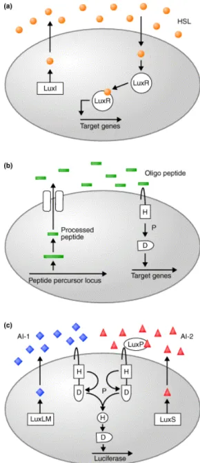
Until now, the QS circuit has been a well-established feature
Fig. 1. Several typical QS systems among bacteria. (a) In most Gram-negative bacteria, LuxI-type enzymes catalyze the formation of species-specific HSLs (circles), which are detected by LuxR-type transcriptional regulators.
(b) Oligopeptides (bars) mediate quorum sensing in Gram-positive bacteria, and two-component phosphory- lation cascades are used for signal transmission. (c) V.
harveyi quorum sensing circuit. LuxLM synthesizes AI-1 (diamonds) and LuxS is required for production of AI-2 (triangles). LuxP is the periplasmic AI-2- binding protein. Signal transduction occurs by two- component phosphorelay. In (b) and (c), the conserved phosphorylation sites on the two- component proteins are indicated as H (histidine) and D (aspartate). P denotes phosphorylation (adapted from Xavier and Bassler, 2003).
of the bioluminescent bacterium Vibrio harveyi (Fig. 1). V.
harveyi makes a typical Gram-negative-type homoserine lactone (HSL) AI, identified as AI-1. Additionally, V. harveyi produces and detects a second AI, identified as AI-2. This alternate type of AI is produced by a remarkably wide variety of Gram-negative and Gram-positive bacteria, and in every case production requires a protein called LuxS. Unlike for AI-1, the biosynthetic pathway and chemical intermediates in AI-2 production, and possibly the AI-2 molecule itself, are identical in all AI-2-producing bacteria studied to date.
These findings have led to the important possibility that AI-2 is a ‘universal’ signal molecule, which functions in inter-species bacteria-to-bacteria communication (Xavier and Bassler, 2003) as compared to AI-1, which functions in intra-species signaling. The structure of V. harveyi AI-2 was recently determined; the molecule is a novel furanosyl borate diester with no similarity to other AIs. Interestingly, EHEC O157:H7 also produces AI-2 in some environmental conditions.
Notably, EHEC O157:H7 could generate another AI named AI-3, whose synthesis depends on the presence of LuxS.
These uncharacterized AIs may indicate the possibility of inter-kingdom communication molecules between the bacteria and host signaling systems (Sperandio et al., 2003).
For bacterial pathogenesis, QS has been suggested to aid the disease process by allowing bacteria such as EHEC O157:H7 to appropriately modulate expression of virulence factors that might stimulate an immune response before the infection has progressed and occurred. As described by De Kievit et al. (2001), utilizing the mechanism of bacterial communication via QS, food-borne pathogens can accumulate a high cell density before virulence factors are fully ex- pressed, and in so doing, the bacteria are able to make a united assault and produce an abundance of virulence factors to overwhelm the host’s defense mechanisms. This simple concept may be suitable to explain the important role of QS in extra-intestinal infections where the pathogen encounters low levels of competing microbiota. On the contrary, under the complex microbial populations of the GI tract, QS may play other roles in illnesses. As described below, QS has been hypothesized to signal transcription of EHEC O157:H7 virulence factors once they arrive in the intestinal tract, as well as to signal transcription of Vibrio environmental survival com- ponents following expression of virulence factors but before environmental release via fecal samples. Therefore, although
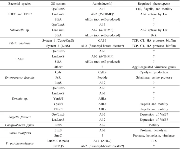
QS has been described in numerous food-borne pathogens of the GI tract, in many cases the role of QS in the patho- genesis of the outbreaks caused by these organisms is still unclear. However, a number of reports for QS in enteric pathogens have been reported that have yielded new insights into the mechanisms of outbreaks and illnesses (Table 1).
The recent discovery of QS in human pathogens has led to considerable interest in developing new concepts for the- rapeutic applications to interfere with these signaling mole- cules involved in bacteria-bacteria communications. Indeed, instead of antibiotics that kill pathogenic bacteria directly, a compound that inhibits the QS mechanism could be em- ployed to suppress the specific regulation of the virulence genes responsible for the food-borne disease. Such a QS- based approach is particularly promising to combat increased resistance of some bacteria to conventional antibiotics, and encouraging results in animal models have been obtained with P. aeruginosa (Camara et al., 2002) and S. aureus (Mayville et al., 1999). However, the development of anti-QS therapy may primarily influence normal microbiota, such as the high numbers of commensal microorganisms present in the GI tract.
4. Biofilms and EHEC virulence
A biofilm is a sessile-type microbial community consisting of cells that are irreversibly attached to a surface via an extracellular polymeric matrix produced by the cells (Donlan and Costerton, 2002). Currently, there is a generalized model for biofilm formation and development. First, microorganisms attach to an abiotic (i.e. glass, steel, or plastic) or biotic surface (i.e. teeth, mucus layer); second, microorganisms transform from planktonic status to a tightly immobilized status; third, the microcolony is formed and a continuous consortium of microorganisms build a three dimensional archi- tecture of biofilm, which is considered a mature biofilm;
finally, biofilms release and disperse cells into the environ- ment to attach to new locations (Branda et al., 2005). The most apparent difference between biofilm and planktonic styles of microorganisms is the mobility and architecture of the microorganisms. Importantly, the biofilm has distinct architectural features (Stoodley et al., 2002), such as water channels that allow the diffusion of necessary nutrients, including oxygen and carbon for cell growth.
Under environmental conditions, many bacteria thrive better
Table 1. Summary of quorum sensing systems described in enteric bacteria (adapted from Kaper and Sperandio, 2005)
Bacterial species QS system Autoinducer(s) Regulated phenotype(s)
EHEC and EPEC
Qse/LuxS AI-3 TTS, flagella, and motility
Lsr/LuxS AI-2 (R-THMF)a AI-2 uptake by Lsr
SdiA AHLs (not self-produced) ?
Salmonella sp.
Qse/LuxS AI-3 ?
Lsr/LuxS AI-2 (R-THMF) AI-2 uptake by Lsr
SdiA AHLs (not self-produced) Rck
Vibrio cholerae System 1 (CqsA/CqsS) CAI-1 TCP, CT, HA protease, biofilm
System 2 (LuxS) AI-2 (furanosyl-borate diester?) TCP, CT, HA protease, biofilm
EAEC
Qse/LuxS AI-3 ?
Lsr/LuxS AI-2 (R-THMF) ?
SdiA AHLs (not self-produced) ?
Other? ? AggR-regulated virulence genes
Enterococcus faecalis
Cyls CyILs Cytolysin production
FsR Peptide Gelatinase, serine protease
LuxS AI-2 ?
Yersinia sp.
Qse/LuxS AI-3 ?
Lsr/LuxS AI-2 ?
YenR/I AHLs ?
YpsR/I AHLs Flagella and motility
YtbR/I AHLs Flagella and motility
Shigella flexneri Qse/LuxS AI-3 Expression of VirB?
Lsr/LuxS AI-2 Expression of VirB?
Campylobacter jejuni LuxS AI-2 Motility
Vibrio vulnificus LuxS AI-2 Protease, hemolysin
SmrC ? Protease, hemolysin, virulence
V. parahaemolyticus LuxMR (OpaR) AI-1 (AHL?) TTS
LuxPQS AI-2 (furanosyl-borate diester?) ?
a R-THMF, (2R,4S)-2-methyl-2,3,3,4-tetrahydroxytetrahyrofuran.
within biofilms rather than as free-living planktonic cells (Watnick and Kolter, 2000). Biofilms are found in diverse locations such as human dental plaques and the GI tract (Probert and Gibson, 2002), leaves and root surfaces of plants (Ramey et al., 2004), biocorrosion or biofouling of industrial surfaces (Videla and Herrera, 2005), and food processing equipment and facilities (Kim et al., 2006). Several advan- tages for living in biofilms are as follows: (1) biofilm cells can survive with limited nutrient resources, due to decreased bacterial growth rates in biofilms; (2) cells in the biofilm are less susceptible to harsh conditions, especially exposure to antibiotics and other antimicrobial agents, due to the
extracellular polymeric matrix of biofilms (Mah and O'Toole, 2001); (3) for pathogens, cells within an biofilm have a higher gene transfer rate than planktonic cells because they are in close contact with other cells (Hausner and Wuertz, 1999).
Some researchers have reported differential gene expression between sessile and planktonic cells (Resch et al., 2005;
Schembri et al., 2003). Early evidence of differential gene expression within a bacterial biofilms using DNA microarray technology, suggested that the expression of up to 38% of the E. coli bacterial genome might be affected by biofilm formation (Prigent-Combaret et al., 1999), although other
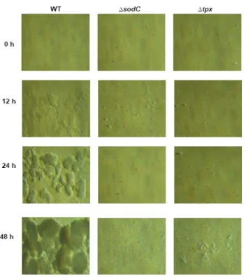
studies reported only a small proportion of the genome was affected (1 to 15%) (Schembri et al., 2003; Beloin et al., 2004). These studies have generated hope that it might be possible to identify a common universal gene-expression pattern within bacterial biofilms. In addition, our two dimen- sional gel electrophoresis showed only 26 genes (5.8%) were differentially up- or down-regulated under biofilm conditions among the 450 detected spots, with some genes encoding periplasmic oxidative defense proteins including copper, zinc superoxide dismutase (SodC), and thiol peroxidase (Tpx) were strongly induced in the EHEC O157:H7 biofilm culture conditions as compared to the planktonic cells (Kim et al., 2006) (Fig. 2).
Up to now, many approaches have been proposed to pre- vent bacterial biofilm formation. The most extensively studied is material surface-coating methods. An example is a photo- catalytic titanium dioxide (TiO2) surface, which has potential as a self-cleaning technology to reduce the adhesion of Deinococcus geothermalis cells (Raulio et al., 2005). Also, antibiotic coating systems are effective in interfering with
Fig. 2. Quantification of EHEH O157:H7 biofilm formation using glass capillary tubes and a continuous flow system.
The biofilms developed by wild-type, sodC, and tpx mutants were observed at different time points, as indicated (adapted from Kim et al., 2006).
biofilm formation, such as the application of ciprofloxacin- releasing polymer in blocking Staphylococcus epidermidis biofilms (Niemela et al., 2006). Application of antimicrobial compounds has also been employed in industry. For example, weak hydrogen peroxide-based disinfectants have had success in inhibiting biofilm formation in dental unit waterlines (Tuttlebee et al., 2002). Physical methods using ultrasound could improve the penetrations of gentamycin into biofilms of P. aeruginosa and E. coli (Carmen et al., 2004). Recently, a research report showed that an efficient way to prevent P.
aeruginosa biofilm formation is by releasing ciprofloxacin from specific coatings materials by ultrasonic control (Norris et al., 2005). Other novel approaches are also being examined, such as utilizing bacteriophages to degrade extracellular poly- meric matrix (Hughes et al., 1998). Interestingly, (5Z)-4-bromo- 5-(bromomethylene)-3-butyl-2(5H)-furanone (furanone), from the marine alga Delisea pulchra, also inhibits E. coli biofilm formation without inhibiting its growth (Ren et al., 2005).
Moreover, released exopolysaccharide (r-EPS) isolated from Lactoabcillus acidophilus could prevent biofilm formation by a wide range of Gram-negative and -positive pathogens including EHEC O157:H7 (Kim et al., 2009). These numerous applications may lead to the development of novel food-grade materials for selective control of microbial biofilms.
In conclusion, the presence of EHEC O157:H7 in milk used in the production of dairy foods including cheese poses a risk to consumers, even though it is present in low amounts.
Therefore, it is imperative to manufacture dairy foods with highest EHEC O157:H7 bacteriological safeguards by taking these new virulence factors into consideration.
Acknowledgments
This work was supported by the Business for Cooperative R&D between Industry, Academy, and Research Institute funded Korea Small and Medium Business Administration (Y.I.M.) and the High Value-Added Food Technology Deve- lopment Program of the Korea Institute of Planning and Evaluation for Technology in Food, Agriculture, Forestry, and Fisheries (iPET), and the Ministry for Food, Agriculture, Forestry, and Fisheries of Republic of Korea (313036-03- 2-HD020; Y.K.).
References
1. Adler, S. and Bollu, R. 1998. Glomerular endothelial cell injury mediated by Shiga-like toxin-1. Kidney Blood Pressure Res. 21:13-21.
2. Bean, N. H., Goulding, J. S., Lao, C. and Angulo, F.
J. 1996. Surveillance for food borne-disease outbreaks- United States, 1988-1992. MMWR. 45(SS-5):1-65.
3. Bidet, P., Mariani-Kurkdjian, P., Grimont, F., Brahimi, N., Courroux, C., Grimont, P. and Bingen, E. 2005.
Characterization of Escherichia coli O157:H7 isolates causing haemolytic uraemic syndrome in France. J. Med.
Microbiol. 54:71-75.
4. Branda, S. S., Vik, S., Friedman, L. and Kolter, R. 2005.
Biofilms: The matrix revisited. Trends Microbiol. 13:20-26.
5. Camara, M., Williams, P. and Hardman, A. 2002. Con- trolling infection by tuning in and turning down the volume of bacterial small-talk. Lancet Infect Dis. 2:667- 676.
6. Carmen, J. C., Nelson, J. L., Beckstead, B. L., Runyan, C. M., Robison, R. A., Schaalje, G. B. and Pitt, W. P.
2004. Ultrasonic-enhanced gentamicin transport through colony biofilms of Pseudomonas aeruginosa and Escherichia coli. J. Infect. Chemother. 10:193-199.
7. D'Amico, D. J., Druart, M. J. and Donnelly, C. W.
2010. Behavior of Escherichia coli O157:H7 during the manufacture and aging of Gouda and Stirred-curd Cheddar cheeses manufactured from raw milk. J. Food Prot. 73:
2217-2224.
8. De Kievit, T. R., Gillis, R., Marx, S., Brown, C. and Iglewski, B. H. 2001. Quorum-sensing genes in Pseudomonas aeruginosa biofilms: Their role and expression patterns.
Appl. Environ. Microbiol. 67:1865-1873.
9. Donlan, R. M. and Costerton, J. W. 2002. Biofilms:
survival mechanisms of clinically relevant microorganisms.
Clin. Microbiol Rev. 15:167-193.
10. Elliot, S., Wainwright, L. A., McDaniel, T. K., MacNamara, B., Donnenberg, M. and Kaper, J. B. 1998. The complete sequence of the locus of enterocyte effacement (LEE) from enteropathogenic Escherichia coli E2348/69. Mol.
Microbiol. 28:1-4.
11. Food Safety and Inspection Service (FSIS). 2004. “Micro- biological results of raw ground beef products analyzed for Escherichia coli O157:H7. [Internet WWW].” ADDRESS:
http://www.fsis.usda.gov/OPHS/ecoltest/tables1.htm.
Accessed 12 Jan 2005.
12. Fraser, M. E., Fujinaga, M., Cherney, M. M., Melton- Celsa, A. R., Twiddy, E. M., O’Brien, A. D. and James, M. N. G. 2004. Structure of Shiga toxin type 2 (Stx2) from Escherichia coli O157:H7. J. Biol. Chem. 279:27511- 27517.
13. Griffin, P. M. and Taxue, R. V. 1991. The epidemiology of infections caused by E. coli O157:H7, other entero- hemorrhagic E. coli, and the associated hemolytic uremic syndrome. Epidemiol. Rev. 13:60-98.
14. Hausner, M. and Wuertz, S. 1999. High rates of con- jugation in bacterial biofilms as determined by quantitative in situ analysis. Appl. Environ. Microbiol. 65:3710-3713.
15. Hoey, D. E., Currie, C., Else, R. W., Nutikka, A., Lingwood, C. A., Gally, D. L. and Smith, D. G. E. 2002.
Expression of receptors for verotoxin 1 from Escherichia coli O157 on bovine intestinal epithelium. J. Med. Microbiol.
51:143-149.
16. Hudson, L. M., Chen, J., Hill, A. R. and Griffiths, M.
W. 1997. Bioluminescence: A rapid indicator of E. coli O157:H7 in selected yogurt and cheese varieties. J.
Food Prot. 60:891-897.
17. Hughes, K. A., Sutherland, I. W. and Jones, M. V. 1998.
Biofilm susceptibility to bacteriophage attack: The role of phage-borne polysaccharide depolymerase. Microbiology 144:3039-3047.
18. Jarvis, K. G., Giron, J. A., Jerse, A. E., McDaniel, T.
K., Donnenberg, M. S. and Kaper, J. B. 1995. Entero- pathogenic Escherichia coli contains a specialized secretion system necessary for the export of proteins involved in attaching and effacing lesion formation. Proc. Natl. Acad.
Sci. USA. 92:7996-8000.
19. Jerse, A. E. and Kaper, J. B. 1991. The eae gene of enteropathogenic Escherichia coli encodes a 94-kilodalton membrane protein, the expression of which is influenced by the EAF plasmid. Infect. Immun. 59:4302-4309.
20. Kaper, J. B. and Sperandio, V. 2005. Bacterial cell-to-cell signaling in the gastrointestinal tract. Infect. Immun.
73:3197-209.
21. Kenny, B., DeVinney, R., Stein, M., Reinscheid, D. J., Frey, E. A. and Finlay, B. B. 1997. Enteropathogenic E.
coli (EPEC) transfers its receptor for intimate adherence into mammalian cells. Cell 91:511-520.
22. Kim, Y., Lee, Y., Kim, S. H., Yeom, J., Kim, B. S., Oh, S., Park, S., Jeon, C. O. and Park, W. 2006. Role of periplasmic antioxidant enzymes (superoxide dismutase and thiol peroxidase) of the Shiga toxin-producing Esche- richia coli O157:H7 in the formation of biofilms. Pro- teomics 6:6181-6193.
23. Kim, Y., Oh, S. and Kim, S. H. 2009. Released exopo- lysaccharide (r-EPS) produced from probiotic bacteria reduce biofilm formation of enterohemorrhagic Escherichia coli O157:H7. Biochemical and Biophysical Research Communications 379:324-329.
24. Knutton, S., Adu-Bobie, J., Bain, C., Phillips, A. D., Dougan, G. and Frankel, G. 1997. Down regulation of intimin expression during attaching and effacing entero- pathogenic Escherichia coli adhesion. Infect Immun. 65:
1644-1652.
25. Lingwood, C. A. 1996. Role of verotoxin receptors in pathogenesis. Trends Microbiol. 4:147-153.
26. Mah, T. F. and O'Toole, G. A. 2001. Mechanisms of biofilm resistance to antimicrobial agents. Trends Microbiol.
9:34-39.
27. Mayville, P., Ji, G., Beavis, R., Yang, H., Goger, M., Novick, R. P. and Muir, T. W. 1999. Structure-activity analysis of synthetic autoinducing thiolactone peptides from Staphylococcus aureus responsible for virulence.
Proc. Natl. Acad. Sci. USA. 96:1218-1223.
28. Mead, P. S., Slutsker, L., Dietz, V., McCaig, L. F., Bresee, J. S., Shapiro, C., Griffin, P. M. and Tauxe, R. V. 1999.
Food-related illness and death in the United States. Emerg.
Infect. Dis. 5:607-625.
29. Mellies, J. L., Elliot, S. J., Sperandio, V., Donnenberg, M. S. and Kaper, J. B. 1999. The Per regulon of entero- pathogenic Escherichia coli: Identification of regulatory cascade and a novel transcriptional activator, the locus of enterocyte effacement (LEE)-encoded regulator (Ler).
Mol. Microbiol. 33:1176-1189.
30. Miller, S. T., Xavier, K. B., Campagna, S. R., Taga, M.
E., Semmelhack, M. F., Bassler, B. L. and Hughson, F.
M. 2004. Salmonella typhimurium recognizes a chemically distinct form of the bacterial quorum-sensing signal AI-2. Mol. Cell 15:677-687.
31. Morbidity and Mortality Weekly Report (MMWR). 2003.
“Preliminary FoodNet Data on the incidence of food- borne illnesses - Selected sites, United States, 2002.”
[Internet WWW]. ADDRESS: http://www.cdc.gov/mmwr/
preview/mmwrhtml/mm5215a4.htm. Accessed 25 March 2005.
32. Nataro, J. P. and Kaper, J. B. 1998. Diarrheagenic Esche- richia coli. Clin. Microbiol. Rev. 11:142-201.
33. Niemela, S. M., Ikaheimo, I., Koskela, M., Veiranto, M., Suokas, E., Tormala, P., Waris, T., Ashammakhi, N. and Syrjala, H. 2006. Ciprofloxacin-releasing bio- absorbable polymer is superior to titanium in preventing Staphylococcus epidermidis attachment and biofilm for- mation in vitro. J. Biomed. Mater Res. B. Appl. Biomater.
76:8-14.
34. Norris, P., Noble, M., Francolini, I., Vinogradov, A. M., Stewart, P. S., Ratner, B. D., Costerton, C. W. and Stoodley, P. 2005. Ultrasonically controlled release of ciprofloxacin from self-assembled coatings on poly (2-hydroxyethyl methacrylate) hydrogels for Pseudomonas aeruginosa biofilm prevention. Antimicrob. Agents Chemother. 49:
4272-4279.
35. Prigent-Combaret, C., Vidal, O., Dorel, C. and Lejeune, P. 1999. Abiotic surface sensing and biofilm dependent regulation of gene expression in Escherichia coli. J.
Bacteriol. 181:5993-6002.
36. Probert, H. M. and Gibson, G. R. 2002. Bacterial biofilms in the human gastrointestinal tract. Curr. Issues Intest.
Microbiol. 3:23-27.
37. Pruimboom-Brees, I. M., Morgan, T. W., Ackermann, M. R., Nystrom, E. D., Samuel, J. E., Cornick, N. A.
and Moon, H. W. 2000. Cattle lack vascular receptors for Escherichia coli O157:H7 Shiga toxins. Proc. Natl.
Acad. Sci. USA 97:10325-10329.
38. Ramey, B. E., Koutsoudis, M., von Bodman, S. B. and Fuqua, C. 2004. Biofilm formation in plant-microbe asso- ciations. Curr. Opin. Microbiol. 7:602-609.
39. Raulio, M., Pore, V., Areva, S., Ritala, M., Leskela, M., Linden, M., Rosenholm, J. B., Lounatmaa, K. and Salkinoja- Salonen, M. 2005. Destruction of Deinococcus geothermalis biofilm by photocatalytic ALD and sol-gel TiO(2) surfaces.
J. Ind. Microbiol. Biotechnol. 1-8.
40. Ren, D., Zuo, R., Gonzalez Barrios, A. F., Bedzyk, L.
A., Eldridge, G. R., Pasmore, M. E. and Wood, T. K.
2005. Differential gene expression for investigation of Escherichia coli biofilm inhibition by plant extract ursolic acid. Appl. Environ. Microbiol. 71:4022-4034.
41. Resch, A., Rosenstein, R., Nerz, C. and Gotz, F. 2005.
Differential gene expression profiling of Staphylococcus aureus cultivated under biofilm and planktonic conditions.
Appl. Environ. Microbiol. 71:2663-2676.
42. Ruggenenti, P. and Remuzzi, G. 1998. Pathophysiology and management of thrombotic microangiopathies. J.
Nephrol. 11:300-310.
43. Schembri, M. A., Kjaergaard, K. and Klemm, P. 2003.
Global gene expression in Escherichia coli biofilms.
Mol. Microbiol. 48:253-267.
44. Smith, D. G. E., Naylor, S. W. and Gally, D. L. 2002.
Consequences of EHEC colonisation in humans and cattle. Int. J. Med. Microbiol. 292:169-183.
45. Sperandio, V., Li, C. C. and Kaper, J. B. 2002. Quorum sensing Escherichia coli regulator A: A regulator of the lysR family involved in the regulation of the locus of enterocyte effacement pathogenecity island in entero- hemorrhagic E. coli. Infect. Immun. 70:3085-3093.
46. Spika, J. S., Parsons, J. E., Nordenberg, D., Wells, J. G., Gunn, R. A. and Blake, P. A. 1986. Hemolytic uremic syndrome and diarrhea associated with E. coli O157:H7 in a day care center. J. Pediatr. 109:287-291.
47. Stoodley, P., Sauer, K., Davies, D. G. and Costerton, J.
W. 2002. Biofilms as complex differentiated communities.
Annu. Rev. Microbiol. 56:187-209.
48. te Loo, D. M. W. M., Heuvelink, A. E., de Boer, E., Nauta, J., van der Walle, J., Schroder, C., van Hinsbergh, V. W. M., Chart, H., van der Kar, N. C. A. J. and van
der Heuvel, L. P. W. J. 2001. Verocytotoxin binding to polymorphonuclear leukocytes among households with children with hemolytic uremic syndrome. J. Infect. Dis.
184:446-450.
49. Tuttlebee, C. M., O'Donnell, M. J., Keane, C. T., Russell, R. J., Sullivan, D. J., Falkiner, F. and Coleman, D. C.
2002. Effective control of dental chair unit waterline biofilm and marked reduction of bacterial contamination of output water using two peroxide-based disinfectants.
J. Hosp. Infect. 52:192-205.
50. Vernozy-Rozand, C., Mazuy-Cruchaudet, C., Bavai, C., Montet, M. P., Bonin, V., Dernburg, A. and Richard, Y.
2005. Growth and survival of Escherichia coli O157:H7 during the manufacture and ripening of raw goat milk lactic cheeses. Int. J. Food Microbiol. 105:83-88.
51. Videla, H. A. and Herrera, L. K. 2005. Microbiologically influenced corrosion: Looking to the future. Int. Microbiol.
8:169-180.
52. Watnick, P. and Kolter, R. 2000. Biofilm, city of microbes.
J. Bacteriol. 182:2675-2679.
53. Xavier, K. B. and Bassler, B. L. 2005. Regulation of up- take and processing of the quorum-sensing autoinducer AI-2 in Escherichia coli. J. Bacteriol. 187:238-248.
54. Xavier, K. B. and Bassler, B. L. 2003. LuxS quorum sensing: More than just a numbers game. Curr. Opin.
Microbiol. 6:191-197.
(Received 1 March 2015 / Accepted 26 March 2015)