Veterinary Science
http://dx.doi.org/10.4142/jvs.2012.13.2.139
Received: 26 Apr. 2011, Revised: 24 Jul. 2011, Accepted: 6 Jan. 2012
Original Article
*Corresponding author: Tel: +55-21-2564-8985; Fax: +55-21-2564-8985; E-mail: [email protected]
ⓒ 2012 The Korean Society of Veterinary Science.
This is an Open Access article distributed under the terms of the Creative Commons Attribution Non-Commercial License (http://creativecommons.org/licenses/by-nc/3.0) which permits unrestricted non-commercial use, distribution, and reproduction in any medium, provided the original work is properly cited.
Molecular characterization of Escherichia coli O157:H7 strains isolated from different sources and geographic regions
Adriana Hamond Regua-Mangia
1,*, Alice Gonçalves M. Gonzalez
2, Aloysio M. F. Cerqueira
3, João Ramos C.
Andrade
41
Departamento de Ciências Biológicas, Fundação Oswaldo Cruz, CEP 21041-210, Rio de Janeiro, Brazil
Departamento de
2Bromatologia e
3Microbiologia e Parasitologia, Universidade Federal Fluminense, CEP 24220-008, Rio de Janeiro, Brazil
4
Departamento de Microbiologia e Imunologia, Universidade do Estado do Rio de Janeiro, CEP 20550-030, Rio de Janeiro, Brazil
Escherichia (E.) coli serotype O157:H7 is a globally
distributed human enteropathogen and is comprised of microorganisms with closely related genotypes. The main reservoir for this group is bovine bowels, and infection mainly occurs after ingestion of contaminated water and food. Virulence genetic markers of 28 O157:H7 strains were investigated and multilocus enzyme electrophoresis (MLEE) was used to evaluate the clonal structure. O157:H7 strains from several countries were isolated from food, human and bovine feces. According to MLEE, O157:H7 strains clustered into two main clonal groups designated A and B. Subcluster A1 included 82% of the O157:H7 strains exhibiting identical MLEE pattern. Most enterohemorrhagic E. coli (EHEC) O157:H7 strains from Brazil and Argentina were in the same MLEE subgroup. Bovine and food strains carried virulence genes associated with EHEC pathogenicity in humans.
Keywords: enterohemorrhagic Escherichia coli, molecular characterization, O157:H7, virulence
Introduction
Enterohemorrhagic Escherichia (E.) coli (EHEC) belong to a subset of Shiga toxin (Stx)-producing E. coli (STEC) serotypes that are associated with bloody diarrhea and hemolytic uremic syndrome [23]. Epidemiologically, O157:H7 is considered a highly pathogenic serotype responsible for severe human diseases, usually occurring as outbreaks, and as a prototypical EHEC [8,9,23]. Besides expressing Stx, EHEC produces a variety of virulence
factors encoded by lysogenic toxigenic bacteriophages along with virulence chromosomal pathogenicity islands and plasmids [2-4,11,23]. Unlike other E. coli pathotypes, cattle are the main reservoirs of EHEC, and are the major source of direct and indirect transmission [23].
Genotypic and phenotypic methods have been employed to investigate the clonal relationship and virulence properties of O157:H7 strains. Epidemiological and phylogenetic studies have revealed that E. coli O157:H7 strains form a clonal complex of related genotypes found worldwide and exhibit differences in virulence characteristics or transmissibility between lineages [10,13,16,21,25,26,28]. Among the molecular systems used, isoenzyme electrophoresis known as multilocus enzyme electrophoresis (MLEE) has been traditionally used for phylogenetic studies of E. coli and provides important information about the virulence background of particular lineages [16,18-20,24].
Isoenzymes play an essential role in housekeeping metabolic activities which make them useful for conducting evolutionary studies of many bacterial populations, particularly among strains belonging to E. coli pathotypes.
Isoenzymes also participate in specific steps of the infection
processes of many pathogens, including colonization,
persistency, and invasion of the host tissue [14]. In the
present study, isoenzymatic type and virulence potential
were examined in order to evaluate the diversity and clonal
relationships of a set of E. coli O157:H7 strains isolated
during distinct periods and from different sources. Of
particular interest was the comparison between O157:H7
strains recovered from healthy cattle in Brazil and strains
isolated from humans.
Table 1. General characteristics of Escherichia coli O157 strains Pathotype
(strain)
Geographic area
Brazilian locations*
/year isolated Serotype Zymovar MLEE group (subgroup)
Virulence genotype
†Source of isolation EHEC
YB20 B1/1 B18/1 GC148
581/1 579//3 691/1 902/1 1728/1 2004/1 1770/1 2228/1 111/1 440/1 678/1 685/1 872/1 869/1 956/1 356/1 ME18/96 TRA 71/96
145/98 ME135/99
E30138 E40705 EDL931 EDL933 EPEC
116I
Brazil Brazil Brazil Brazil Brazil Brazil Brazil Brazil Brazil Brazil Brazil Brazil Brazil Brazil Brazil Brazil Brazil Brazil Brazil Brazil Argentina Argentina Argentina Argentina
UK UK USA USA Brazil
SP/1997 RJ/1997 RJ/1997 RJ/1997 RJ/1999 RJ/1999 RJ/1999 RJ/1999 RJ/2000 RJ/2000 RJ/2000 RJ/2000 RS/2001 RS/2001 RS/2001 RS/2001 RS/2001 RS/2001 RS/2001 RS/2001 1996 1996 1998 1999
RS
O157:H7 O157:H7 O157:H7 O157:H7 O157:H7 O157:H7 O157:H7 O157:H7 O157:H7 O157:H7 O157:H7 O157:H7 O157:H7 O157:H7 O157:H7 O157:H7 O157:H7 O157:H7 O157:H7 O157:H7 O157:H7 O157:H7 O157:H7 O157:H7 O157:H7 O157:H7 O157:H7 O157:H7 O157:H-
5 1 3 6 1 1 2 1 1 1 1 1 1 1 1 1 1 1 1 1 1 1 1 1 1 1 1 4 7
B (B2) A (A1) A (A3) B (B2) A (A1) A (A1) A (A2) A (A1) A (A1) A (A1) A (A1) A (A1) A (A1) A (A1) A (A1) A (A1) A (A1) A (A1) A (A1) A (A1) A (A1) A (A1) A (A1) A (A1) A (A1) A (A1) A (A1) B (B1)
C
stx1, stx2c, eae γ, ehlyA stx2c, eae γ, ehlyA stx2c, eae γ, ehlyA stx2, eae γ, ehlyA stx2c, eae γ, ehlyA stx2c, eae γ, ehlyA stx2c, eae γ, ehlyA,
stx2c, eae γ, ehlyA stx2c, eae γ, ehlyA stx2c, eae γ, ehlyA stx2c, eae γ, ehlyA stx2c, eae γ, ehlyA stx2c, eae γ, ehlyA stx2c, eae γ, ehlyA stx2c, eae γ, ehlyA stx2c, eae γ, ehlyA stx1, stx2c, eae γ, ehlyA stx1, stx2c, eae γ, ehlyA
stx2c, eae γ, ehlyA stx1, stx2, eae γ, ehlyA stx2, stx2c, eae γ, ehlyA stx2, stx2c, eae γ, ehlyA
stx2c, eae γ, ehlyA stx1, stx2, stx2c, eae γ, ehlyA
stx2, stx2c, eae γ, ehlyA stx1, eae γ, ehlyA stx1, stx2, eae γ, ehlyA stx1, stx2, eae γ, ehlyA
eae α
Bovine Bovine Bovine Bovine Bovine Bovine Bovine Bovine Bovine Bovine Bovine Bovine Bovine Bovine Bovine Bovine Bovine Bovine Bovine Bovine Human Food Bovine Human Human Human Human Food Human
*RJ: Rio de Janeiro, SP: São Paulo, RS: Rio Grande do Sul. †stx1: Shiga toxin type 1, stx2: Shiga toxin type 2, stx2c: Shiga toxin type 2c, eae γ: gamma intimin, eae α: alpha intimin, ehlyA: enterohemolysin. MLEE: multilocus enzyme electrophoresis, EHEC: enterohemorrhagic E. coli, EPEC: enteropathogenic E. coli.
Materials and Methods Bacterial strains
The study included 28 E. coli O157:H7 strains isolated from diverse geographic regions. These included Argentina (n = 4), UK (n = 2), USA (n = 2), and three Brazilian states [São Paulo (n = 1), Rio Grande do Sul (n = 8), and Rio de Janeiro (n = 11)]. Among the total number of strains, two were isolated from food, five from patients, and 21 from bovine feces (Table 1). Brazilian bovine strains were isolated between 1999 and 2001 from distinct farms located in 13 rural counties. Only one strain from each farm was included in this analysis. EHEC strains from Argentina, the
UK, and USA were kindly donated by Dr. Marta Rivas (Administración Nacional de Laboratorios e Institutos de Salud, Argentina) and Dr. Sylvia Scotland (Central Public Health Laboratory, UK). EPEC strain 116I (O157:H-, sorbitol fermenting, stx-, eae+, EAF+, bfpA+) was included in this study for comparison [17] .
Detection of virulence genes
Multiplex PCR assays were used to detect Shiga toxin
(stx), intimin (eae), and enterohemolysin (E-hlyA) genes as
previously described [5,25].
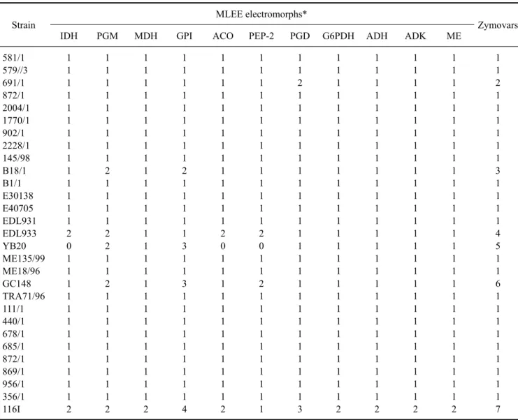
Table 2. Numerical representation of electromorphs and zymovars according to MLEE analysis of E. coli O157 strains
Strain MLEE electromorphs*
Zymovars
IDH PGM MDH GPI ACO PEP-2 PGD G6PDH ADH ADK ME
581/1 579//3 691/1 872/1 2004/1 1770/1 902/1 2228/1 145/98 B18/1 B1/1 E30138 E40705 EDL931 EDL933 YB20 ME135/99 ME18/96 GC148 TRA71/96 111/1 440/1 678/1 685/1 872/1 869/1 956/1 356/1 116I
1 1 1 1 1 1 1 1 1 1 1 1 1 1 2 0 1 1 1 1 1 1 1 1 1 1 1 1 2
1 1 1 1 1 1 1 1 1 2 1 1 1 1 2 2 1 1 2 1 1 1 1 1 1 1 1 1 2
1 1 1 1 1 1 1 1 1 1 1 1 1 1 1 1 1 1 1 1 1 1 1 1 1 1 1 1 2
1 1 1 1 1 1 1 1 1 2 1 1 1 1 1 3 1 1 3 1 1 1 1 1 1 1 1 1 4
1 1 1 1 1 1 1 1 1 1 1 1 1 1 2 0 1 1 1 1 1 1 1 1 1 1 1 1 2
1 1 1 1 1 1 1 1 1 1 1 1 1 1 2 0 1 1 2 1 1 1 1 1 1 1 1 1 1
1 1 2 1 1 1 1 1 1 1 1 1 1 1 1 1 1 1 1 1 1 1 1 1 1 1 1 1 3
1 1 1 1 1 1 1 1 1 1 1 1 1 1 1 1 1 1 1 1 1 1 1 1 1 1 1 1 2
1 1 1 1 1 1 1 1 1 1 1 1 1 1 1 1 1 1 1 1 1 1 1 1 1 1 1 1 2
1 1 1 1 1 1 1 1 1 1 1 1 1 1 1 1 1 1 1 1 1 1 1 1 1 1 1 1 2
1 1 1 1 1 1 1 1 1 1 1 1 1 1 1 1 1 1 1 1 1 1 1 1 1 1 1 1 2
1 1 2 1 1 1 1 1 1 3 1 1 1 1 4 5 1 1 6 1 1 1 1 1 1 1 1 1 7
*IDH: isocitrate dehydrogenase, PGM: phosphoglucomutase, MDH: malate dehydrogenase, GPI: glucose phosphate isomerase, ACO:
aconitase, PEP-2: leucyl amino peptidase, PGD: 6-phosphogluconate dehydrogenase, G6PDH: glucose-6-phosphate dehydrogenase, ADH:
alcohol dehydrogenase, ADK: adenylate kinase, ME: malic enzyme.