165
광바이오센서를 이용한 우유 및 유제품의 식중독균 신속검출법
최은영․장진희․홍성욱․김소영․배효주․박범영․오미화*
농촌진흥청 국립축산과학원
Rapid Detection Methods for Food-Borne Pathogens in Milk and Dairy Products using an Optical Biosensor
Eun-Young Choi, Jin Hee Chang, Sung Wook Hong, So-Young Kim, Hyo Ju Bae, Beom Young Park and Mi-Hwa Oh*
National Institute of Animal Science, RDA, Suwon 441-706, Korea
Abstract
Milk and dairy products are not only excellent foods for humans, providing plentiful varied nutrients, but are also a good medium for detrimental food-borne pathogens. Although the food safety field has stabilized due to standardization of food processing, such as the hazard analysis critical control point (HACCP), outbreaks and cases caused by food-borne pathogens still occur at high rates. In approximately 30% of cases, the disease-causing pathogenic organism is undetermined. Recently, a biosensor was developed that has a simple and fast response and overcomes the problems of conventional methods such as cultivation, immuno-assay, polymerase chain reaction, and microarray. Due to the high selectivity and sensitivity of optical biosensors, it is a suitable method for the immediate detection of food-borne pathogens in milk and dairy products.
Keywords: Rapid detection, food-borne pathogen, optical biosensor
* Corresponding author: Mi-Hwa Oh, National Institute of Animal Science, RDA, Suwon 441-350, Korea. Tel: +82-31-290-1689, Fax:
+82-31-290-1697, E-mail: [email protected]
서 론
광바이오센서는 분석물질과 인식분자 사이의 특정한 상 호작용을 광학적인 신호로 유도한 후, 이를 전기적인 신호 로 변화시켜 검출을 가능하게 하는 장치이다(Turner, 2000).
광바이오센서는 저분자 펩타이드 분자에서부터 미생물에 이르기까지 다양한 타겟물질을 검출할 수 있다(Baeumner
et al., 2003).
전통적인 검출방법과 비교하여, 바이오센서는특히 숙련된 기술노동력을 필요하지 않고, 높은 민감성과 선택성이 있으므로 식품산업체에서 손쉽게 적용이 가능한 장점이 있다.
우유와 유제품은 생산과정 중에 다양한 식중독균에 오염 될 수 있으며(Table 1), 미생물이 증식하기에 필요한 영양
성분으로 구성되어 있어 위생학적 주의가 필요하다. 또한 우유내 미생물의 오염은 축사 규모와 위생상태, 착유 방법 과 계절에 따라 달라져 예측하기가 어려우며, 우유를 저온 살균(pasteurization)한 후에도 Listeria monocytogenes와 같 은 미생물이 잔류하여 최종제품에 검출이 된 보고가 있다 (Wong, 1998).
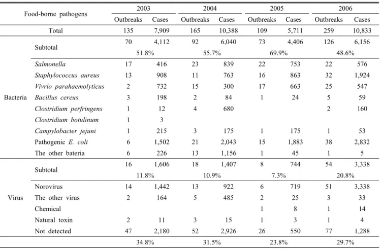
세계보건기구(World Health Organization, WHO)는 식중 독을 “식품 또는 물의 섭취에 의해 발생되었거나, 발생된 것으로 생각되는 감염성 또는 독소형 질환”으로 규정 및 관리하고 있다. 식중독은 식품의 안전성 확보를 위한 시스 템이 운영되고 있음에도 여전히 인간에게 가장 높은 빈도 로 발생하는 위해 요인이다. 식중독 사건이 발생하였을 때, 그 원인 병원체를 찾지 못하는 경우가 발생건수의 상당비 율(약 30%)을 차지하고 있다(Table 2). 이에 따라 신속하고 정확한 역학조사와 원인 병원체 검출기술의 개발에 힘쓸 필요가 있다(권과 이, 2007).
Table 1. Surveys on the isolation of Camphylobacter jejuni, Shiga toxin-producing Escherichia coli, Listeria monocytogenes and Salmonella spp. from bulk tank milk
Foodborne pathogens
Isolation rate
(%) Reference
Campylobacter jejuni
12.3 Rohrbach et al. (1992) 0.5 Steele et al. (1997)
9.2 Jayarao and Henning (2001) Shiga toxin-
producing Escherichia coli
0.9 Steele et al. (1997)
3.8 Jayarao and Henning (2001) 0.8 Murinda et al. (2002b)
Listeria monocytogenes
4.6 Jayarao and Henning (2001) 12.6 Hassan et al. (2000) 1.0 Waak et al. (2002) 6.5 Van Kessel et al. (2004)
Salmonella spp.
6.1 Jayarao and Henning (2001) 1.5 Hassan et al. (2000) 2.6 Van Kessel et al. (2004) Note: Oliver et al. (2005)
Table 2. Recent food-borne disease outbreaks and cases in Korea according to cause of occurrence
Food-borne pathogens 2003 2004 2005 2006
Outbreaks Cases Outbreaks Cases Outbreaks Cases Outbreaks Cases
Total 135 7,909 165 10,388 109 5,711 259 10,833
Bacteria
Subtotal 70 4,112 92 6,040 73 4,406 126 6,156
51.8% 55.7% 69.9% 48.6%
Salmonella 17 416 23 839 22 753 22 576
Staphylococcus aureus 13 908 11 763 16 863 32 1,924
Vivrio parahaemolyticus 2 732 15 300 17 663 25 547
Bacillus cereus 3 198 2 84 1 24 5 59
Clostridium perfringens 1 12 4 680 2 160
Clostridium botulinum 1 3
Campylobacter jejuni 1 215 3 175 1 175 1 53
Pathogenic E. coli 6 1,502 21 2,043 15 1,883 38 2,832
The other bateria 6 226 13 1,156 1 45 1 5
Virus
Subtotal 16 1,606 18 1,407 8 744 54 3,338
11.8% 10.9% 7.3% 20.8%
Norovirus 14 1,442 13 922 6 719 51 3,338
The other virus 2 164 5 485 2 25 3 33
Chemical 1 8 1 14
Natural toxin 2 11 3 15 1 3 1 4
Not detected 47 2,180 52 2,926 26 550 77 1,288
34.8% 31.5% 23.8% 29.7%
Note: KFDA, http://www.mfda.go.kr/open_content/foodpoison
이에 본 총설에서 우유 및 유제품에서 식중독균 신속검 출에 적용이 가능한 바이오센서에 대해 전반적으로 살펴보 고자 한다.
본 론
1. 바이오센서
바이오센서는 적절한 변환장치를 가지고 생물학적으로 항 체, 파지, 압타머, 단일가닥 DNA와 같은 인지(derived mole- cular recognition) 분자를 통합하는 분석장치이다. 일반적인 바이오센서는 검출영역, 변환영역, 해독영역의 세 부분으로 구성되어 있다(Fig. 1). 검출영역은 인지분자와 타겟물질과 의 상호작용으로 신호를 발생시키는 부분이며, 보통 항체가 사용된다. 변환영역은 일반적으로 광학적, 전기화학적, 온 도적, 자성적 등의 방법으로 이루어지며, 분석물질과 인식 분자 사이의 특정한 상호작용에 대한 적절한 전기적 또는 광학적인 신호를 만드는 부분에 해당된다(Iqbal et al., 2000, Turner, 2000). 마지막으로 해독영역은 그 반응을 기록하게 된다. 즉, 바이오센서는 생물학적 상호작용을 쉽게 측정되
Fig. 1. Schematic of a biosensor. The three main parts of the biosensors are : (1) detector that has the recognition molecules and interacts with the target antigen to produce a signal, (2) transducer that converts the signal received from the detector to a useful electronic output and (3) the display or readout system to record the response (Harsh and Raj, 2013).
고 기록할 수 있는 전기적인 신호로 변화시키는 장치이다.
2. 바이오센서를 이용한 식중독균 검출
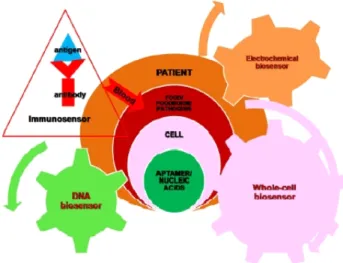
식중독균 검출을 위한 전통적인 방법들은 매우 민감하고 값이 저렴하지만, 노동력과 변이가 가능한 박테리아의 특 성에 의존한다(Biswas, 2005). 또한 결과에 대한 혈청학적 확인뿐만 아니라, 동시에 전배양, 선택배양, 생화학적 스크 리닝 중 하나는 추가로 더 진행할 필요가 있다(Vunrcrzant and Pllustoesser, 1987). 반면, 바이오센서는 검출대상이 다 양하므로(Stroot et al., 2012; Bae et al., 2004) 식중독균의 현장분석 및 동시분석을 가능하게 한다. 식중독균 확인을 위한 바이오센서 검출 가능 대상을 살펴보면 균의 특정 염 기서열, 대사물질 변화, 균과 진핵세포와의 상호작용 분석 등이 있다(Fig. 2). 이로 인해 착유에서부터 제품제조와 소 비자에게로의 유통시간이 짧은 유제품의 경우, 바이오센서 를 적용시 완제품뿐만 아니라, 원재료에 존재 가능한 식중 독균에 대해 검출할 수 있는 장점이 있다.
Fig. 2. Multiple biosensor configurations for pathogen detection (Arora et al., 2011).
3. 광바이오센서
바이오센서 중 광바이오센서는 선택성과 민감도 때문에 산업에 가장 많이 적용되고 있다. 특히 섬유광학 바이오센서 는 가장 먼저 상용되었으며, 기본원리는 병원성균 또는 독소 가 센서 표면에 부착하면 특정 레이저에 의해 자극(excitation) 되어 광신호가 발생하고, 이를 감지하게 되는 것이다(Bhunia, 2008; Taitt et al., 2005).
오염물질(Willardson et al., 1998; Tschmelak et al., 2004), 독소(Bae et al., 2004), 그리고 병원성 세균(Baeumner et al., 2003)을 빠르게 검출하는 광바이오센서 연구는 항체에 붙는 형광물질 연구와 변환기 개발 연구가 주로 진행되고 있다. 광 바이오센서에서 항체에 붙은 형광물질은 병원성 세균을 검 출하는데 도움을 주는데, 일반적으로 사용하는 형광 마커는 fluorescein isothiocyanate(FITC)이며, 란타나이드(lanthanide) 등이 있다(Selvin, 2002). 이 형광물질은 다른 검출반응 PCR, ELISA에도 적용이 가능하다. 형광물질이 다양한 항체와 결합 된 섬유광학 바이오센서가 botulinum toxin, Staphylococcal enterotoxin, E. coli O157:H7, Listeria, Salmonella 등을 검 출하기 위해 개발되고 있다. 또한 광변환기(Optical transducer) 는 직접적 또는 간접적인 병원성 세균과 독소 검출에 적용 가능하다. 광변환기는 목표하는 분석물질이 변환기 표면에 고정화된 수용기에 붙었을 때 나타나는 굴절률이나 두께 같은 분광광도 기준으로 미세한 변화를 감지할 수 있다(Li
et al., 2004).
광바이오센서는 형광과 표면 플라즈몬 공명으로 분류된다(Fig. 3).
Fig. 3. Classification of biosensors.
4. 형광 기반 광바이오센서
형광은 형광발색단(fluorophore)이 빛이나 전자기 방사선 (electro-magnetic radiation)을 흡수하고, 가시범위에서 빛을 내보내는 방출현상이다. 때때로, 방출된 방사선은 들뜬 상 태의 방사선보다 짧은 파장 또는 같은 파장에 있기도 한다.
여기에서 후자를 공명형광(resonance fluorescence)이라고 한다. 이것은 방사 에너지의 손실과 함께 일어나기 때문에, 형광의 빛은 항상 흡수되는 빛보다 더 긴 파장에 존재하며, 이때의 에너지 차이를 ‘stokes shift’라 한다(Velusamy et al., 2010; Lazcka et al., 2007). 형광 샌드위치 측정법을 이용하 여 Listeria species가 생산한 내독소(endotoxin)를 10 ng/mL 까지 검출하였고(Leung et al., 2007; James et al., 1996),
Escherichia coli와 Staphylococcus aureus가 혼합되어 있는
상태에서는 동시에 102 CFU/mL까지도 검출능을 나타내었 다(Xeu et al., 2009). 프레넬 반사 분광기(fresnel reflection spectroscopy)에 결합한 형광으로 200 cells/mL 정도로 낮은 수준의 Legionella pneumolhilia를 검출하였다(Li et al., 2013).더 즉각적인 검출로는 포획항체 표적 검출(capture antibody targeted detection, CAT-FISH)을 통해 E. coli O157:H7과 S.
aureus를 각각 검출하였다(Stroot et al., 2012). 화학발광(a
chemi-luminiscence)기반 방법으로 0.017~1.6 EU/mL의 내 독소를 검출(Romaschin et al., 1998)하였고, 이후 2004년Hafnial alvei PCM 1186
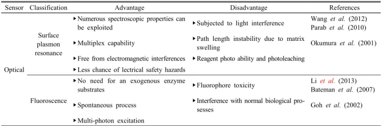
균주를 검출하였다고 보고하였다 (Hreniak et al., 2002). 녹색 형광성의 단백질(enhanced green fluorescent protein, EGFP)을 이용하여 세균성의 내독소를 검출하는 바이오센서도 등장하였다(Goh et al., 2002). 내독 소 농도 0.0005 EU/mL까지 검출 가능한 새로운 발광센서 는 돌연변이 루시페라아제(luciferase) 효소를 이용하였으며 (Noda et al., 2002), 전체적으로 새로운 비색적 형광센서는Table 3. Comparison in terms of general advantages and disadvantages of optical individual biosensor
Sensor Classification Advantage Disadvantage References
Optical
Surface plasmon resonance
‣Numerous spectroscopic properties can
be exploited ‣Subjected to light interference Wang et al. (2012) Parab et al. (2010)
‣Multiplex capability ‣Path length instability due to matrix
swelling Okumura et al. (2001)
‣Free from electromagnetic interferences ‣Reagent photo ability and photoleaching
‣Less chance of lectrical safety hazards
Fluoroscence
‣No need for an exogenous enzyme
substrates ‣Fluorophore toxicity Li et al. (2013)
Bateman et al. (2007)
‣Spontaneous process ‣Interference with normal biological pro-
sesses Goh et al. (2002)
‣Multi-photon excitation Note: Das et al. (2014)
색 변화를 통해 육안으로 270 pM까지 검출 가능하다고 보 고하였다(Lan et al., 2012).
5. 표면 플라즈몬 공명 기반 광바이오센서
표면 플라즈몬 공명(SPR)에 기반을 둔 변환은 얇은 필름 금속 표면 부근에서 구조적인 변화로 인한 굴절률에 따른 측 정 가능한 변화를 감지하는 표면민감성방법(surface sensitive method)이다. 프리즘 인터페이스에서, 높은 굴절률로부터 나온 빛이 임계각보다 더 큰 각도의 낮은 굴절률에 있는 인 터페이스와 만났을 때, 전반사(total internal reflection, TIR) 가 일어난다. 빛의 전반사는 표면장파가 새어나감으로 인 해 점점 커지는 플라즈몬 파장을 끌어낸다(Lotierzo et al., 2004). 표면 플라즈몬 공명을 이용한 바이오센서는 살모넬 라를 검출하는 것으로 이용되어져 왔다(Barlen et al., 2007).
Chlamydia trachomatis
병원성균 진단을 위하여 금 나노막대를 기반으로 한 광학 DNA 바이오센서가 보고되었다(Parab
et al., 2010).
바이오 리셉터로서 박테리오파지를 이용한 표면 플라즈몬 공명은 E. coli O157:H7과 항생제 내성 황색포 도상구균(methicillin-resistant Staphylococcus aureus)을 진 단하는데 성공적으로 사용되었다(Tawil et al., 2012). 자성 의 나노입자 분석법(magnetic nanoparticle assay, MNP)과 결합한 long-range surface plasmons(LRSPs)로 불리는 일반 적인 표면 플라즈몬 공명의 변형은 50 CFU/mL 정도의 E. coli O157:H7의 검출이 가능하였다(Wang et al., 2012). E. coli K-12 균주와 포자 및 단백질 등을 포함한 다양한 박테리아의 부산물, 그리고 바이러스들의 실시간 모니터링도 성공적으로 보고되었다(Zibaii et al., 2010; Bhatta et al., 2010). 표면 플라 즈몬 공명 바이오센서는 독점적으로 Bacillus thuringiensis의 δ-내독소의 스크리닝에 이용된다(Okumura et al., 2001).
결 론
식중독균 및 독소를 검출하는 전통적인 방법은 정확하지 만, 병원성 미생물 오염 시 증균 속도가 빠르고, 가식기간 이 짧은 유제품을 대상으로 하기에는 부족하다. 현재 민감 도와 선택성 개선 필요성에도 불구하고, 경제성, 휴대성 뿐 아니라 사전진단을 가능하게 하는 광바이오센서가 대안이 될 수 있을 것으로 보인다.
감사의 글
본 논문은 농촌진흥청 공동연구사업(과제번호: PJ009329) 의 지원에 의해 이루어진 것입니다.
참고문헌
1. Arora, P., Sindhu, A., Dilbaghi, N. and Chaudjury, A.
2011. Biosensors as innovative tools for the detection of food borne pathogens. Biosens. Bioelectron. 28:1-12.
2. Bae, Y. M., Oh, B. K., Lee, W., Lee, W. H. and Choi, J.
W. 2004. Detection of insulin-antibody binding on a solid surface using imaging ellipsometry. Biosens. Bioelectron.
20:895-902.
3. Baeumner, A. J., Cohen, R. N., Miksic, V. and Min, J.
2003. RNA biosensor for the rapid detection of viable
Escherichia coli in drinking water. Biosens. Bioelectron.
18:405-413.
4. Barlen, B., Mazumdar, S. D., Lezrich, O., Kampfer, P. and Keusgen, M. 2007. Detection of Salmonella by surface plasmon resonance. Sensors 7:1427-1446.
5. Bhunia, A. K. 2008. Biosensors and bio-based methods for the separation and detection of foodborne pathogens. Adv.
Food Nutr. Res. 54:1-44.
6. Biswas, A. K. 2005. Evaluation of export Buffalo meat for some chemical residues and microbial contaminants. Ph.D.
thesis. Indian Research Institue, Izatnagar, Bareilley.
7. Das, A. P., Kumar, P. S. and Swain, S. 2014. Recent advances in biosensor based endotoxin detection. Biosens. Bioelectron.
51:62-75.
8. Elaine, S., Robert, M. H., Frederick, J. A., Robert, V. T., Marc-Alain, W., Sharon, L. R., Jeffery, L. J. and Patricia, M. G. 2011. Foodborne illness acquired in the United States-major pathogens. Emerg. Infect. Dis. 17:7-15.
9. Goh, Y. Y., Frecer, V., Ho, B. and Ding, J. L. 2002. Rational
desing of green fluorescent protein mutants as biosensor for bacterial endotoxin. Protein Eng. Des. Sel. 15:493-502.
10. Hreniak, A., Maruszewski, K., Rybka, J., Galmian, A. and Czyewski, J. 2004. A luminescence endotoxin biosensor prepared by the sol-gel method. Optical Materials 26:
141-144.
11. Iqbal, S. S., Mayo, M. W., Bruno, J. G., Bronk, B. V., Batt, C. A. and Chambers, J. P. 2000. A review of molecular recognition technologies for detection of biological threat agents. Biosens. Bioelectron. 15:549-578.
12. James, E. A., Schmeltzer, K. and Ligler, F. S. 1996. De- tection of endotoxin using an evanescent wave fiber-optic biosensor. Appl. Biochem. Biotechnol. 60:189-202.
13. KFDA(http://www.kfda.go.kr/open_content/foodpoison/) 14. Lazcka, O., Campo, F. J. D. and Munoz, F. X. 2007.
Pathogen detection: A perspective of traditional methods and biosensors. Biosens. Bioelectron. 22:1205-1217.
15. Leung, A., Shankar, P. M. and Mutharasan, R. 2007. A review of fiber-optic biosensors. Sens. Actuators B. Chem.
125:688-703.
16. Li, Y., Dick, W. A. and Tuovinen, O. H. 2004. Fluore- scence microscopy for visualization of soil microorganisms- a review. Biol. Fert. Soils 39:301-311.
17. Lotierzo, M., Henry, O. Y. F., Piletsky, S., Tothill, I., Cullen, D., Kania, M., Hock, B. and Turner, A. P. F.
2004. Surface plasmon resonance sensor for domoic acid based on grafted imprinted polymer. Biosens. Bioelectron.
20:145-152.
18. Lu, L., Chee, G., Yamada, K. and Jun, S. 2013. Micro- fluidic characterization of specific membrane capacitance and cytoplasm conductivity of single cells. Biosens. Bio- electron. 42:492-495.
19. Noda, K., Goto, H., Murakami, Y., Ahmed, A. B. F. and Kuroda, A. 2010. Endotoxin assay by bioluminescence using mutant firefly luciferase. Anal. Biochem. 397:152- 155.
20. Okumura, S., Akao, T., Mizuki, E., Ohba, M. and Inouye, K. 2001. Screening of the Bacillus thuringiensis Cry1Ac δ-endotoxin on the artificial phospholipid monolayer incorporated with brush border membrane vesicles of
Plutella xylostella by optical biosensor technology. Methods
47:177-188.21. Parab, H. J., Jung, C., Lee, J. H. and Park, H. G. 2010.
A gold nanorod-based optical DNA biosensor for the
diagnosis of pathogens. Biosens. Bioelectron. 26:667-673.
22. Selvin, P. R. 2002. Principles and biophysical applications of lanthanide-based probes. Annu. Rev. Biophys. Biomol.
Struct. 31:275-302.
23. Stroot, J. M., Leach, K. M., Stroot, P. G. and Lim, D.
V. 2012. Capture antibody targeted fluorescence in situ hybridization (CAT-FISH): Dual labeling allows for increased specificity in complex samples. J. Microbiol.
Methods 88:275-284.
24. Taitt, C. R., Anderson, G. P. and Ligler, E. S. 2005. Evane- scent wave fluorescence biosensors. Biosens. Bioelectron.
20:2470-2487.
25. Tawil, N., Sacher, E., Mandeville, R. and Meunier, M.
2012. Surface plasmon resonance detection of E. coli and methicillin-resistant S. aureus using bacteriophages. Biosens.
Bioelectron. 37:24-29.
26. Turner, A. P. F. 2000. Biosensors - sense and sensitivity.
Science. 290:1315-1317.
27. Vunrcrzant, C. and Pllustoesser, D. F. 1987. Compeodium of methods for microbial examination of food, third ed.
American Public Health Association, New York.
28. Wang, J. 2002. Electrochemical nucleic acid biosensors.
Anal. Chim. Acta. 469:63-71.
29. Wang, Y., Knoll, W. and Dostalek, J. 2012. Long range surface plasmon resonance biosensors for magnetic nano- particle - Enhanced detection of bacterial pathogens. Anal.
Chem. 84:8345-8350.
30. Willardson, B. M., Wilkins, J. F., Rand, T. A., Schupp, J. M., Hill, K. K., Keim, P. and Jackson, P. J. 1998.
Development and testing of a bacterial biosensor for toluene-based environmental contaminants. Appl. Environ.
Microbiol. 64:1006-1012.
31. Wong, A. C. 1998. Biofilms in food processing environ- ments. J. Dairy Sci. 81:2765-2770.
32. Xue, X., Pan, J., Xie, H., Wang, J. and Zhang, S. 2009.
Fluorescence detection of total count of Escherichia coli and Staphylococcus aureus on water-soluble cdse quantum dots coupled with bacteria. Talanta. 77:1808-1813.
33. Zibaii, M. I., Kazemi, A., Latifi, H., Azar, M. K., Hosseini, S. M. and Ghezelaiagh, M. H. 2010. Measuring bacterial growth by refractive index tapered fiber optic biosensor.
J. Photochem. Photobiol. B. 101:313-320.
34. 권준욱, 이철헌. 2007 최근 우리나라 식중독 발생 현황 고찰. 대한의사협회지. 50:573-581.
(Received: November 5, 2013 / Accepted: November 25, 2013)