Study of tumor transglutaminase 2 expression in gallbladder cancer – Is it a novel predictor of survival?
Sameer Gupta1, Sudeep Garg1, Vijay Kumar1, Arun Chaturvedi1, Sanjeev Misra1,2, Naseem Akhtar1, Shiv Rajan1, Jatinder Kaur3, Manikandan Lakshmanan1, and Kavitha Jain1
1Department of Surgical Oncology, King George’s Medical University, Lucknow,
2AIIMS, Jodhpur, Rajasthan, 3Molecular Quest Healthcare Pvt. Ltd., Gurgaon, India
Backgrounds/Aims: Transglutaminase 2 (TG2) is known to be an important mediator of inflammation induced carcinogenesis pathway. Chronic inflammation is the most important causative factor in Gallbladder cancer (GBC) carcinogenesis. We ana- lyzed the expression of TG2 in GBC and its role as potential prognostic marker, first of its kind study. Methods: We analyzed TG2 expression in 100 cases of GBC and 28 cases of non-cancer gallbladder specimen (calculus cholecystitis). We studied TG2 expression in GBC in comparison to control group and evaluated its role as a potential prognostic marker.
Results: TG2 score (1-9) was calculated by multiplying percentage cytoplasmic expression (P) with intensity of ex- pression (I) in tumor cells. Positive TG-2 expression was observed in 62% of GBC patients compared to only 21%
(n=6) in control group (p=0.001). In curative resection subgroup (n=54), TG2 positive patients showed shorter disease free survival rate (p=0.04) and higher rate of recurrence (p=0.03) compared to TG2 negative patients. TG2 positive expression was observed in 15/16 of patients with recurrent disease. In palliative treatment subgroup, patients with strong TG2 positive expression had poorer disease specific survival (p=0.01) as compared to weakly positive group.
On multivariate analysis, lymph node status (p=0.03) and TG2 expression (p=0.037), were found to be significant pre- dictor of recurrence and eventual survival. Conclusions: Positive TG2 expression was related to higher recurrence rates post curative surgery, shorter disease free and overall survival and ultimately portended poor prognosis. It may be helpful in better prognostication and tailoring therapeutic approach for better management of GBC. (Ann Hepatobiliary Pancreat Surg 2020;24:460-468)
Key Words: Transglutaminase 2; Gallbladder cancer; Inflammation; Prognostic factor
Received: June 10, 2020; Revised: July 10, 2020; Accepted: July 12, 2020 Corresponding author: Sameer Gupta
Department of Surgical Oncology, King George’s Medical University (KGMU), Shah Mina Road, Lucknow 226003, India Tel: +91-9794361103, Fax: +91-522-2258991, E-mail: [email protected]
Copyright Ⓒ 2020 by The Korean Association of Hepato-Biliary-Pancreatic Surgery
This is an Open Access article distributed under the terms of the Creative Commons Attribution Non-Commercial License (http://creativecommons.org/
licenses/by-nc/4.0) which permits unrestricted non-commercial use, distribution, and reproduction in any medium, provided the original work is properly cited.
Annals of Hepato-Biliary-Pancreatic Surgery ∙ pISSN: 2508-5778ㆍeISSN: 2508-5859
INTRODUCTION
Gallbladder cancer (GBC) is the most common malig- nancy of the biliary tract and the fifth most common gas- trointestinal (GI) cancer.1 The disease is characterized by late onset of symptoms, advanced stage at presentation and a rapidly progressive disease with a median survival of 6 months in advanced disease.2 In early-stage disease, a 5-year survival rate up to 75% can be achieved if stage-adjusted treatment is given. Chronic calculous chol- ecystitis and gall stones are associated with GBC in 68 to 98% cases.3,4 Chronic inflammation induced by gall stones is recognized as an important factor in gallbladder carcinogenesis and cancer progression. The gallbladder
epithelium undergoes a recurrent cycle of gallstone in- duced damage and repair leading to an inflammatory envi- ronment that promotes carcinogenesis. Since no prog- nostic or predictive markers are known for GBC, we ana- lyzed the expression of TG2 and its role as novel prog- nostic marker in GBC. It plays an important role in in- flammation and is known to facilitate cancer growth and progression. Evaluation of TG2 expression in GBC pa- tients and its correlation with survival characteristics in GBC has not been studied till date.
TG2 is a trans-peptidase with a wide distribution in various tissues. It is known to mediate cross-linking of proteins and participate in signal transduction via activat- ing and hydrolyzing guanidine tri-phosphate (GTP) enzyme.
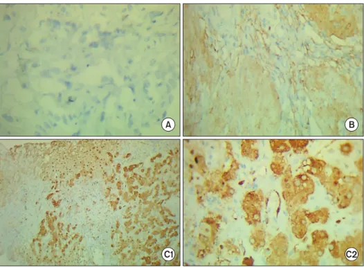
Fig. 1. Immunohistochemical TG2 expression in gallbladder cancer specimen (20×/40× mag- nification). (A) Negative expre- ssion (TG2 score <3), (B) weak- ly positive expression (TG2 score 3-6), (C1) strongly positive TG2 expression by tumor cells (TG2 score >6), (C2) strongly posi- tive TG2 expression shown by tumor cells with minimal cyto- plasmic staining seen in 40× mag- nification (TG2 score >6).
Table 1. Calculation of TG2 score based on cytoplasmic ex- pression and intensity of TG2 expression
Transglutaminase-2 (TG-2) score Cytoplasmic expression of
TG2 (P)
<30%=1 31-50%=2
>50%=3 Intensity of expression (I)- Nil to mild=1
Moderate=2 Strong=3 TG2 score=P×I (range-1-9)
It is also an active participant in promoting malignant cell mobility and invasion mainly through epithelial mesen- chymal transition (EMT) induction, and in inducing che- mo-resistance of cancer cells.5,6
Role of TG2 in various inflammatory processes like fibrosis, celiac disease, atherosclerosis and autoimmune disease are well known.7 Many studies have demonstrated a close link between inflammation and tumor.8 TG2 in- volved in chronic inflammation is known to play an im- portant role in tumorigenesis and cancer progression.9 Role of TG2 in cancer progression has been studied in various cancers including ovary,8 colorectal,10 breast,11 re- nal12 and lung.13 These studies showed that high TG2 ex- pression group had poorer overall survival rate than those in the low expression group. We aim to analyze the ex- pression of TG2 in GBC and its role as potential prog-
nostic marker, a first of its kind study.
MATERIALS AND METHODS
Patients
The present study evaluated TG2 expression in patients of GBC and the control group calculous cholecystitis (non-cancer Gallbladder) by immunohistochemistry (IHC).
We studied the correlation of molecular markers ex- pression of TG2 with clinco-pathological features of the tumor as histological type, grade of differentiation, lymph node status, level of invasion of the tumor (T stage), dis- ease free and disease specific survival. Patients presenting in our Department from October 2013-October 2017 (4 years) with histological diagnosis of GBC and tissue blocks available for immunohistochemical analysis were considered. Both prospective and retrospective patients were included in the study. Non-cancer gallbladder speci- mens were obtained from patients operated in General Surgery department for gallstone disease. Informed con- sent was obtained from all individual participants included in the study in a prescribed format. This study was grant- ed Ethical approval by Institutional Ethics Committee.
IHC analysis was performed at Molecular Quest Healthcare Pvt. Ltd. (MolQ) Gurgaon, Haryana. Tumor stage was determined according to the pTNM staging
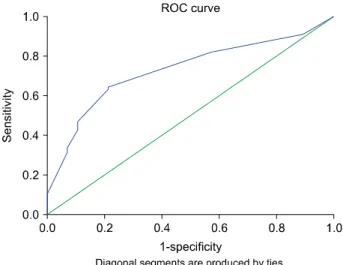
Fig. 2. Receiver operator characteristic (ROC) curve for de- termining cut off value of TG2 in GBC patients (maximum sensitivity (69%) and specificity (79%) were obtained with cut-off value of 2.7 (AUC=0.72, 95% confidence interval:
0.63-0.82, p<0.001).
Table 2. General characteristic of GBC patients (n=100) General characteristics
(GBC group) Distribution
M:F 1:3
Mean age (years) 50 years
Rural:urban distribution 73:27 Dietary habits
Vegeterian 75
Non-vegeterian 25
Cholelithiasis
Present 70
Absent 30
Curative surgery subgroup 54 (stage I-IVA) Palliative treatment subgroup 46 (stage IVB) Median follow up (months) 21 months (13-52) guideline published by the 2017 American Joint Committee
on Cancer (8th Edition AJCC). Three monthly follow up was done by clinical examination and USG abdomen in operated patient group while symptomatic improvement (like pain, appetite, jaundice)was assessed in palliative pa- tient group for response assessment.
Immunohistochemical staining
Following a review of the tumor slides, a representative area from each tumor block was selected and sectioned.
Immunohistochemical staining was performed on 5-m-thick sections. Polyclonal rabbit anti-TG2 antibody (Neomarkers, CUB7401) was used for IHC staining.
Evaluation and scoring
Cytoplasmic expression of TG2 intensity was studied and evaluated. Percentage of TG2 expression (P) was cate- gorized in three categories <30%=1, 31-50%=2, >50%=
3 while intensity of expression (I) were nil to mild (1), moderate (2) and strong (3) expression. TG2 score was calculated by multiplying percentage expression category (P) with intensity (I) [TG2 score=P×I] (Table 1, Fig. 1).
Scores in GBC and non-cancer gallbladder group (control group) were compared to generate cut-off value for pos- itive score in GBC group by plotting Receiver Operator Characteristic (ROC) curve. Maximum sensitivity (69%) and specificity (79%) were obtained with cut-off value of
2.7 (AUC=0.72, 95% confidence interval: 0.63-0.82, p<
0.001) (Fig. 2). Thus, we considered score ≥3 (Range–
3-9) as positive and <3 as negative. Positive TG2 ex- pression was classified as strongly positive if score is >6 and weakly positive if score is between 3 to 6 (Range of TG-2 expression score–0-9) (Fig. 1).
Statistical analysis
SPSS version 23 was used for statistical analysis. The association between clinic-pathologic findings and TG ex- pression was analyzed using the linear association. Disease free survival (DFS) and disease specific survival (DSS) were analyzed using the Kaplan-Meier method supported by the log rank test. All statistical analyses were 2-tailed, and a p<0.05 was regarded as statistically significant.
RESULTS
Clinico-pathologic characteristics
Total of 100 patients of GBC were included in the study. All GBC cases were divided in two groups–curative resection group (n=54) and palliative treatment group (n=46) (Table 2). Curative group comprised of patients who underwent curative resection (Radical cholecystectomy with or without CBD or adjacent organ resection), while palliative group comprised of patients with advanced stage (metastatic or locally advanced/inoperable disease) and were kept on palliative chemotherapy (Gemcitabine and Cisplatin Day 1 and 8, 3 weekly regimen). Curative re- section group was further divided into two subgroup–
Early stage including stage I and II (n=17) and locally
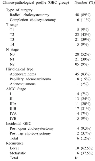
Table 3. Clinical characteristics of GBC patients in curative surgery group (n=54)
Clinico-pathological profile (GBC group) Number (%) Type of surgery
Radical cholecystectomy 48 (89%) Completion cholecystectomy 6 (11%) T stage
T1 5 (9%)
T2 23 (43%)
T3 21 (39%)
T4 5 (9%)
N stage
N0 28 (52%)
N1 21 (39%)
N2 05 (9%)
Histological type
Adenocarcinoma 45 (83%)
Papillary adenocarcinoma 8 (15%)
Adenosquamous 1 (2%)
AJCC Stage
I 4 (7%)
II 13 (24%)
IIIA 11 (20%)
IIIB 17 (31%)
IVA 4 (7%)
IVB 5 (9%)
Incidental GBC
Post open cholecystectomy 4 (9.3%) Post lap cholecystectomy 2 (3.7%)
Total 6 (12%)
Recurrence
Local 10 (62.5%)
Metastatic 6 (37.5%)
Total 16
advanced (stage III and IVA) group (n=37) (Table 3).
Females (n=73, 73%) outnumbered males (n=27, 27%).
The age of patients ranged from 20 to 80 years with peak incidence in fourth and fifth decade of life with mean age at diagnosis of 50 years. 70% of GBC patients in our study population had coexistent gallstones (Table 2).
Control group - A total of 28 patients of cholelithiasis who underwent Laparoscopic/open Cholecystectomy for symptomatic gallstone disease were recruited as control group. Out of 28 patients, 7 were male (25%) and 21 fe- male (75%). Majority (71%) 20/28 of patients were in the age range of 31-50 years with median age of patients be- ing 42 years.
TG2 expression
In the control group, TG2 expression was seen only in
21% (n=6) and none of them showed strong TG2 expression.
In GBC group, TG2 expression could not be assessed in two patients due to insufficient tissue block, thus excluded from study. In stark comparison to only 6 patients show- ing positive TG2 expression in the control group (none in strongly positive group), positive TG2 expression was demonstrated in 62 GBC patients (63%), this differential expression of TG-2 being statistically significant (p=0.001). In 62 GBC patients with TG2 positive ex- pression, 56 patients (90%) showed weakly positive ex- pression (score 3-6) while rest 6 patients (10%) were strongly positive (score 7-9).
Of the 62 GBC patients with positive TG2 expression, 40/54 were in curative resection group while 22/46 were in palliative treatment group (p=.31). In curative resection group (n=54), more patients (28/37; 76%) with locally ad- vanced disease showed positive TG2 expression as com- pared to early stage disease (12/17; 70%), though the dif- ference was not statistically significant (p=.69).
In curative resection group, median follow up was 21 months (13-52 months). Following curative surgery, local or metastatic recurrence was reported in 29.6% (16/54) of patients. TG2 positive expression was present in 93.8%
(15/16) of patients with recurrent disease (local or meta- static) indicating that TG2 expression may be a marker of inherent aggressive biological behavior and thus asso- ciated with higher likelihood of disease recurrence (p=0.03). TG2 positive patients had mean DFS of 21.3 months (95% confidence interval 17.02-25.60) as com- pared to 33.3 months (95% confidence interval 26.7-39.8) in TG2 negative patients with a difference of about 12 months which was statistically significant (p=0.04).
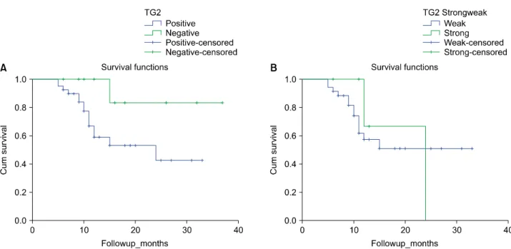
Correlation of recurrence with TG2 expression demon- strated TG2 negative patients had better survival as com- pared to TG2 positive patients, difference being statisti- cally significant (log rank test, p=0.04) (Fig. 3A). Weakly positive patients (21.7 months; 95% confidence interval 17.1-26.3) had slightly better DFS as compared to strong- ly TG2 positive patients (20 months; 95% confidence in- terval 10.9-29) though this difference was not statistically significant (p=0.84) (Table 4, Fig. 3B).
Univariate analysis was performed for TG2 expression and its correlation with seven clinic-pathological factors like patient age, sex, T stage, N stage, tumor histological grade, AJCC stage, association with cholelithiasis was
Table 4. Correlation of TG2 expression with disease free sur- vival (DFS) in curative surgery group demonstrating statisti- cally significant difference in survival based on TG-2 ex- pression (log rank test)
Marker Marker status Median survival
95%
confidence interval
p value
TG2 Positive 21.3 17.20-25.6 0.04*
Negative 33.33 26.77-39.8 TG2 Strongly positive 20 10.9-29 0.84
Weakly positive 21.7 17.1-26.3
*Statistically significant difference in median survival be- tween TG2 positive and negative group
Fig. 3. Kaplan Meier curves showing TG2 expression and its correlation with recurrence affecting survival in GBC patients in curative surgery group (n=54). (A) Kaplan Meier curve showing TG2 negative patients had better survival compared to TG2 positive patients (log rank test, p=0.04). (B) Recurrence affecting survival in weakly (score: 3-6) and strongly TG2 (score: 6-9) positive patients.
evaluated in both curative and palliative subgroup. In Univariate analysis, none of these factors were sig- nificantly associated with TG2 expression except for its association with cholelithiasis. TG2 positive patients were associated with cholelithiasis in 60% (24/40) and 100%
(22/22) of curative and palliative GBC group respectively.
These findings were statistically significant (p=0.04 &
0.01).
On Univariate analysis, various factors were analyzed for predicting Overall Survival (OS). Out of the five clin- ic-pathological variables studied for correlation with OS, (AJCC stage, T stage, LN status, Histological grade, TG2 status), AJCC stage (p=0.017), LN status (p=0.019),
Histological grade (p=0.015) and TG2 status (p=0.04) were found to illustrate significant correlation with OS in Univariate analysis. On multivariate analysis, only two of these four variables, lymph node status (p=0.03) and TG2 expression (p=0.037), were found to be significant pre- dictor of recurrence and eventual survival (Table 5).
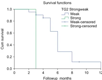
In Palliative group, median follow up was 6 months (4-24 months). Weakly TG2 positive had mean Disease Specific Survival (DSS) of 6.3 months (95% confidence interval 5.3-6.6) as compared to 3 months in strongly pos- itive patients (95% confidence interval 3-3), this differ- ence being statistically significant (log rank test, p=0.01).
Survival in relation to weakly and strongly TG2 positive expression was plotted by Kaplan Meier curve and demon- strated that weakly TG2 positive patients had better surviv- al as compared to strongly TG2 positive patients (Fig. 4).
DISCUSSION
Currently, there are no specific biomarkers or ther- apeutic molecular targets that can be used as predictive or prognostic indicators to either predict the risk or for use as therapeutic targets in metastatic GBC. Lack of ef- fective chemotherapy agents for treatment of GBC in neo- adjuvant/adjuvant setting further accentuates this clinical
Fig. 4. Weak and strong TG2 positive expression effecting survival in palliative group (n=46).
Table 5. Univariate and multivariate analysis of clinico- pathological variables with OS in curative surgery group (n=54)
Clinical parameters
Univariate analysis (p-value)
Multivariate analysis (p-value)
AJCC stage 0.017 NS
T stage 0.019 NS
Lymph node (LN) positive status
0.015 0.03
Histological grade 0.019 NS
TG2 expression 0.04 0.037
problem. Transglutaminase 2 (TG2) is the most widely distributed and most abundantly expressed member of transglutaminase family of enzymes, which are known to catalyze the Ca2+-dependent post-translational modification of proteins.14 TG2 is a multifunctional protein and its en- zymatic and non-enzymatic activities have been im- plicated in wide gamut of patho-physiological processes such as wound healing, cell growth and survival, ex- tracellular matrix modification, apoptosis and autophagy:
all of these attributes are known to play an important role in wound healing and inflammation. Thereby, TG2 may have a facilitator role in inflammation mediated cancer growth and progression.15
TG2 mediated signaling pathways enable the cancer cells to proliferate, resist cell death, invade, to reprogram glucose metabolism and to metastasize: all these charac- teristics are considered important hallmarks of aggressive cancer.16,17 Role of TG2 expression in inflammation in- duced carcinogenesis is well established in various can- cers including ovary, colorectal, breast, renal and lung.8,10-13 Its role in GBC has not yet been studied. Our study, comprising 100 patients of GBC and 28 patients of cholelithiasis (control group) is the first study to eval- uate expression of TG2 in a large cohort of GBC patients.
We attempt to analyze TG2 expression and its correlation with various clinic-pathological features and to evaluate its role as a potential predictive/prognostic marker.
On Univariate analysis, only cholelithiasis was shown to have statistically significant correlation with TG2 ex- pression in our series. It underlines the important role of TG2 in inflammation induced carcinogenesis which is known to be the main carcinogenic pathway in GBC.
Higher TG2 expression correlated with higher recurrence
rates and shorter DFS post curative resection. On similar lines, median survival was shorter in strong TG2 ex- pression cohort as compared to weak TG2 positive pa- tients in the palliative treatment group (3 versus 6.3 months, p=0.01).
Various studies have demonstrated TG2 as a potential negative prognosticator, and is often associated with ad- vanced disease stage, early recurrence, and chemo-resistance.
Erdem et al.12 studied the role of TG2 expression in meta- static (n=33) and non-metastatic (n=33) renal cell carcino- ma (RCC) and patients were stratified into two groups us- ing median primary tumor staining score as the cutoff val- ue: Group 1 (high risk, n=41) and Group 2 (low risk, n=22). The percentage of metastatic patients was sig- nificantly higher in Group 1 compared to Group 2 (68.3 vs. 18.2%, p<0.001). 5-year disease-free (34.9 vs. 92.9%, p=0.001) and cancer-specific (47.4 vs. 86.5%, p=0.04) sur- vival rates were significantly lower in high-risk group thus demonstrated increased expression of TG2 in primary tumor associated with a decrease in disease-free and can- cer-specific survival outcomes in RCC.12 Similar results were observed in our study with patients with TG2 pos- itive expression showed higher likelihood of developing recurrence as compared to those with TG-2 negative ex- pression, in curative surgery group (p=0.03). It was also noted that TG2 positive patients had shorter survival as compared to TG2 negative patients (21.3 vs 33.3 months, p=0.04), in our series.
Similarly in colorectal cancer, Miyoshi et al.10 analyzed TG2 expression in 91 paired cases of colorectal cancer
(CRC) and noncancerous regions. They showed poorer overall survival rate in high TG2 expression group than those in the low expression group (p=.001), indicating that increased TG2 expression was an independent poor prog- nostic factor.10
Chihong et al.13 have demonstrated the role of TG2 in cell survival and cancer progression in 194 patients diag- nosed with non-small cell lung cancer (NSCLC) and found that TG2 expression was significantly higher in lung cancer tissues as compared to paired marginal tissues or normal tissues. They also postulated that patients with low TG2 expression levels had longer disease free surviv- al and overall survival as compared those with higher TG2 expression and also found that high TG2 expression correlated with NSCLC recurrence.13 Findings in our study corroborated the above findings with high TG-2 ex- pression demonstrated in GBC group (63%) as compared to control (21%) and this differential expression was stat- istically significant (p=0.001).
Study by Fisher et al.18 demonstrated elevated TG2 levels in epidermal cancer stem cells (ECS cells) and important role TG2 plays in maintaining cancer stem cell survival, invasive, and metastatic behavior. They also showed that inhibitors induced TG2 knockdown or suppression of TG2 function can markedly reduce ECS cell survival. Thus TG2 inhibition can be an important therapeutic anti-cancer target by facilitating reduced survival of cancer stem cells in epidermal squamous cell carcinoma.18
Few studies have tried to study the correlation of some other biomarkers (EGFR, VEGFR, HER-2neu, p53) ex- pression and its clinico-pathological correlation in GBC.
Most of these studies are characterized by small number of GBC patients with incomplete clinical data and limited follow up details. Also most of these studies were per- formed on biliary tract cancers in general, with GBC com- prising only a small proportion. Misra et al.19 studied the correlation of p53 expression in operated GBC patients (n=20) and observed significant correlation between gall stone, T stage, grade of tumor and liver invasion with p53 over expression. In contrast, Ajiki et al.20 found no sig- nificant correlation between p53 expression and prognosis or recurrence.
In a study of EGFR and HER-2neu expression in 124 advanced biliary tract cancer patients, GBC patients ac- counted for only 27% of the study cohort (34/124) and
found no statistical association between grade, stage, over- all survival and marker expression thus having no prog- nostic significance.21 Tian et al.22 found that positive VEGFR expression was present in 63.3% of GBC patients and revealed that positive VEGFR expression were more common in higher Nevin stage group as compared to low- er Nevin stage group (p<0.05).
Despite various advancements in molecular biology, GBC treatment continues to be hampered by lack of effec- tive chemotherapy options and targeted agents. Therefore, development of agents inhibiting TG2 may offer a novel therapeutic approach for treatment of GBC.
Strength of our study is the sample size of GBC pa- tients undertaken for evaluation with all relevant clin- ic-pathological details and follow up. Our study has some limitations. First, the use of core biopsy samples in meta- static patients for immunohistochemistry, the small speci- mens may not represent the accurate TG2 status. Second, our sample size is small to show the association between drug resistance and the expression of TG2.
Present study is the first to demonstrate the clin- ic-pathological significance of TG2 expression in a large cohort of GBC patients (n=100). Positive TG2 expression was related to higher recurrence rates post curative sur- gery, shorter disease free and overall survival and ulti- mately portended poor prognosis. Further studies may be undertaken to better evaluate its role as novel prog- nostic/predictive marker in GBC and may be helpful in better prognostication and tailoring therapeutic approach for better management of GBC.
Novelty and impact
TG2 is a marker for inflammation induced carcino- genesis pathway. Strength of our study is large sample size (n=100) of GBC patients with all relevant clin- ic-pathological details and follow up. We studied TG2 ex- pression and its correlation with various clinic-patho- logical factors. In our study, Positive TG2 expression cor- related with higher recurrence rates post curative surgery, shorter disease free/overall survival and ultimately por- tended poor prognosis.
ETHICS APPROVAL
This study was performed in accordance with the
Ethical Principles for Medical Research Involving Human Subjects outlined in the Helsinki Declaration in 1975 (revised in 2000). Approval was granted by the Ethics Committee of King George’s Medical University vide let- ter no 3176/Ethics/R. Cell-15 dated 7/01/15.
ACKNOWLEDGEMENTS
We express our gratitude to Prof Nuzhat Husain and Miss Swati for their support in arranging blocks and slides of specimen for immunohistochemical analysis. Source of funding is from Department of Biotechnology, Government of India, under Pilot project Grant for Young Investigators.
Permanent Project Registration Number of this project is BT/PR9400/MED/30/1178/2013. This funding source had no role in the design of this study and during its ex- ecution, analyses, interpretation of the data, or decision to submit results.
CONFLICT OF INTEREST
The authors declare that there is no conflict of interest.
ORCID
Sameer Gupta: https://orcid.org/0000-0002-1865-1640 Sudeep Garg: https://orcid.org/0000-0002-4012-4505 Vijay Kumar: https://orcid.org/0000-0001-6477-8274 Arun Chaturvedi: https://orcid.org/0000-0002-4448-7039 Sanjeev Misra: https://orcid.org/0000-0002-0641-0946 Naseem Akhtar: https://orcid.org/0000-0001-8724-3248 Shiv Rajan: https://orcid.org/0000-0003-2513-6281 Jatinder Kaur: https://orcid.org/0000-0003-1650-0707 Manikandan Lakshmanan:
https://orcid.org/0000-0001-5673-7392
Kavitha Jain: https://orcid.org/0000-0002-4134-9829
AUTHOR CONTRIBUTIONS
Conceptualization: Sameer Gupta, SM. Data curation:
Sameer Gupta, Sudeep Garg, ML, KJ. Formal analysis:
Sameer Gupta, Sudeep Garg, AC, JK. Methodology: VK, NA. Project administration: Sameer Gupta, Sudeep Garg, SR, JK. Visualization: Sameer Gupta, VK, NA. Writing - original draft: Sameer Gupta, Sudeep Garg. Writing - re-
view & editing: Sameer Gupta, AC, SM.
REFERENCES
1. Misra S, Chaturvedi A, Misra NC. Gallbladder cancer. Curr Treat Options Gastroenterol 2006;9:95-106.
2. Misra S, Chaturvedi A, Misra NC, Sharma ID. Carcinoma of the gallbladder. Lancet Oncol 2003;4:167-176.
3. Silk YN, Douglass HO Jr, Nava HR, Driscoll DL, Tartarian G.
Carcinoma of the gallbladder. The Roswell Park experience. Ann Surg 1989;210:751-757.
4. Perpetuo MD, Valdivieso M, Heilbrun LK, Nelson RS, Connor T, Bodey GP. Natural history study of gallbladder cancer: a re- view of 36 years experience at M.D. Anderson hospital and tu- mor institute. Cancer 1978;42:330-335.
5. Begg GE, Carrington L, Stokes PH, Matthews JM, Wouters MA, Husain A, et al. Mechanism of allosteric regulation of trans- glutaminase 2 by GTP. Proc Natl Acad Sci U S A 2006;103:
19683-19688.
6. Kumar A, Xu J, Brady S, Gao H, Yu D, Reuben J, et al. Tissue tran- sglutaminase promotes drug resistance and invasion by inducing mesenchymal transition in mammary epithelial cells. PLoS One 2010;5:e13390.
7. Szondy Z, Korponay-Szabó I, Király R, Sarang Z, Tsay GJ.
Transglutaminase 2 in human diseases. Biomedicine (Taipei) 2017;7:15.
8. Shao M, Cao L, Shen C, Satpathy M, Chelladurai B, Bigsby RM, et al. Epithelial-to-mesenchymal transition and ovarian tumor progression induced by tissue transglutaminase. Cancer Res 2009;69:9192-9201.
9. Coussens LM, Werb Z. Inflammation and cancer. Nature 2002;420:860-867.
10. Miyoshi N, Ishii H, Mimori K, Tanaka F, Hitora T, Tei M, et al. TGM2 is a novel marker for prognosis and therapeutic target in colorectal cancer. Ann Surg Oncol 2010;17:967-972.
11. Agnihotri N, Kumar S, Mehta K. Tissue transglutaminase as a central mediator in inflammation-induced progression of breast cancer. Breast Cancer Res 2013;15:202.
12. Erdem S, Yegen G, Telci D, Yildiz I, Tefik T, Issever H, et al. The increased transglutaminase 2 expression levels during ini- tial tumorigenesis predict increased risk of metastasis and de- creased disease-free and cancer-specific survivals in renal cell carcinoma. World J Urol 2015;33:1553-1560.
13. Chihong Z, Yutian L, Danying W, Ruibin J, Huaying S, Linhui G, et al. Prognostic value of Transglutaminase 2 in non-small cell lung cancer patients. Oncotarget 2017;8:45577-45584.
14. Mehta K, Kumar A, Kim HI. Transglutaminase 2: a multi-task- ing protein in the complex circuitry of inflammation and cancer.
Biochem Pharmacol 2010;80:1921-1929.
15. Odii BO, Coussons P. Biological functionalities of trans- glutaminase 2 and the possibility of its compensation by other members of the transglutaminase family. ScientificWorldJournal 2014;2014:714561.
16. Agnihotri N, Mehta K. Transglutaminase-2: evolution from pe- destrian protein to a promising therapeutic target. Amino Acids 2017;49:425-439.
17. Gundemir S, Colak G, Tucholski J, Johnson GV. Transglutaminase 2: a molecular Swiss army knife. Biochim Biophys Acta 2012;
1823:406-419.
18. Fisher ML, Keillor JW, Xu W, Eckert RL, Kerr C. Transgluta- minase is required for epidermal squamous cell carcinoma stem cell survival. Mol Cancer Res 2015;13:1083-1094.
19. Misra S, Chaturvedi A, Goel MM, Mehrotra R, Sharma ID, Srivastava AN, et al. Overexpression of p53 protein in gall- bladder carcinoma in North India. Eur J Surg Oncol 2000;26:
164-167.
20. Ajiki T, Onoyama H, Yamamoto M, Asaka K, Fujimori T, Maeda S, et al. p53 protein expression and prognosis in gall- bladder carcinoma and premalignant lesions. Hepatogastroenter- ology 1996;43:521-526.
21. Harder J, Waiz O, Otto F, Geissler M, Olschewski M, Weinhold B, et al. EGFR and HER2 expression in advanced biliary tract cancer. World J Gastroenterol 2009;15:4511-4517.
22. Tian Y, Ding RY, Zhi YH, Guo RX, Wu SD. Analysis of p53 and vascular endothelial growth factor expression in human gall- bladder carcinoma for the determination of tumor vascularity.
World J Gastroenterol 2006;12:415-419.