대한남성과학회지:제 29 권 제 3 호 2011년 12월 Korean J Androl. Vol. 29, No. 3, December 2011 h t t p : / / d x . d o i . o r g / 1 0 . 5 5 3 4 / k j a . 2 0 1 1 . 2 9 . 3 . 2 5 1
251
접수일자: 2011년 9월 15일, 수정일자: 2011년 10월 12일, 게재일자: 2011년 10월 19일
Correspondence to: Chang Hee Han
Department of Urology, Uijongbu St. Mary’s Hospital, 65-1, Keumoh-dong, Uijongbu 480-130, Korea
Tel: 031-820-3546, Fax: 031-847-6133 E-mail: [email protected]
Laparoscopic Excision of a Congenital Seminal Vesicle Cyst and Coexisting Ipsilateral Renal Agenesis
Yun Seok Jung, Jang Ho Wee, Jin Bong Choi, Myung Sun Choi, Seol Kim, Jun Ho Sohn, Joon Ho Lee, Sung Hak Kang, Yong Seok Lee, Chang Hee Han Department of Urology, The Catholic University of Korea College of Medicine, Seoul, Korea
= Abstract =
Seminal vesicle cyst (SVC) with ipsilateral renal agenesis is a rare congenital anomaly. When the patient is symptomatic, surgical treatment may be necessary. The open surgical approach, traditionally considered the definite form of treatment, has been associated with a high rate of morbidity. The laparoscopic approach for the management of SVCs has recently been described. A 18-year-old man presented with a 2-year history of dysuria and perineal pain. The diagnostic evaluation revealed a 45×35×48 mm sized left seminal vesicle cyst. In addition, he had a solitary, right, functioning kidney, with left renal agenesis. Transperitoneal laparoscopic excision of the cyst was performed successfully. The patient was discharged from the hospital on the fourth postoperative day and did not present with any complaints or complications.
Key Words: Seminal vesicle, Cyst, Renal agenesis
Most seminal vesicle cysts (SVCs) are congenital, and two thirds are associated with renal dysplasia or agenesis with an ectopic ureter opening into the semi- nal vesicle. Surgical treatment is indicated for sympto- matic patients. However, the seminal vesicle is diffi- cult to access surgically, and all transrectal, transure- thral, or open surgical approaches have inherent short- comings. Recently, laparoscopy has become an alter- native approach to seminal vesicle disease that offers an excellent intraoperative approach and minimal post- operative morbidity. We report a case of laparoscopic excision of a left seminal vesicle cyst in a young pa- tient with ipsilateral renal agenesis.
Case Report
An 18-year-old male patient was referred to the ur- ology clinic with a 2-year history of perineal pain and dysuria. Conservative treatment did not provide symp- tomatic relief. Digital rectal examination revealed a large cystic mass in the left prostate region. Routine biochemical examination of the blood and urine was unremarkable.
Pelvic CT and MRI revealed a 45×35×48 mm left seminal vesicle cyst (Fig. 1). In addition, the patient had a solitary right functioning kidney, with no evi- dence of a left renal unit.
Transperitoneal laparoscopic excision of the cyst was performed. Four laparoscopic ports were used: a 10-mm port at the umbilicus, a 5-mm port in the mid- dle in the proximity of the pubis, and a 5-mm and a 10-mm port on the left and right sides lateral to the rectus muscle, respectively.
252 대한남성과학회지: 제 29 권 제 3 호 2011
Fig. 1. Coronal view of pelvis MRI showing a left seminal vesicle cyst.
Fig. 2. Operative view under laparoscopy. The dilated seminal vesicle cyst is exposed.
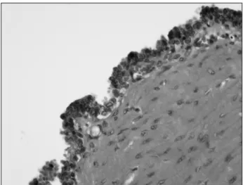
Fig. 3. Histopathological findings of the seminal vesicle cyst.
The cyst is lined by cuboidal epithelium with a fibrous wall of variable thickness (H&E, ×400).
The patient was placed in the Trendelenburg position.
The bladder was retracted anteriorly, and an incision was made in the retrovesical peritoneum. The dilated left vas deferens was easily identified and dissected medially to the ampulla, which was used as a guide to the seminal vesicle. The dilated seminal vesicle was punctured and drained (Fig. 2). A blunt dissection was used to expose the anterior and posterior aspects of the seminal vesicle with Hem-o-lok clips applied close to the seminal vesicle to control the vascular supply. The entire cystic specimen was extracted through the 12-mm port.
The total operative time was 160 minutes, and blood loss was minimal. The patient was discharged from the hospital on the fourth postoperative day and did not present with any complications thereafter. The patient has been asymptomatic during 1 year of follow-up care. Histopathological examination confirmed the di- agnosis of SVC (Fig. 3).
Discussion
SVCs are exceedingly rare lesions that may be ei- ther congenital or acquired in origin. Most of them have congenital causes, which are believed to be sec- ondary to the obstruction of the ejaculatory duct, and two-thirds are associated with renal agenesis or dys- plasia. The formation of a congenital SVC with ipsi- lateral renal agenesis is believed to result from the ab- normal development of the distal portion of the meso- nephric duct.1 However, in the absence of associated genitourinary abnormalities, acquired cysts can be caused by ejaculatory duct obstruction as a result of genitourinary inflammation, ejaculatory duct lithiasis, or transurethral prostatectomy.2
Most SVCs are diagnosed in adults during the third to fifth decade of life.3 Symptoms may develop due to the irritation of adjacent organs by an enlarged and inflamed SVC. Cyst distension may cause perineal or pelvic pain, hemospermia, postcoital pain, or painful
Yun Seok Jung, et al: Congenital Seminal Vesicle Cyst 253
defecation.4 Bladder irritation can cause frequency, ur- gency, dysuria, and hematuria. These patients have fre- quently been misdiagnosed and thus treated with mul- tiple courses of antibiotics without symptom resolution.5 The diagnostic workup includes physical examina- tion, transabdominal and/or transrectal ultrasonography, computed tomography, and/or magnetic resonance imaging. Additional studies include intravenous urog- raphy, retrograde cystourethrography, cystoscopic ex- amination, and vesiculography.6
Treatment is indicated for symptomatic cases.
However, conservative management has not usually been effective, and surgical management may be nec- essary for the relief of symptoms. Surgical treatment of symptomatic SVCs has included transrectal ultra- sonography-guided fine needle cyst aspiration, tran- surethral unroofing, transurethral resection of the ejac- ulatory duct, transurethral endoscopic aspiration, or open surgical excision.
The open surgical approach, traditionally considered to be the definitive form of treatment, has been asso- ciated with a high rate of morbidity. Significant com- plications have been reported, for example, rectal wall laceration, ureteral injury, injury to the erectile neuro- vascular bundle, and pelvic urinoma.7
Recently, laparoscopy has been advocated as an op- timal, minimally invasive technique for surgical treat- ment. Laparoscopic SVC excision provides excellent visualization of the retrovesical space compared to open resection. The vas deferens can be easily identi- fied and used as a surgical guide to locate the seminal vesicle. Also, without damaging the bladder and rec- tum, the seminal vesicle can be dissected from the per- itoneum that is covering the bladder and prostate.
Recently, many medical centers have reported success- ful laparoscopic excisions of seminal vesicle cysts.8,9 The advantages of the laparoscopic approach are the simplicity of convalescence, minimal pain, the lack of need for a prolonged bladder catheter, and the possi- bility of immediate feeding. The patient had minimal postoperative pain, quick recovery, and complete symptom resolution. The laparoscopic approach af-
fords minimal postoperative morbidity, effective surgi- cal extirpation, short hospitalization, and rapid recov- ery for the patient. In conclusion, laparoscopy is likely to be the optimal surgical approach for treating most symptomatic seminal vesicle space-occupying lesions.
REFERENCES
1) Patel B, Gujral S, Jefferson K, Evans S, Persad R.
Seminal vesicle cysts and associated anomalies. BJU Int 2002;90:265-71
2) Cherullo EE, Meraney AM, Bernstein LH, Einstein DM, Thomas AJ, Gill IS. Laparoscopic management of congenital seminal vesicle cysts associated with ipsilateral renal agenesis. J Urol 2002;167:1263-7 3) van den Ouden D, Blom JH, Bangma C, de Spiegeleer
AH. Diagnosis and management of seminal vesicle cysts associated with ipsilateral renal agenesis: a pooled analysis of 52 cases. Eur Urol 1998;33:433-40 4) King BF, Hattery RR, Lieber MM, Berquist TH,
Williamson B Jr, Hartman GW. Congenital cystic disease of the seminal vesicle. Radiology 1991;178:
207-11
5) Roehrborn CG, Schneider HJ, Rugendorff EW, Hamann W. Embryological and diagnostic aspects of seminal vesicle cysts associated with upper urinary tract malformation. J Urol 1986;135:1029-32 6) Ikari O, Castilho LN, Lucena R, D'Ancona CA,
Netto NR Jr. Laparoscopic excision of seminal vesi- cle cysts. J Urol 1999;162:498-9
7) Williams RD, Sandlow JL. Surgery of the seminal vesicles. In: Walsh PC, Retik AB, Vaughan ED Jr, Wein AJ, editors. Campbell’s Urology. 7th ed. Phila- delphia: Saunders; 1998;3299-315
8) Sohn DW, Kim HW, Cho WY, Sung GT. Laparo- scopic Excision of a Seminal Vesicle Cysts Associated with Ipsilateral Renal Agenesis. Korean J Urol 2004;
45:732-4
9) Jang KD, Choi KH, Yang SC, Jang WS, Jang JY, Han WK. Laparoendoscopic single-site surgery (LESS) for excision of a seminal vesicle cyst associated with ipsilateral renal agenesis. Korean J Urol 2011;52:
431-3