53 DOI: 10.4196/kjpp.2011.15.1.53
ABBREVIATIONS: [Ca2+]i, intracellular calcium concentration;
CICR, calcium-induced calcium release; RyRs, ryanodine receptors;
CPA, cyclopiazonic acid; CCh, carbamylcholine.
Received February 11, 2011, Revised February 14, 2011, Accepted February 16, 2011
Corresponding to: Hyung Seo Park, Department of Physiology, College of Medicine, Konyang University, 685, Gasoowon-dong, Seo-gu, Daejeon 302-718, Korea. (Tel) 82-42-600-6474, (Fax) 82-42-600-6314, (E-mail) [email protected]
Ca
2+-induced Ca
2+Release from Internal Stores in INS-1 Rat Insulinoma Cells
Kyung Jin Choi1, Dong Su Cho1, Ju Young Kim2, Byung Joon Kim2, Kyung Moo Lee1, Shin Hye Kim1, Dong Kwan Kim1, Se Hoon Kim1, and Hyung Seo Park1
Departments of 1Physiology, 2Internal Medicine, College of Medicine, Konyang University, Daejeon 302-718, Korea
The secretion of insulin from pancreatic β -cells is triggered by the influx of Ca2+ through voltage-dependent Ca2+ channels. The resulting elevation of intracellular calcium ([Ca2+]i) triggers additional Ca2+ release from internal stores. Less well understood are the mechanisms involved in Ca2+ mobilization from internal stores after activation of Ca2+ influx. The mobilization process is known as calcium-induced calcium release (CICR). In this study, our goal was to investigate the existence of and the role of caffeine-sensitive ryanodine receptors (RyRs) in a rat pancreatic β -cell line, INS-1 cells. To measure cytosolic and stored Ca2+, respectively, cultured INS-1 cells were loaded with fura-2/AM or furaptra/AM. [Ca2+]i was repetitively increased by caffeine stimulation in normal Ca2+ buffer. However, peak [Ca2+]i was only observed after the first caffeine stimulation in Ca2+ free buffer and this increase was markedly blocked by ruthenium red, a RyR blocker. KCl-induced elevations in [Ca2+]i were reduced by pretreatment with ruthenium red, as well as by depletion of internal Ca2+
stores using cyclopiazonic acid (CPA) or caffeine. Caffeine-induced Ca2+ mobilization ceased after the internal stores were depleted by carbamylcholine (CCh) or CPA. In permeabilized INS-1 cells, Ca2+
release from internal stores was activated by caffeine, Ca2+, or ryanodine. Furthermore, ruthenium red completely blocked the CICR response in permeabilized cells. RyRs were widely distributed throughout the intracellular compartment of INS-1 cells. These results suggest that caffeine-sensitive RyRs exist and modulate the CICR response from internal stores in INS-1 pancreatic β-cells.
Key Words: INS-1, Caffeine, Ryanodine, Calcium release, CICR
INTRODUCTION
Elevation of the intracellular free calcium concentration ([Ca2+]i) is a key event in insulin secretion by pancreatic β-cells. Increased glucose fueling, a major stimulus to β-cells, ultimately results in elevation of the ATP/ADP ratio, which causes plasma membrane depolarization through the clos- ing of ATP-sensitive K+ channels (KATP), and in turn stim- ulates Ca2+ entry through the opening of voltage-gated Ca2+
channels (CaV), which are located on plasma membranes [1-3]. Increases in [Ca2+]i due to sequential activation of KATP and CaV channels triggers Ca2+ release from the endo- plasmic reticulum (ER), an internal Ca2+ store, and this process is called calcium-induced calcium release (CICR) [4-6]. Mobilization of Ca2+ from internal stores can result from the opening of Ca2+ channels on the ER membranes such as inositol 1,4,5-trisphosphate receptors (InsP3Rs) or ryanodine receptors (RyRs) [7-10]. While it is well estab- lished that InsP3R-mediated Ca2+ release is operative in insulin secreting β-cells, the presence and role of RyRs in
pancreatic β-cell signaling remains controversial.
RyRs are named because a plant alkaloid, ryanodine, binds to this channel. They are expressed abundantly in muscle cells and neurons. A critical property of RyRs is that increased cytosolic Ca2+, cyclic ADP ribose, or nicotinic acid adenine dinucleotide phosphate (NAADP) can activate this channel physiologically, resulting in even greater amplifica- tion of Ca2+ signals elicited by Ca2+ entry or release [11-13]. Another key property of RyRs is that caffeine at millimolar concentrations can pharmacologically cause channel opening. In some studies, caffeine did not activate Ca2+ release from the internal stores of β-cells [14,15], whereas Ca2+ release from the internal stores was acti- vated if caffeine-sensitive RyRs were present in intact β-cells [16,17]. In fact, the presence of RyRs in insulin se- creting β-cells is well-accepted at the present time, al- though the expression of each type of RyRs is variable, de- pending on the species studied. The mRNAs for the three types of RyRs have been detected in human β-cells, where- as mRNA for RyR2 but not RyR1 was detected in INS-1 cells and rat islets using RT-PCR methods [18-20]. Since there is still no clear consensus on the role of RyR in stim- ulus-secretion coupling in β-cells, we determined, using in-
Cell culture
INS-1 cells, a rat insulinoma cell line, were grown in RPMI1640 medium containing 11.1 mM glucose supple- mented with 10% heat-inactivated FBS, penicillin (100 U/ml), streptomycin (100 μg/ml), 0.6 mM sodium bicar- bonate, 2 mM L-glutamate, 1 mM sodium pyruvate, 10 mM HEPES, and 50 μM β-mercaptoethanol. The cells were subcultured every 7 days and were maintained at 37oC in a humidified atmosphere containing 5% CO2. All experi- ments were performed with cells in passages 30∼50. To stabilize them after Ca2+-sensitive dye loading, cells were resuspended in HEPES-buffered physiological saline sol- ution (HEPES-PSS) containing 2.5 mM glucose, 137 mM NaCl, 0.56 mM MgCl2, 4.7 mM KCl, 1 mM Na2HPO4, 10 mM HEPES (pH 7.4), 1.28 mM CaCl2, and 1% (w/v) bovine serum albumin.
Cytosolic Ca2+ measurement in intact cells
For measurement of [Ca2+]i, cultured INS-1 cells were loaded with 2 μM fura-2/AM, a Ca2+-sensitive dye, for 30 min at room temperature. Fura-2 loaded cells were mount- ed on a glass coverslip at the bottom of perfusion chambers.
Cells were continuously superfused with HEPES-PSS at a flow rate of 1 ml/min using an electronically controlled per- fusion system (Warner Instrument, Hamden, CT, USA).
Cytosolic Ca2+ imaging was performed using an inverted Olympus IX71 microscope through a 40× fluorescence ob- jective lens. Cells were excited alternately with light at 340 nm and 380 nm, using a Polychrome V monochrometer (Till Photonics, Pleasanton, CA, USA). Fluorescence images were captured at an emitted wavelength of 510 nm using a cooled charged-coupled device, Cool-SNAP HQ2 camera (Photometrics, Tucson, AZ, USA).
Measurement of stored Ca2+ in permeabilized cells INS-1 cells were loaded with 10 μM furaptra/AM for 30 min at room temperature. Cells were permeabilized by su- perfusion with 20 μM β-escin for 1∼2 min in intracellular medium (ICM) containing 125 mM KCl, 19 mM NaCl, 10 mM HEPES, and 1 mM EGTA (pH 7.3) as described pre- viously [21,22]. Permeabilized cells were washed in ICM without β-escin for 15 min to facilitate removal of cytosolic dye. Cells were superfused in intracellular medium contain- ing 0.650 μM CaCl2 (free [Ca2+]=200 nM), 1.4 mM MgCl2, and 3 mM Na2ATP to activate sarco/endoplasmic reticulum Ca2+-ATPase (SERCA) and to load intracellular Ca2+
stores. Prior to activation of stored calcium release, cells were superfused without MgCl2 to inactivate SERCA. The free Ca2+ concentration was changed from 0 to 10 μM ac- cording to the experimental maneuvers. We recorded fluo-
ing a polyclonal RyR (N-19) primary antibody (Santa Cruz Biotechnology, Santa Cruz, CA, USA) and Cy-2 conjugated rabbit anti-goat secondary antibody (Jackson Immuno- Research, PA, USA). After overnight incubation in primary antibody (1:100 dilution) at 4oC, cells were incubated with secondary antibody (1:100 dilution) for 1 hr at 37oC.
Immunofluorescence images for RyRs were collected using a confocal microscope (Carl Zeiss, Germany), and processed using Photoshop 7.0 software (Adobe, Mountain View, CA, USA).
Drugs
To activate calcium mobilization in intact cells, 30 mM caffeine, 10 μM carbamylcholine (CCh), or 45 mM KCl was added to HEPES-PSS. To activate calcium release from in- ternal stores in permeabilized cells, 10 mM caffeine, 10 μM calcium or 1 μM ryanodine was added to ICM. Carbamyl- choline, caffeine, ruthenium red, β-escin, and other chem- icals for making buffers were purchased from Sigma- Aldrich Chemical Co. (St Louis, MO, USA). Ryanodine and cyclopiazonic acid (CPA), a SERCA inhibitor, were pur- chased from Calbiochem (San Diego, CA, USA). Fura 2/AM and furaptra/AM were purchased from TefLabs Inc.
(Austin, TX, USA). RPMI 1640, fetal bovine serum (FBS), trypsin-EDTA, and penicillin/streptomycin were purchased from Gibco BRL (Grand Island, NY, USA).
Statistical analysis
Results are presented as mean±S.E. Data were analyzed using the Student t-test. Rates of Ca2+ release were esti- mated from these responses by fitting the initial 10 sec peri- od of decreasing fluorescence to a single exponential func- tion using the Origin program as described previously [21].
Differences were considered significant when the p value was less than 0.05.
RESULTS
Caffeine mobilizes calcium from internal stores in intact INS-1 cell
First, to detect the presence of RyRs, we determined them functionally by testing the effects of 30 mM caffeine, a RyR activator, on Ca2+ mobilization in intact INS-1 cells. In the presence of extracellular Ca2+, repetitive caffeine perfusion resulted in reiterative elevation of [Ca2+]i even if those re- sponses were slightly reduced (Fig. 1A). However, in Ca2+
free solution, INS-1 cells only responded to the first caffeine stimulation, and this response was transient (Fig. 1B). [Ca2+]i
peaks were not significantly different from each other when the same cells were stimulated with caffeine in the presence
Fig. 1. Caffeine stimulated calcium mobilization from internal stores in intact INS-1 cells. The representative traces show the effects of repetitive 30 mM caffeine stimulation on [Ca2+]i increases in the presence (A) and absence (B) of extracellular Ca2+. The data were obtained from 5 and 7 separate experiments, respectively.
INS-1 cells were responsive to repetitive caffeine stimulation in normal extracellular Ca2+ buffer, but only responded to the first caffeine stimulation in Ca2+ free solution. (C) A 50 μM of ruthenium red markedly reduced the [Ca2+]i peak in the absence of extracellular Ca2+. Data were normalized to control values and expressed as mean %±S.E. Asterisk indicates the value is significantly different from the corresponding value of caffeine alone (p<0.05).
Fig. 2. KCl triggered Ca2+ release from internal stores in intact INS-1 cells. (A) The representative trace shows the effect of 45 mM KCl on [Ca2+]i increases in the presence and absence of extra- cellular Ca2+. The data were obtained from 6 separate experiments.
[Ca2+]i elevation was not observed in Ca2+ free medium. (B) Effects of CPA plus caffeine, CPA alone or caffeine alone on KCl-induced [Ca2+]i peaks in the presence of extracellular Ca2+. The data were obtained from at least 5 separate experiments. Data were normalized to the initial [Ca2+]i peak and expressed as mean %
± S.E. Asterisks indicate that the values are significantly different from the corresponding value for control (p<0.05). Intracellular Ca2+
store depletion reduced depolarization-induced Ca2+ mobilization.
(C) Representative trace shows the effect of ruthenium red on KCl-induced [Ca2+]i elevations. The data were obtained from 6 separate experiments. A 50 μM of ruthenium red significantly reduced depolarization-induced Ca2+ mobilization; the effect was restored after washout of the ruthenium red.
and the absence of extracellular Ca2+ (data not shown).
These results mean that caffeine can mobilize Ca2+ from internal stores without the presence of extracellular Ca2+, and can easily deplete internal Ca2+ stores by repetitive stimulation. To determine the involvement of RyRs, we added ruthenium red, a RyRs blocker, to the perfusion medium. Ruthenium red markedly reduced the caffeine-in- duced [Ca2+]i peak to 11.60±2.09% of control value in the absence of extracellular Ca2+ (Fig. 1C). These results sug- gest that caffeine can mobilize Ca2+ from internal stores through RyRs in intact INS-1 cells.
Depolarization-induced calcium entry triggers calcium release from internal stores
Next, the experiments were performed to clarify whether
membrane depolarization can mobilize Ca2+ from internal stores through RyRs in INS-1 cells. A 45 mM KCl solution was perfused to mimic membrane depolarization. The re- petitive depolarization by KCl perfusion resulted in a con- stant [Ca2+]i rise in normal Ca2+ buffer; the response ceased when we changed to a Ca2+ free solution (Fig. 2A).
However, INS-1 cells did not respond to KCl in the absence of extracellular Ca2+. These results mean that KCl initially mobilizes Ca2+ from extracellular spaces via voltage-oper- ated Ca2+ channels. To confirm whether the Ca2+ that en- tered due to depolarization can trigger Ca2+ release from internal stores, we examined the effects of KCl in the store-depleted condition by pretreatment cells with cyclo- piazonic acid (CPA), a sarcoplasmic/endoplasmic reticulum Ca2+ ATPase (SERCA) inhibitor, or caffeine, a RyRs
Fig. 3. Effects of internal calcium store depletion on caffeine- induced calcium release. (A) The representative trace shows the 30 mM caffeine-induced [Ca2+]i rise after internal store depletion by 10 μM carbamylcholine (CCh) in Ca2+ free solution. (B) The representative trace shows the 10 μM CCh-induced [Ca2+]i rise under store depleted conditions induced by pretreatment with 30 mM caffeine in the absence of extracellular Ca2+. (C) A represen- tative trace of the caffeine effect under store depleted condition induced by pretreatment of cells with 10 μM cyclopiazonic acid (CPA) in Ca2+ free solution. All data were obtained from at least 5 separate experiments. Caffeine failed to increase [Ca2+]i after internal Ca2+
store depletion induced by pretreatment with CCh or CPA.
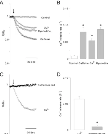
Fig. 4. Caffeine, Ca2+ or ryanodine-induced calcium release in permeabilized INS-1 cells. (A) 10 mM caffeine (□), 10 μM Ca2+
(◇) or 1 μM ryanodine (△) significantly stimulated Ca2+ release from internal stores in permeabilized INS-1 cells. Arrows indicate the starting point of each drug perfusion. (B) Summarized Ca2+
release rates (S−1) induced by caffeine, Ca2+ or ryanodine. Data were summarized from at least 5 experiments. Asterisks indicate that the values are significantly different from the corresponding value for control (p<0.05). (C) The blocking effect of 50 μM ruthenium red on 10 μM Ca2+-induced Ca2+ release in permea- bilized INS-1 cells. (D) Summarized data showing the effects of ruthenium red on Ca2+-induced Ca2+ release rates in permea- bilized cells. Asterisk indicates that the value is significantly different from the corresponding value of Ca2+ (p<0.05). Ca2+
release induced by elevated Ca2+ was completely blocked by ruthenium red, a RyR blocker, in permeabilized INS-1 cells.
Caffeine and carbamylcholine activate the same intra- cellular calcium stores
We tested whether RyRs and InsP3Rs were expressed on the same membranes of intracellular Ca2+ stores. Pretreat-
Stored calcium is released by caffeine, calcium, and ryanodine in permeabilized INS-1 cells
We employed luminal Ca2+ measurements of ER to exam- ine direct effects of RyRs activation on Ca2+ release in per- meabilized INS-1 cells. Perfusion of 10 mM caffeine, 10 μM free Ca2+, or 1 μM ryanodine dramatically initiated Ca2+
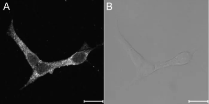
Fig. 5. The expression of ryanodine receptors in INS-1 rat insulinoma cells. The fluorescence image (A) and the bright image (B) show the expression and distribution of RyRs in the intra- cellular compartments. The images were obtained from 4 separate experiments. Immunocytochemistry was done using primary RyR antibody as described under experimental procedures. The scale bar is 10 μm.
release from internal stores (Fig. 4A). As shown in Fig. 4B, calcium release rates (S−1) were 0.080±0.014, 0.059± 0.005, and 0.088±0.005 by perfusion of caffeine, Ca2+, or ryano- dine, respectively, which were markedly different from the value for control cells (0.008±0.002). Ca2+-induced Ca2+ re- lease (CICR) from ER was completely blocked by ruthenium red in permeabilized INS-1 cells (Fig. 4C). Ca2+ release rate was reduced to 0.006±0.002 by pretreatment with ruthe- nium red (Fig. 4D). These results strongly indicate that caf- feine-sensitive RyRs modulate Ca2+ signals through a CICR response from the ER of INS-1 cells.
Cellular distribution of RyRs in INS-1 cells
We utilized immunocytochemical imaging to analyze cel- lular expression and distribution of RyRs. As shown in Fig. 5, RyRs were strongly expressed and widely distributed in in- tracellular compartment, and no immunoreactive signals were detected in the nuclear compartment. We clearly con- firmed the existence of RyRs in the intracellular organelle of INS-1 cell.
DISCUSSION
The obvious findings of the present study, which used immunocytochemistry and calcium imaging, are that caf- feine-sensitive RyRs are expressed in the subcellular com- partments and that depolarization-induced Ca2+ entry trig- gers RyR-mediated CICR responses in INS-1 insulinoma cells. There are two main families of intracellular Ca2+
channels, InsP3Rs and RyRs in insulin secreting β-cells [7-10]. Among these channels, the critical properties of RyR are that cytosolic Ca2+ physiologically and caffeine pharma- cologically can activate this channel [11]. In the current study, we determined the role of RyRs on Ca2+ mobilization in the INS-1 insulinoma cell line. To stimulate RyRs, we used caffeine, which is known to increase both the open time and the open probability of RyRs in a cooperative man- ner with Ca2+ [23]. Caffeine at 30 mM markedly stimulated Ca2+ mobilization in both normal and Ca2+ free medium of intact INS-1 cells, while failed to increase [Ca2+]i after repetitive stimuli without the extracellular presence of Ca2+
because of internal Ca2+ store depletion. In the absence of
external Ca2+, ruthenium red, a RyR blocker, completely inhibited the caffeine-induced initial rise in [Ca2+]i. In some studies, caffeine increased cytosolic Ca2+ through activa- tion of voltage-gated Ca2+ channels (CaV) [14,16]. However, in our study, caffeine still elicit Ca2+ mobilization in the absence of extracellular Ca2+, and these increases were blocked by ruthenium red in intact INS-1 cells. In addition, 10 mM caffeine and 1 μM ryanodine directly released stor- ed Ca2+ from ER in permeabilized INS-1 cells. These re- sults provide evidence that caffeine induces a rise in [Ca2+]i
through Ca2+ release from RyR-sensitive internal stores.
Cytosolic Ca2+ is known to have a dual effect on RyRs;
nanomolar to micromolar concentrations of Ca2+ increase but millimolar concentrations decrease the open probability of RyRs [24-26]. Similar to caffeine and ryanodine, 10 μM of Ca2+ clearly released stored Ca2+, and this CICR was completely blocked by ruthenium red in permeabilized cells.
Some studies, however, failed to detect caffeine-sensitive RyR in permeabilized β-cells despite successful detection of Ca2+ release from the ER of intact cells [14-16]. These discrepancies may be due to differences in methods used for the permeabilization, which can cause the loss of regu- latory molecules. In our study, we detected caffeine-sensi- tive Ca2+ release in both intact and permeabilized cells, which indicate that caffeine-sensitive RyRs are present and are fully operational in INS-1 cells.
The depolarization of plasma membranes for activation of voltage-gated Ca2+ channels (CaV) is one of the most critical events leading to a rise in [Ca2+]i and insulin secre- tion in β-cell [1-3,27,28]. The depolarization-induced in- crease in [Ca2+]i is not considered as being due just to Ca2+
entry through CaV. There is some evidence that Ca2+ sig- nals can be modulated by CICR though Ca2+ release from the ER in β-cells [29,30]. One important function of CICR in β-cells is to amplify Ca2+ signaling for insulin secretion.
To clarify the involvement of the CICR response, we eval- uated KCl-induced Ca2+ mobilization from internal stores that have been depleted. Pretreatment of CPA, caffeine, or a combination of both resulted in a substantial reduction in depolarization-induced [Ca2+]i peaks in intact INS-1 cells. In this condition, ER was completely depleted by SERCA inhibition by CPA and by pre-activation of RyRs by caffeine. These results indicate that Ca2+ has entered into the cells by membrane depolarization through CaV
triggers additional Ca2+ mobilization from internal Ca2+
stores. InsP3Rs are also involved in the CICR response of insulin secreting β-cells [7,8,30]. Therefore, we used ruthe- nium red to distinguish RyR-mediated CICR responses from InsP3R-mediated responses. A blockage of RyRs by ruthenium red significantly reduced depolarization-induced elevations in [Ca2+]i in normal Ca2+ buffer, whereas ruthe- nium red had no effect on CCh-induced Ca2+ mobilization, which is mediated by InsP3Rs (data not shown). As men- tioned above, the CICR response was completely attenuated by pretreatment with ruthenium red in permeabilized cells.
This does not mean that the CICR response is fully medi- ated by RyRs. Actually, two types of Ca2+ channels are dif- ferently involved in responses to increased cytosolic Ca2+. RyRs can be directly activated by cytosolic Ca2+, but InsP3Rs can be potentiated by Ca2+ in the presence of cyto- solic InsP3 [24,31]. In permeabilized conditions, InsP3Rs, therefore, might not be operative due to the loss of cytosolic InsP3 because the plasma membrane became permeable to this molecule. Based on these results, we suggest that the Ca2+ that entered through CaV can trigger additional Ca2+
medium. Moreover, pretreatment with caffeine completely eliminated the CCh-induced increase in [Ca2+]i. Although we did not clearly confirm the co-localization of InsP3R and RyR on ER membranes, unfortunately, our results do show that InsP3Rs and RyRs regulate the same internal Ca2+
stores, or at least they are functionally cross-linked. One critical role of the CICR in β-cells is that it amplifies Ca2+- dependent insulin exocytosis. Further studies are now needed to clarify the roles of InsP3Rs and RyRs based on localized Ca2+ signals, because CICR generates large local Ca2+ transients [35], and their functions depend on sub- cellular localization of the receptors.
ACKNOWLEDGEMENTS
This work was supported by Basic Research Program through the National Research Foundation (NRF) of Korea funded by the Ministry of Education, Science and Technology (2010-0012568), and Myung-Gok Research Fund of Konyang University (2009).
REFERENCES
1. Islam MS. Calcium signaling in the islets. Adv Exp Med Biol.
2010;654:235-259.
2. Hiriart M, Aguilar-Bryan L. Channel regulation of glucose sensing in the pancreatic β-cell. Am J Physiol Endocrinol Metab. 2008;295:E1298-E1306.
3. Mears D. Regulation of insulin secretion in islets of Langerhans by Ca2+ channels. J Membr Biol. 2004;200:57-66.
4. Varadi A, Rutter GA. Ca2+-induced Ca2+ release in pancreatic islet β-cells: critical evaluation of the use of endoplasmic reticulum-targeted "cameleons". Endocrinology. 2004;145:4540- 4549.
5. Lemmens R, Larsson O, Berggren PO, Islam MS. Ca2+-induced Ca2+ release from the endoplasmic reticulum amplifies the Ca2+
signal mediated by activation of voltage-gated L-type Ca2+
channels in pancreatic β-cells. J Biol Chem. 2001;276:
9971-9977.
6. Graves TK, Hinkle PM. Ca2+-induced Ca2+ release in the pancreatic β-cell: direct evidence of endoplasmic reticulum Ca2+
release. Endocrinology. 2003;144:3565-3574.
7. Hagar RE, Ehrlich BE. Regulation of the type III InsP3 receptor and its role in β cell function. Cell Mol Life Sci.
2000;57:1938-1949.
8. Dyachok O, Tufveson G, Gylfe E. Ca2+-induced Ca2+ release by activation of inositol 1,4,5-trisphosphate receptors in primary pancreatic β-cells. Cell Calcium. 2004;36:1-9.
9. Johnson JD, Kuang S, Misler S, Polonsky KS. Ryanodine receptors in human pancreatic β cells: localization and effects on insulin secretion. FASEB J. 2004;18:878-880.
10. Bruton JD, Lemmens R, Shi CL, Persson-Sjögren S, Westerblad H, Ahmed M, Pyne NJ, Frame M, Furman BL, Islam MS.
Ryanodine receptors of pancreatic β-cells mediate a distinct context-dependent signal for insulin secretion. FASEB J. 2003;
14. Islam MS, Larsson O, Nilsson T, Berggren PO. Effects of caffeine on cytoplasmic free Ca2+ concentration in pancreatic β-cells are mediated by interaction with ATP-sensitive K+ channels and L-type voltage-gated Ca2+ channels but not the ryanodine receptor. Biochem J. 1995;306:679-686.
15. Rutter GA, Theler JM, Li G, Wollheim CB. Ca2+ stores in insulin-secreting cells: lack of effect of cADP ribose. Cell Calcium. 1994;16:71-80.
16. Chen TH, Lee B, Yang C, Hsu WH. Effects of caffeine on intracellular calcium release and calcium influx in a clonal β-cell line RINm5F. Life Sci. 1996;58:983-990.
17. Gamberucci A, Fulceri R, Pralong W, Bánhegyi G, Marcolongo P, Watkins SL, Benedetti A. Caffeine releases a glucose-primed endoplasmic reticulum Ca2+ pool in the insulin secreting cell line INS-1. FEBS Lett. 1999;446:309-312.
18. Dror V, Kalynyak TB, Bychkivska Y, Frey MH, Tee M, Jeffrey KD, Nguyen V, Luciani DS, Johnson JD. Glucose and endoplasmic reticulum calcium channels regulate HIF-1β via presenilin in pancreatic β-cells. J Biol Chem. 2008;283:9909-9916.
19. Takasawa S, Kuroki M, Nata K, Noguchi N, Ikeda T, Yamauchi A, Ota H, Itaya-Hironaka A, Sakuramoto-Tsuchida S, Takahashi I, Yoshikawa T, Shimosegawa T, Okamoto H. A novel ryanodine receptor expressed in pancreatic islets by alternative splicing from type 2 ryanodine receptor gene. Biochem Biophys Res Commun. 2010;397:140-145.
20. Li F, Zhang ZM. Comparative identification of Ca2+ channel expression in INS-1 and rat pancreatic β cells. World J Gastroenterol. 2009;15:3046-3050.
21. Park HS, Betzenhauser MJ, Won JH, Chen J, Yule DI. The type 2 inositol (1,4,5)-trisphosphate (InsP3) receptor determines the sensitivity of InsP3-induced Ca2+ release to ATP in pancreatic acinar cells. J Biol Chem. 2008;283:26081-26088.
22. Choi KJ, Kim KS, Kim SH, Kim DK, Park HS. Caffeine and 2-aminoethoxydiphenyl borate (2-APB) have different ability to inhibit intracellular calcium mobilization in pancreatic acinar cell. Korean J Physiol Pharmacol. 2010;14:105-111.
23. Rousseau E, Ladine J, Liu QY, Meissner G. Activation of the Ca2+ release channel of skeletal muscle sarcoplasmic reticulum by caffeine and related compounds. Arch Biochem Biophys.
1988;267:75-86.
24. Hamilton SL. Ryanodine receptors. Cell Calcium. 2005;38:253- 260.
25. Ehrlich BE, Kaftan E, Bezprozvannaya S, Bezprozvanny I. The pharmacology of intracellular Ca2+-release channels. Trends Pharmacol Sci. 1994;15:145-149.
26. Zalk R, Lehnart SE, Marks AR. Modulation of the ryanodine receptor and intracellular calcium. Annu Rev Biochem. 2007;76:
367-385.
27. Drews G, Krippeit-Drews P, Düfer M. Electrophysiology of islet cells. Adv Exp Med Biol. 2010;654:115-163.
28. Braun M, Ramracheya R, Bengtsson M, Zhang Q, Karanau- skaite J, Partridge C, Johnson PR, Rorsman P. Voltage-gated ion channels in human pancreatic β-cells: electrophysiological characterization and role in insulin secretion. Diabetes.
2008;57:1618-1628.
29. Kang G, Holz GG. Amplification of exocytosis by Ca2+-induced Ca2+ release in INS-1 pancreatic β cells. J Physiol. 2003;546:
175-189.
30. Dyachok O, Gylfe E. Ca2+-induced Ca2+ release via inositol
1,4,5-trisphosphate receptors is amplified by protein kinase A and triggers exocytosis in pancreatic β-cells. J Biol Chem.
2004;279:45455-45461.
31. Foskett JK, White C, Cheung KH, Mak DO. Inositol trisphosp- hate receptor Ca2+ release channels. Physiol Rev. 2007;87:
593-658.
32. Islam MS, Larsson O, Berggren PO. Cyclic ADP-ribose in β cells. Science. 1993;262:584-586.
33. Islam MS, Rorsman P, Berggren PO. Ca2+-induced Ca2+
release in insulin-secreting cells. FEBS Lett. 1992;296:287-291.
34. Takasawa S, Nata K, Yonekura H, Okamoto H. Cyclic ADP-ribose in insulin secretion from pancreatic β cells.
Science. 1993;259:370-373.
35. Mitchell KJ, Lai FA, Rutter GA. Ryanodine receptor type I and nicotinic acid adenine dinucleotide phosphate receptors mediate Ca2+ release from insulin-containing vesicles in living pancreatic β-cells (MIN6). J Biol Chem. 2003;278:11057-11064.