Veterinary Science
http://dx.doi.org/10.4142/jvs.2014.15.1.91
Received: 8 Mar. 2013, Revised: 23 May 2013, Accepted: 9 Jul. 2013
Original Article
*Corresponding author: Tel: +82-41-550-1939, Fax: +82-41-550-1998, E-mail: [email protected]
†The first two authors contributed equally to this work.
ⓒ 2014 The Korean Society of Veterinary Science.
This is an Open Access article distributed under the terms of the Creative Commons Attribution Non-Commercial License (http://creativecommons.org/licenses/by-nc/3.0) which permits unrestricted non-commercial use, distribution, and reproduction in any medium, provided the original work is properly cited.
Development and clinical evaluation of a rapid diagnostic kit for feline leukemia virus infection
Won-Shik Kim
1,†, Chom-Kyu Chong
2,†, Hak-Yong Kim
2, Gyu-Cheol Lee
3, Wooseog Jeong
4, Dong-Jun An
4, Hye-Young Jeoung
4, Jae-In Lee
5, Young-Ki Lee
6,*
1
Department of Clinical Pathology Laboratory Science, Daejeon Health Science College, Daejeon 300-711, Korea
2
Department of Biochemistry and CBITRC, College of Natural Sciences, Chungbuk National University, Cheongju 361-763, Korea
3
Water Analysis and Research Center, K-water, Daejeon 306-711, Korea
4
Bacteriology and Parasitology Division, Animal and Plant Quarantine Agency, Anyang 430-824, Korea
5
Seoul Metropolitan Government Research Institute of Public Health and Environment, Gwacheon 427-070, Korea
6
Department of Public Health, Graduate School of Public Health & Social Welfare, Dankook University, Cheonan 330-714, Korea
Feline leukemia virus (FeLV) causes a range of neoplastic and degenerative diseases in cats. To obtain a more sensitive and convenient diagnosis of the disease, we prepared monoclonal antibodies specific for the FeLV p27 to develop a rapid diagnostic test with enhanced sensitivity and specificity. Among these antibodies, we identified two clones (hybridomas 8F8B5 and 8G7D1) that specifically bound to FeLV and were very suitable for a diagnostic kit. The affinity constants for 8F8B5 and 8G7D1 were 0.35 × 10
9and 0.86 × 10
9, respectively. To investigate the diagnostic abilities of the rapid kit using these antibodies, we performed several clinical studies. Assessment of analytical sensitivity revealed that the detection threshold of the rapid diagnostic test was 2 ng/mL for recombinant p27 and 12.5 × 10
4IU/mL for FeLV.
When evaluating 252 cat sera samples, the kit was found to have a kappa value of 0.88 compared to polymerase chain reaction (PCR), indicating a significant correlation between data from the rapid diagnostic test and PCR. Sensitivity and specificity of the kit were 95.2% (20/21) and 98.5% (257/261), respectively. Our results demonstrated that the rapid diagnostic test would be a suitable diagnostic tool for the rapid detection of FeLV infection in cats.
Keywords: anti-FeLV p27 monoclonal antibody, feline leukemia virus, polymerase chain reaction, rapid diagnostic test, recombinant FeLV p27
Introduction
Feline leukemia virus (FeLV) is one of the most important infectious pathogens that causes death in cats and is broadly spread worldwide [12,17,24]. FeLV was discovered among cats that lived in a cluster-household where several animals had developed lymphosarcoma [19]. The virus is an enveloped, positive single-stranded RNA retrovirus. Cats are usually infected by direct contact with infected cats [12,31,33], mostly via oronasal exposure to saliva and nasal secretions containing high levels of the virus especially through mutual grooming and sharing food dishes or water bowls [11,12,17]. Vertical transmission occasionally occurs [35] but is of secondary importance. FeLV-associated diseases include a variety of neoplastic disorders, anemia, leucopenia, thrombocytopenia, neurological disorders, reproductive failure in female cats, and numerous secondary infections caused by FeLV-induced immunosuppression [12,34]. Although the period of disease progression is highly variable, 83% of FeLV-infected cats die within 3 years [29].
Given these findings, the accurate diagnosis of FeLV infection is very important to break the cycle of horizontal and vertical transmission in feline populations. Several diagnostic tools have been introduced in veterinary clinics:
the passive hemagglutination test [37], complement
fixation test [36], immunofluorescent assay [13], enzyme
linked-immunosorbent assay (ELISA) [25-28], saliva test
[10], and rapid diagnostic test (RDT) [7]. The RDT, also
known as the lateral flow rapid test, has several advantages such as quick turnaround, cost-effectiveness, and usability in locations far removed from laboratories. Consequently, the RDT has been widely used in clinics and elsewhere [30,39]. In this report, we describe the preparation of monoclonal antibodies specific for the p27 of FeLV. We also provide details about the development of an RDT system using these antibodies and clinical characteristics of the assay.
Materials and Methods
Viruses, host cells, and clinical samples
Feline leukemia virus (VR-719), feline immunodeficiency virus (FIV, VR-1312), feline panleukopenia virus (FPV, VR-2017), feline coronavirus (FCoV, VR-2004), feline calicivirus (FCaV, VR-782), canine adenovirus (CAV, VR-293), canine coronavirus (CCV, VR-809), and canine distemper virus (CDV, VR-1587) were purchased from the American Type Culture Collection (USA). Canine parvovirus Type 2a VI (CPV, KVCC-VR0900161) was kindly provided by the National Veterinary Research and Quarantine Service (Korea). Additionally, 282 sera samples from 155 household felines and 127 stray cats were provided by the National Veterinary Research and Quarantine Service as well as four different animal hospitals in Chungbuk province (Korea): Jeonju Animal Hospital, Woori Animal Hospital, Soo Animal Hospital, and Juju Animal Hospital. The samples were collected from December 2009 to March 2012.
Virus culture and purification
Vero cells (kindly provided by Professor Chan Hee Lee, Chungbuk National University, Korea) were used for FeLV culture. The cells were cultured in Dulbecco’s modified Eagle’s medium (DMEM; Invitrogen, USA) supplemented with 10% fetal bovine serum (FBS; Invitrogen) at 37
oC in 5% CO
2. Before infection with the virus, the cells were washed with phosphate-buffered saline (PBS) and inoculated with the viruses for 6 h in DMEM containing 5%
FBS at 37
oC in 5% CO
2. After 4 days, the culture media was changed and the infection progressed until 80∼90% of the cells were floating or lightly attached to the T-75 flask (typically 10 days post-infection) (Thermo Scientific, USA). The viruses were harvested and purified by density gradient ultracentrifugation (3% sucrose) as previously described [32].
Preparation of recombinant FeLV p27 (rec. p27) To clone the genes encoding p27 in FeLV, two primers were designed and synthesized (Cosmogenetech, Korea).
The forward primer was 5´-gaattccccttgagggagggcccca acaac-3´ (the BamHI site is underlined) and the reverse primer was 5´-ctcgagcagaactttagtcatctccttgtg-3´ (the XhoI
site is underlined). Reverse transcription was carried out using the viral genome under the following conditions: 90 min at 37
oC, 5 min at 94
oC, and holding at 4
oC.
Polymerase chain reaction (PCR) amplification of the p27 gene was performed under the following conditions: 35 cycles consisting of 1 min 30 sec at 94
oC, 1 min 30 sec at 59
oC, and 1 min 30 sec at 72
oC. Each reaction mixture contained 100 ng of template, 10 pmole of two primers, 100 mM KCl, 200 mM Tris (pH 8.8), 100 mM (NH
4)
2SO
4, 20 mM MgSO
4. The 756 base pair (bp) PCR product was cloned into a pGEM-T vector (Promega, USA) for sequencing by Cosmogenetech and the data were analyzed by ClustalW ver. 2.0 [23].
The gene insert in the pGEM-T vector was subcloned into a pET28a expression vector (Merk Millipore, USA) and used to transformed CaCl
2-competent E.coli BL21(DE3) cells (Promega). The transformants were grown at 37
oC in Luria-Bertani medium (Difco; Becton Dickinson and Company, USA) to an optical density at 600 nm (OD
600) of 0.7∼0.8 by DU UV/Vis Spectrophotometer (Beckman Coulter, USA).
Expression of the recombinant protein was induced with 1 mM isopropyl- β-D-thiogalatoside (Thermo Scientific) for 5 h at 30
oC and the cells were then harvested by centrifugation at 5,000 × g 20 min at 4
oC. The cell pellets were resuspended in a standard buffer (50 mM Tris-HCl (pH 7.9) and 500 mM NaCl) containing 1 mM phenylmethylsulfonyl fluoride (Sigma-Aldrich, USA).
The suspension was then lysed by sonication (Vibra-Cell;
Sonics & Materials, USA) under ice-cold conditions (15 min with 5 sec on/5 sec off cycles). The homogenate was centrifuged at 20,000 × g for 20 min at 4
oC and the supernatant was then subjected to metal affinity chromatography using Ni-NTA agarose (Qiagen, Germany). The cell lysate was added into a column that had been equilibrated with standard buffer. Unbound proteins were removed by washing with a sufficient volume of washing buffer (standard buffer containing 20 mM imidazole (Sigma-Aldrich). Next, 6xHis-tagged recombinant protein was recovered from the column with elution buffer (standard buffer containing 500 mM imidazole). The isolated protein was dialyzed at 4
oC with 10 mM sodium carbonate (pH 9.0) (Sigma-Aldrich) and identified by SDS-PAGE analysis [22]. The protein concentration was measured by the method developed by Bradford [21].
Preparation of monoclonal antibodies against FeLV p27 Hybridoma technology was used to manufacture anti-FeLV p27 antibodies as previously described [20].
Hybridomas producing monoclonal antibodies (mAbs) against the core p27 protein were generated as follows.
Spleen cells from BALB/c mice (7-week-old females;
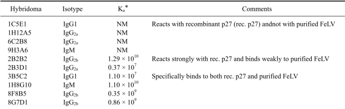
Table 1. Properties of hybridomas produced in this study
Hybridoma Isotype K
a* Comments
1C5E1 1H12A5 6C2B8 9H3A6 2B2B2 2B3D1 3B5C2 1H8G10 8F8B5 8G7D1
IgG1 IgG
2aIgG
2aIgM IgG
2bIgG
2aIgG1 IgM IgG
2bIgG
2bNM NM NM NM 1.29 × 10
100.37 × 10
71.10 × 10
71.10 × 10
100.35 × 10
90.86 × 10
9Reacts with recombinant p27 (rec. p27) andnot with purified FeLV
Reacts strongly with rec. p27 and binds weakly to purified FeLV Specifically binds to both rec. p27 and purified FeLV
*Affinity constant = 1/dissociation constant (Kd) where Kd was determined by a Klotz plot according to the method described by Friguet et al. [9]. NM: not measured.