128 CASE REPORT
Korean Circ J 2008;38:128-130
Print ISSN 1738-5520 / On-line ISSN 1738-5555 Copyright ⓒ 2008 The Korean Society of Cardiology
Successful Pacemaker Revision Through Sustained Right Superior Vena Cava in a Patient With Situs Inversus Totalis
Hack-Lyoung Kim, MD, Min-Kyung Kim, MD, Hee-Suk Min, MD, Byoung-Yong Choi, MD, Eue-Keun Choi, MD, Jae-Jin Kwak, MD, Yun-Shik Choi, MD and Seil Oh, MD
Department of Internal Medicine, Seoul National University College of Medicine, Seoul, Korea ABSTRACT
In patients with situs inversus totalis, the superior vena cava is normally positioned on the left side and drains into a left-sided right atrium (RA). If right-side superior vena cava (RSVC) is also present, it should be thought of as a combined congenital anomaly. Here, we report a case of successful pacemaker lead insertion through the RSVC in a patient with situs inversus totalis. The left-side superior vena cava (LSVC) had been already used as a route for the first pacemaker lead insertion 15 years earlier. During the pacemaker lead revision, we found that the LSVC was obliterated, and used the RSVC as a route for a new pacemaker lead insertion. (Korean Circ J 2008;
38:128-130)
KEY WORDS: Vena cava, superior; Situs inversus; Pacemaker, artificial.
Introduction
In patients with situs inversus totalis, the superior vena cava is normally positioned on the left side and drains into a left-sided right atrium (RA). If these pa- tients also have right superior vena cava (RSVC), it should be thought of as a combined congenital anomaly.
This venous anomaly can often be recognized during the implantation of a pacemaker or implantable car- dioverter defibrillator (ICD), and it also can be used as a route for lead access.1) Here, we present a case of suc- cessful pacemaker lead revision through the RSVC in a patient with situs inversus totalis.
Case
A 44-year-old woman was admitted to our hospital for pulse generator revision. She had previously been diagnosed with congenitally corrected transposition of the great arteries with situs inversus totalis. She had undergone atrial septal defect patch closure and aortic valve replacement (AVR) due to severe aortic regurgita-
tion 18 years earlier. In the operation field, both right and left superior vena cava had been observed. The left superior vena cava (LSVC) was 0.5 cm in size and drained directly to a left-sided right atrium (RA); the RSVC was 3 cm in size and drained to the RA via an enlarged coronary sinus. Fifteen years earlier, she com- plained of dizziness during the postoperative care of redo AVR. Her electrocardiography (ECG) showed com- plete atrioventricular (AV) blockage. She underwent permanent VVI pacemaker implantation through the LSVC (Fig. 1).
During the follow-up, pacemaker interrogation re- vealed that replacement of the pulse generator was in- dicated, although the pacing was still working (Fig. 2).
Echocardiography showed corrected transposition of great arteries (TGA) and slightly depressed function of the right-sided left ventricle, with an estimated ejection fraction of 45%. There were no intra-cardiac shunts and the prosthetic aortic valve had normal function.
The coronary sinus was enlarged, and its diameter was 1.6 cm. During the revision procedure, we noticed that the pacing threshold of the ventricular lead was greater than 5 V and the lead impedance was 750 ohm. Thus, we planned to insert a new pacemaker lead. After punc- turing the left axillary vein, we attempted to insert a guide wire into the LSVC. However, the LSVC was obstructed (Fig. 3A), so we decided to use the RSVC for the insertion of a new lead. In spite of its long distance and tortuosity, we were able to insert a lead
Received: October 24, 2007 Accepted: November 21, 2007
Correspondence: Seil Oh, MD, Department of Internal Medicine, Seoul National University College of Medicine, 28 Yeongeon-dong, Jongno-gu, Seoul 110-744, Korea
Tel: 82-2-2072-2088, Fax: 82-2-762-9662 E-mail: [email protected]
Hack-Lyoung Kim, et al.·129
through the RSVC (arrowheads in Fig. 3B) and suc- cessfully placed the new lead on the left-sided RV apex (Fig. 4). Pacemaker interrogation showed that the im- pedance and threshold were 742 ohm and 0.3 V, re- spectively. The patient was discharged 4 days after the procedure. At one month follow-up, the pacing status was good (impedance 850 ohm; threshold <0.5 V), and the follow-up ECG showed that the pacemaker was functioning well (Fig. 5).
Discussion
Situs inversus is a rare congenital condition in which major visceral organs are reversed or mirrored from their normal position. If the heart is swapped to the right side of the thorax, it is known as situs inversus totalis. Compared to LSVC with situs solitus, there have been few reports on RSVC in patients with situs in- versus.2) In patients with situs solitus, LSVC is an asymp- tomatic and infrequently observed anomaly. Its prev- alence in the general population is approximately 0.3%,2) while it is much higher in patients with congenital cardiac abnormalities, ranging from 2.8% to 4.3%.3)4) The incidence was not different (0.47%) in patients undergoing implantation of a pacemaker or implanta- ble cardioverter defibrillator.5) LSVC is the most com- mon form of anomalous venous drainage involving SVC, which results from the failure of the left com- mon cardinal vein to become occluded.6) In most cases, blood drains into the right atrium via an enlarged coronary sinus,6) but in 8% of patients, it drains di- rectly into the left atrium through a defect in the form of an unroofed coronary sinus.7) The RSVC was absent in 1% of patients with persistent LSVC, and is fre-
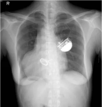
Fig. 1. Initial chest X-ray on admission. The chest X-ray showed situs inversus totalis.
Fig. 3. Pacemaker revision through RSVC. In Figure A, the previously inserted pacemaker lead is shown in the LSVC (arrows). However, the LSVC was not visualized below the level of the most inferior arrow in this figure, and we could not pass a guide wire in order to insert a new pacemaker lead. However, the RSVC drained into the right atrium via the coronary sinus, and we were able to insert a guide wire into the left-sided right ventricle (B). Arrowheads indicate the venous drainage route to the RSVC. LSVC: left superior vena cava, RSVC: right superior vena cava.
A B
Fig. 2. Initial ECG on admission. Tall pacing spikes resulted from ventricular pacing with high output. ECG: electrocardiography.
130·Pacemaker Lead Insertion Through Right SVC in Situs Inversus
quently associated with alteration of cardiac situs.8) Associated cardiac anomalies include stenosis or atresia of the pulmonary artery, D-transposition, complete at- rioventricular septal defects, anomalous pulmonary vein drainage, and cor triatriatum.9-12)
Persistence of LSVC in patients with situs solitus is almost always found incidentally during an invasive procedure such as central catheter insertion or pace- maker and ICD implantation. In pacemaker implanta- tion, the most common problems related to the unusual anatomic access to the heart were reaching a conven- ient pacing site and ensuring stable lead placement.13-15) Once the RA is reached, further placement of the lead to the RV is not easy because of acute angulation bet-
ween the coronary sinus and the tricuspid valve open- ing. During the pacemaker revision for this case, the pacemaker lead could not be easily placed in the right ventricle using LSVC. Fortunately, the RSVC drained into the right atrium, and we were able to successfully insert pacemaker leads into the apex of the RV through the RSVC and the coronary sinus.
To the best of our knowledge, this is the first report on pacemaker lead insertion through the RSVC in a patient with situs inversus totalis.
REFERENCES
1) Biffi M, Boriani G, Frabetti L, Bronzetti G, Branzi A. Left supe- rior vena cava persistence in patients undergoing pacemaker or cardioverter-defibrillator implantation: a 10-year experience.
Chest 2001;120:139-44.
2) Murayama H, Maeda M, Sakurai H, Watanabe T. Absent left superior vena cava with persistent right superior vena cava in visceroatrial situs inversus. Pediatr Cardiol 2006;27:293-6.
3) Son JW, Lee CS, Han SW, Lee SW, Kim SK, Kwon YJ. A case of persistent left SVC associated with tricuspid regurgitation.
Korean Circ J 1993;23:609-13.
4) Campbell M, Deuchar DC. The left-sided superior vena cava. Br Heart J 1954;16:423-39.
5) Morgan DR, Hanratty CG, Dixon LJ, Trimble M, O’Keeffe DB.
Anomalies of cardiac venous drainage associated with abnor- malities of cardiac conduction system. Europace 2002;4:281-7.
6) Nsah EN, Moore GW, Hutchins GM. Pathogenesis of persistent left superior vena cava with a coronay sinus connection. Pediatr Pathol 1991;11:261-9.
7) Rigatelli G. Congenitally persistent left superior vena cava: a possible unpleasant problem during invasive procedures. J Car- diovasc Med 2007;8:483-7.
8) Raghib G, Ruttenberg HD, Anderson RC, Amplatz K, Adams P Jr, Edwards JE. Termination of left superior vena cava in left atrium, atrial septal defect, and absence of coronary sinus: a de- velopmental complex. Circulation 1965;31:906-18.
9) Jeong SY, Sin PJ, Cho SY, et al. A case of situs inversus (I.D.D.) with corrected TGA. Korean Circ J 1993;23:296-301.
10) Gonzalez-Juanatey C, Testa A, Vidan J, et al. Persistent left superior vena cava draining into thecoronary sinus: report of 10 cases and literature review. Clin Cardiol 2004;27:515-8.
11) Zaglavara T, Hamilton JR, Kenny A. A combination of persistent left superiorvena cava and a large secundumatrial septal defect in a 34 year old woman. Heart 2001;85:406.
12) Gheissari A, Malm JR, Bowman FO Jr, Bierman FZ. Cor triatri- atumsinistrum: one institution’s 28-year experience. Pediatr Cardiol 1992;13:85-8.
13) Krukal JC. Transvenous pacemaker failure due to anomalous venous return to the heart. Chest 1971;59:458-61.
14) Garcia L, Levine R, Kosowsky W, Lyon AF. Persistent left supe- rior vena cava complicating pacemaker insertion. Chest 1972;
61:396-7.
15) Rubenfire M, Evangelista J, Wajszczuk WJ, Kantrowitz A. Im- plication of a persistent left superior vena cava in transvenous pacemaker therapy and cardiac hemodynamic monitoring. Chest 1974;65:145-7.
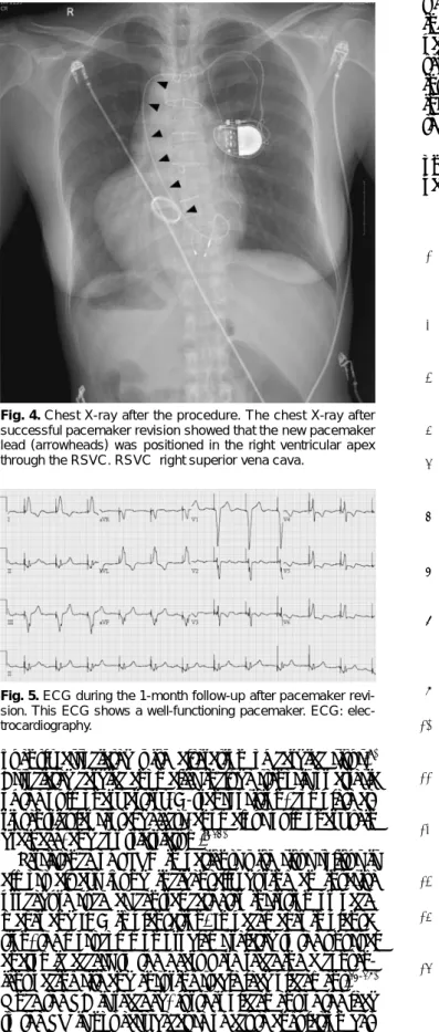
Fig. 4. Chest X-ray after the procedure. The chest X-ray after successful pacemaker revision showed that the new pacemaker lead (arrowheads) was positioned in the right ventricular apex through the RSVC. RSVC: right superior vena cava.
Fig. 5. ECG during the 1-month follow-up after pacemaker revi- sion. This ECG shows a well-functioning pacemaker. ECG: elec- trocardiography.