ABSTRACT
The efficacies of a supraglottic airway device (SGAD) and an endotracheal tube (ETT) in cats under general anesthesia with volume-controlled ventilation (VCV) were compared. Thirty healthy cats were randomly allocated for airway control using either an SGAD or an ETT. Five tidal volumes (6, 8, 10, 12, and 14 mL/kg) were randomly tested, and respiratory rates were adjusted to achieve a minute ventilation of 100 mL/kg/min. The dose of propofol necessary to insert the SGAD or ETT, the static respiratory pressure, leakage during VCV, and end tidal CO
2(ETCO
2) were recorded. Dosages of propofol and static respiratory measurements for the SGAD and ETT groups were compared using a t-test. The distribution of leakages and hypercapnia (ETCO
2> 45 mmHg) were compared using Fisher's exact test. A significance level of p < 0.05 was established. No significant difference in dose of propofol was observed between the SGAD and ETT groups (7.1 ± 1.0, 7.3 ± 1.7 mg/kg; p = 0.55). Static resistance pressure of the SGAD (22.0 ± 8.1 cmH
2O/L/sec) was significantly lower than that of the ETT (36.6 ± 12.9 cmH
2O/L/sec; p < 0.01). Of the 75 trials, leakage was more frequent when using an SGAD (8 events) than when using an ETT (1 event; p = 0.03). Hypercapnia occurred more frequently with SGAD (18 events) than with ETT (3 events; p < 0.01). Although intubation with an ETT is the gold standard in small animal anesthesia, the use of an SGAD can reduce airway resistance and the work of breathing. Nonetheless, SGAD had more dead space and the tidal volume for VCV needs adjustment.
Keywords: Airway; cats; hypercapnia; hypoventilation; lung
INTRODUCTION
Airway management is one of the most important elements of general anesthesia. Intubation with an endotracheal tube (ETT) is considered the gold standard in small animal anesthesia [1]. Intubation with an ETT maintains a patent airway [2], prevents aspiration to an oropharynx, and allows the option to apply controlled mechanical ventilation (CMV) [3].
Despite the benefits of orotracheal intubation, various complications have been associated with ETT, including malpositioning or tube displacement, occlusion of the tube by thickened secretions, soft tissue swelling, laryngeal edema [4,5], arytenoid tears [2], and tracheal mucosal damage [2,6]. Furthermore, lethal complications related to the use of ETT include
Original Article
Received: Mar 16, 2019 Revised: Jul 19, 2019 Accepted: Nov 30, 2019
*Corresponding author:
Naris Thengchaisri
Department of Companion Animal Clinical Sciences, Kasetsart University, 50 Thanon Ngam Wong Wan, Lat Yao, Chatuchak, Bangkok 10900, Thailand.
E-mail: [email protected]
© 2020 The Korean Society of Veterinary Science
This is an Open Access article distributed under the terms of the Creative Commons Attribution Non-Commercial License (https://
creativecommons.org/licenses/by-nc/4.0) which permits unrestricted non-commercial use, distribution, and reproduction in any medium, provided the original work is properly cited.
ORCID iDs
Nutawan Niyatiwatchanchai
https://orcid.org/0000-0001-9680-8335 Naris Thengchaisri
https://orcid.org/0000-0003-0815-0743
FundingThis study was supported by a grant from the Faculty of Veterinary Medicine, Kasetsart University.
Conflict of Interest
The authors declare no conflicts of interest.
Author Contributions
Conceptualization: Thengchaisri N; Data curation: Niyatiwatchanchai N; Formal analysis: Thengchaisri N; Funding acquisition:
Nutawan Niyatiwatchanchai
1, Naris Thengchaisri
2,*1
Surgery Unit, Kasetsart University Veterinary Teaching Hospital, Bangkok 10900, Thailand
2
Department of Companion Animal Clinical Sciences, Kasetsart University, Bangkok 10900, Thailand
Clinical assessment of the efficacy of supraglottic airway devices compared with endotracheal tubes in cats
during volume-controlled ventilation
Anesthesiology &
Pain Medicine
Niyatiwatchanchai N; Investigation:
Niyatiwatchanchai N, Thengchaisri N;
Methodology: Niyatiwatchanchai N, Thengchaisri N; Project administration:
Niyatiwatchanchai N; Resources: Thengchaisri N; Software: Thengchaisri N; Supervision:
Thengchaisri N; Validation: Niyatiwatchanchai N; Visualization: Niyatiwatchanchai N, Thengchaisri N; Writing - original draft:
Niyatiwatchanchai N, Thengchaisri N; Writing - review & editing: Niyatiwatchanchai N, Thengchaisri N.
tracheal rupture [7] and pneumothorax. A novel supraglottic airway device (SGAD) has been developed for use in cats. Since an SGAD is placed outside the trachea, the complications due to the intubation process can be avoided. The information on the use of SGAD in cats is limited; thus, more studies are needed to evaluate the effectiveness and ventilatory mechanics of an SGAD during CMV.
The resistance of the respiratory tract to airflow is referred to as airway resistance, and it can be calculated from the driving pressure and airflow during breathing. During mechanical ventilation, airway resistance can be influenced by various factors, including contraction or relaxation of smooth muscles, lung volume, length and radius of the airways, and the type of airflow. Moreover, diseases affecting the airway such as asthma can cause difficulty breathing by increasing airway resistance due to a temporary restriction of the airways [8]. During general anesthesia, application of a small ETT facilitates the intubation process, especially in cats with a small trachea. By contrast, the SGAD is inflatable and allows a seal around the laryngeal [9]; thus, it is possible to use an SGAD instead of an ETT during mechanical ventilation. The use of an SGAD allows anesthesia of cats without the need for drugs and equipment for intubation, such as muscle relaxants and a laryngoscope [10].
The most commonly used modes of mechanical ventilation in small animal practice are volume-controlled ventilation (VCV) and pressure-controlled ventilation (PCV). In VCV, the tidal volume is calculated, and the ventilator provides a constant volume of air to the patient, despite alterations in compliance and resistance of the airway during general anesthesia [11]. With a pressure-limited ventilator, the peak inspiratory pressure (PIP) preset is predetermined to limit the tidal volume delivered to the patient [12]. A recent study compared the use of SGAD and ETT in cats during PCV [3]. The leakage of gas during ventilation with PCV in cats occurred more often in the ETT group than in the SGAD and laryngeal mask airway (LMA) groups [3]. However, the effectiveness of an SGAD in cats during mechanical ventilation with VCV has not been reported. Moreover, the internal diameter of the SGAD is larger than that of the ETT. Since the internal diameter of the tube correlates inversely with airway resistance [8,13], the use of an SGAD possibly provides less airway resistance than that of a small ETT. Nonetheless, SGAD use may increase the dead space in cats, and an adjustment of the tidal volume setting for VCV may be needed. In the present study, SGAD and ETT efficacies were compared using different tidal volume setting in cats under general anesthesia with VCV. Other parameters including the total volume of anesthetic drugs required for intubation, airway resistance, occurrence of gas leakage, and end tidal CO
2(ETCO
2) measured during VCV in cats undergoing general anesthesia were also compared between SGAD and ETT in cats.
MATERIALS AND METHODS
Animals
This study was approved by the Kasetsart University Institutional Animal Care and Use
Committee and by the Ethical Review Board of the Office of National Research Council of
Thailand (NRCT license U1-07457-2561). Thirty client-owned cats (29 Domestic Shorthair
and 1 Scottish Fold), consisting of 15 males and 15 females that were undergoing professional
dental-scoring examinations [14], were included in the study. Each cats' owner provided
informed consent. All animals were clinically healthy and no abnormalities were detected in
their hematogram and serum biochemistry results. Cats with severe stomatitis, pharyngitis,
faucitis, and glossitis were excluded in the study since an oral lesion may increase airway resistance during SGAD ventilation. The physical status of the cats was classified according to the American Society of Anesthesiologists (ASA) as ASA I or II. Food was withheld for 12 h and water for 8 h before general anesthesia was administered. The cats were equally and randomly assigned to either the ETT or the SGAD group. The 15 cats in the ETT group were intubated with an ETT with an internal diameter of 3.5–4.0 mm (Tuoren, Henan Tuoren Medical Device Co., Ltd., China), and the tube was lubricated with Xylocaine
®Jelly 2% (Lidocaine Hydrochloride 30 g, AstraZeneca AB, Sweden). The ETT size used was selected according to the hospital's guidelines (ETT size: 3.5 mm for 3.5–4.0 kg cats; 4.0 mm for 4.0–5.0 kg cats).
Size C3 and C4 SGADs (v-gel Supraglottic Airway Device, Docsinnovent Ltd., UK) were used for the other 15 cats. A water-based lubricant (VetLube, Docsinnovent Ltd.) was applied to the SGAD before insertion to allow a tight seal between the SGAD and the larynx.
Anesthesia
All cats underwent the same anesthetic protocol applied by the same veterinarian (NN).
Prior to undergoing anesthesia, the results of a physical examination and the cat's body temperature, heart rate, and electrocardiogram (EKG) results were recorded. An intravenous (IV) catheter was placed in a cephalic vein, and normal saline 0.9% solution (NSS, General Hospital Products Public Co., Ltd., Thailand) was administered at a rate of 5 mL/kg/h. The cats were preoxygenated for 5 min before induction. Anesthesia was induced with a slow IV infusion of propofol (Troypofol, Troikaa Pharmaceuticals Ltd., India), and the amount of propofol needed for induction in each cat was recorded. The depth of anesthesia was assessed before intubation based on five criteria [15]: palpebral reflex, jaw tone, protraction of tongue, laryngoscope on tongue, and reaction of the larynx. Before inserting the SGAD or the ETT, Xylocaine 10% spray (Lidocaine 10 mg/puff, AstraZeneca AB) was applied to desensitize the larynx. A laryngoscope was used during ETT insertion, and its cuff was inflated to 20 cm H
2O using a pressure gauge. The SGAD was inserted without a laryngoscope, and the dorsal cuff was inflated with 1 mL of air. All airway devices were secured with gauze. Using capnography, the airway connector was placed between the airway device and the Y-piece of the anesthesia machine (FLOW-i, Maquet Critical Care AB, Sweden). Anesthesia was maintained with a sevoflurane vaporizer (SEVO, Singapore Pharmawealth Lifesciences, Inc., Philippines) and an oxygen/air mixture (FiO
2targeted at 90%) at a flow of 2 L/min using an infant circle rebreathing system. The end tidal concentration of sevoflurane in each cat was set at 2.5%
(approximately 1 minimum alveolar concentration).
Mechanical controlled ventilation and monitoring
Baseline values for pulmonary and cardiovascular measurements were recorded during spontaneous ventilation after intubation. When breathing and the depth of anesthesia were stable, VCV was initiated by the anesthesia machine (FLOW-i, Maquet Critical Care AB). The inspiratory-to-expiratory time ratio (I:E ratio) was set at 1:2. Five inspiratory tidal volumes (VTi) were fixed during the study, from 6 mL/kg to 14 mL/kg with increments of 2 mL/kg.
The respiratory rates (6 to 20 breaths/min) were adjusted to achieve a minute ventilation
of 100 mL/kg/min. The VTi was randomly changed every 3 min. Oxygen saturation (SpO
2),
heart rate, EKG, body temperature, and non-invasive blood pressure were recorded every
minute by the monitoring machine (CARESCAPE Monitor B650, GE Healthcare Finland Oy,
Finland). The ETCO
2, respiratory rate, VTi, expiratory tidal volume (VTe), PIP, sevoflurane
concentration, and gas leakage were monitored every minute by the anesthesia machine
(FLOW-i, Maquet Critical Care AB). To detect leakage, the difference between VTi and
VTe was monitored. Static respiratory measurements including static compliance, static
resistance, and static elastance were recorded at a VTi of 10 mL/kg. All static measurements were measured by the FLOW-i ventilator. Hypothermia was monitored and prevented with a water-circulating blanket (Warm pad TP700, Soar Medical-Tech. Co., Ltd., Taiwan) placed under the cat's body and a Bair Hugger warming blanket (Breeze, Be Hos group Ltd., Thailand).
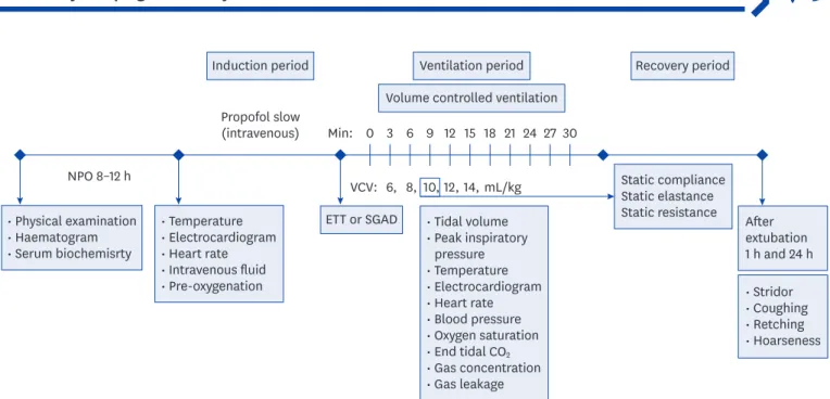
A diagram illustrating the timeline of the protocol used in this study was shown in Fig. 1.
Recovery
All cats were monitored for 1 h after extubation for upper respiratory airway discomfort, including stridor, coughing, retching, and hoarse voice. After full recovery from the general anesthesia, the cats were returned to their owners. The owners were instructed to record any abnormal signs in the first 24 h at home.
Statistical analysis
STATA12 (StataCorp, USA) was used to estimate the required sample size using t-tests for paired samples with a power of 80% and an alpha error of 0.05 to detect a difference of 2 mg in propofol need for anesthetic induction and comparing the data for the SGAD and ETT groups. All data were tested for normality using a Shapiro-Wilk test. All parameters, including dosage of propofol and static respiratory measurements of cats in the SGAD and ETT groups, were analyzed using a t-test. The association between leakage and hypercapnia was determined using Fisher's exact test. The significant level was set at p < 0.05.
RESULTS
Patients' characteristics and the doses of propofol used to insert the SGAD and the ETT in the cats are compared in Table 1. No statistically significant differences were identified between the SGAD and ETT groups, including age (SGAD: 2.3 ± 0.5 years, ETT: 2.5 ± 0.5 years; p = 0.49), sex (SGAD: 7 males and 8 females, ETT: 8 males and 7 females; p = 1.00), body weight
• Physical examination
• Haematogram
• Serum biochemisrty
• Temperature
• Electrocardiogram
• Heart rate
• Intravenous fluid
• Pre-oxygenation • Stridor
• Coughing
• Retching
• Hoarseness After extubation 1 h and 24 h Static compliance
Static elastance Static resistance
• Tidal volume
• Peak inspiratory pressure
• Temperature
• Electrocardiogram
• Heart rate
• Blood pressure
• Oxygen saturation
• End tidal CO
2• Gas concentration
• Gas leakage Induction period Ventilation period
Propofol slow (intravenous) Min:
VCV:
0 3 6 9 12 15 18 6, 8, 10, 12, 14, mL/kg
21 24 27 30 NPO 8–12 h
Volume controlled ventilation
Recovery period
ETT or SGAD
Fig. 1. Diagram illustrating the timeline of the protocol used in this study. Cats were randomly allocated to airway control groups using an ETT or a SGAD. Five tidal
volumes (6, 8, 10, 12, and 14 mL/kg) were randomly applied during VCV, and respiratory rates were adjusted to achieve a minute ventilation of 100 mL/kg/min.
ETT, endotracheal tube; SGAD, supraglottic airway device; VCV, volume-controlled ventilation.
(SGAD: 3.8 ± 0.7 kg, ETT: 3.7 ± 0.7 kg; p = 0.17), body condition score (SGAD: 3.2 ± 0.5, ETT:
3.4 ± 0.7; p = 0.20), and propofol dose needed for induction (SGAD: 7.1 ± 1.0 mg/kg, ETT: 7.3
± 1.7 mg/kg; p = 0.62).
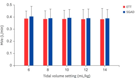
The minute ventilation at the five different tidal volumes used in the present study were comparable between the SGAD and ETT groups (p > 0.05; Fig. 2). For each tidal volume of controlled ventilation, different respiratory rates were adjusted in each cat to allow a minute ventilation of 100 mL/kg/min (p > 0.05; Fig. 3). There was no significant difference in respiratory rates between the SGAD group and the ETT group (p > 0.05; Fig. 3). Airway resistance was compared between the SGAD and ETT groups at a tidal volume of 10 mL/kg and a minute ventilation of 100 mL/kg/min (Table 2). The static resistance of the SGAD (22.0 ± 8.1 cmH
2O/L/sec) was significantly lower than that of the ETT (36.6 ± 12.9 cmH
2O/L/sec; p < 0.01).
In addition, airway leakage (> 20% of the baseline tidal volume) was compared between the SGAD and ETT groups (Table 3). There was no significant difference in the number of leakages at different tidal volume settings between the SGAD and ETT groups. However, of the 75 total trials, there were significantly more leakages in the SGAD group (8 events) than in the ETT group (1 event; p = 0.03) (Table 3).
Table 1. Age, sex, body weight, and body condition score of the sample and dose of propofol needed to insert the
ETT or SGAD
Parameters ETT SGAD
p-valueNumber 15 15 -
Age (yr) 2.5 ± 0.5 2.3 ± 0.5 0.49
Sex 1.00
Male 8 7
Female 7 8
Body weight (kg) 3.7 ± 0.7 3.8 ± 0.7 0.17
Body condition score 3.4 ± 0.7 3.2 ± 0.5 0.20
Dose of propofol (mg/kg) 7.3 ± 1.7 7.1 ± 1.0 0.62
Values are presented as number or mean ± SD.
ETT, endotracheal tube; SGAD, supraglottic airway device.
0.5
0.4
MV e (L/ min)
Tidal volume setting (mL/kg) 0.2
0.3
0.1
0 6 8 10 12 14
ETT SGAD
Fig. 2. Comparison of average minute ventilation (MVe) in cats during VCV when using a SGAD or an ETT. There
was no significant difference between the SGAD and ETT groups at the different tidal volume settings.
ETT, endotracheal tube; SGAD, supraglottic airway device; VCV, volume-controlled ventilation.
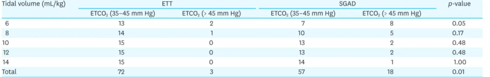
The occurrence of hypercapnia (ETCO
2> 45 mmHg) was compared between the ETT and SGAD groups. No significant difference in the number of hypercapnia occurrences at different tidal volume settings between the SGAD and ETT groups was detected.
Nonetheless, of the 75 total trials, there were significantly more occurrences of hypercapnia in the SGAD group (18 events) than in the ETT group (3 events; p < 0.01) (Table 4).
20
15
Respir at ory r at e ( br eath/ min)
Tidal volume setting (mL/kg) 5
10
0 6 8 10 12 14
ETT SGAD
Fig. 3. Tidal volume (mL/kg) and respiratory rate (times/min) when using a SGAD or an ETT. No significant
difference between the SGAD and ETT groups at different tidal volume settings was detected.
ETT, endotracheal tube; SGAD, supraglottic airway device.
Table 2. Static respiratory measurements of the ETT and SGAD groups*
Static respiratory measurements ETT SGAD
p-valuePIP (cm H
2O) 7.8 ± 1.6 6.3 ± 2.0 0.04
Static compliance (mL/cm H
2O) 6.1 ± 2.0 7.0 ± 6.0 0.17
Static resistance (cm H
2O/L/sec) 36.6 ± 12.9 22.0 ± 8.1 < 0.01
Static elastance (cm H
2O/mL) 180.8 ± 62.6 151.4 ± 34.8 0.12
ETT, endotracheal tube; SGAD, supraglottic airway device; PIP, peak inspiratory pressure.
*
Static respiratory measurements were recorded using a tidal volume of 10 mL/kg and minute ventilation of 100 mL/kg/min.
Table 3. Number of airway leakages (> 20% of the baseline tidal volume) in the ETT group vs. the SGAD group
Tidal volume (mL/kg) ETT SGAD
p-valueNo leak Leak > 20% No leak Leak > 20%
6 15 0 13 2 0.48
8 15 0 15 0 1.00
10 15 0 14 1 1.00
12 15 0 13 2 0.48
14 14 1 12 3 0.60
Total 74 1 67 8 0.03
ETT, endotracheal tube; SGAD, supraglottic airway device.
Table 4. Comparison of the occurrence of hypercapnia (ETCO2
> 45 mmHg) between the ETT and SGAD groups
Tidal volume (mL/kg) ETT SGAD
p-valueETCO
2(35–45 mm Hg) ETCO
2(> 45 mm Hg) ETCO
2(35–45 mm Hg) ETCO
2(> 45 mm Hg)
6 13 2 7 8 0.05
8 14 1 10 5 0.17
10 15 0 13 2 0.48
12 15 0 13 2 0.48
14 15 0 14 1 1.00
Total 72 3 57 18 0.01
ETT, endotracheal tube; SGAD, supraglottic airway device.
DISCUSSION
In the present study, the efficacies of SGAD and ETT for airway management in cats during VCV were compared. The propofol dose required for cats in the SGAD group and those in the ETT group did not differ. However, cats in the SGAD group had lower airway resistance than that of the cats in the ETT group. Nonetheless, gas leakage and hypercapnia events were more common in cats in the SGAD group than in the cats of the ETT group.
As noted above, the amount of propofol used in cats with an SGAD (7.1 ± 1.0 mg/kg) was not different from the amount used in cats with an ETT (7.3 ± 1.7 mg/kg; p = 0.55).
These results differ from those in previous research that suggested a lower propofol dose was required in cats with an SGAD than in cats with an ETT [3]. This difference may be explained by the effects of the premedication applied in the previous study [3]. In this study, premedication was not used in order to avoid any possible sedative effect that may cause cortical suppression, thus preventing a direct comparison of the effective dose of propofol.
It should be noted that the total propofol dose required for insertion of an SGAD or an ETT in the present study was higher than the total dose of propofol (2–5 mg/kg) used in the previous study [3]. Nonetheless, the amount of propofol used in the present study did not exceed 8 mg/kg (non-premedicated dose), and no side effects such as apnea [16], cyanosis, or bradycardia [17] were detected.
Inserting an ETT in cats should be done carefully because cats have a small and delicate trachea and larynx [18]. An ideal cuff pressure of 20–30 cm H
2O has been recommended to minimize damage to tracheal mucosa as well as to prevent trachea rupture [19,20].
Intubation with underinflation of the cuff can increase the risk of aspiration [20] and interferes with mechanical ventilation. The first LMA was developed for use in humans in 1981 [21]. At present, the LMA is the most common type of SGAD used in human adult, child, and infant patients due to the ease of its use [22]. LMAs have been used in many animal species, but their design may not match the oropharyngeal anatomy, size, species, and breed of animals [12], thereby resulting in gas leakage and trauma to the oropharyngeal area [1].
Recently, a veterinary specific SGAD has been develop for use in cats and rabbits [23].
The use of an SGAD can help maintain airway patency for routine procedures as well as during emergency procedures. In the present study, SGADs were used successfully during professional dental scoring in cats, since caudal teeth are affected with more severe gingivitis [14]. However, it remains unclear whether SGAD can be used in cats for dental scaling since that procedure involves the use of a large fluid volume. Furthermore, in patients with the ASA physical status as ASA 3 or higher, the use of an SGAD is not recommended [23].
In the present study, static resistance was compared between the SGAD and ETT groups based on ventilator-derived measurements. Static resistance in the ETT group was significantly higher than that in the SGAD group (Table 2). These finding can be explained in part by the fact that an SGAD has a notably larger internal diameter than that of an ETT;
there may have been a significant increase in airway resistance due to ETT use because airway resistance varies inversely with the tube radius to the fourth power. If the radius is reduced by half, the resistance will increase by up to sixteen-fold [8,13].
The leakage during VCV was similar in the SGAD and ETT groups, but leakage events were
identified more often in the SGAD group than in the ETT group. In contrast, in the previous
study described earlier, significantly more cats in the ETT group had leakages during CMV at a PIP of > 8 cm H
2O compared with those of the study's SGAD and LMA groups [3]. Both that study and the present study detected leakages by measuring the difference between VTi and VTe, whereas other previous studies detected leakages by measuring the peak concentration of anesthetic gas in the mouth and detecting audible leakages [1,18]. The results of the present study also differed from another previous study that reported a higher incidence of leakages in their ETT group than in their SGAD group after device placement and during the surgical procedure [24]. It should be noted that PCV has allowed a lower PIP while maintaining equal ventilation compared with that of VCV during general anesthesia in children when using an LMA [25]. Ventilation using VCV, therefore, may lead to a higher PIP, resulting in more leakages. Moreover, it is possible that an ETT provides a better air seal than that of an SGAD.
The physiological dead space is that part of the tidal volume that does not participate in the gas exchange and is comprised of the anatomical dead space and the alveolar dead space [26]. Significant increases in alveolar dead space may occur in animals under anesthesia, and these may be due to a drop in cardiac output (Q) and/or pulmonary artery blood pressure [12]. Moreover, the volume of dead space is also influenced by alteration of tidal volume and the frequency of ventilation. Alveolar ventilation is the portion of ventilation that participates in the gas exchange with pulmonary blood, and alveolar ventilation has a positive association with CO
2production (VCO
2) and a negative association with the arterial CO
2partial pressure (PaCO
2). Thus, if the alveolar ventilation is reduced by half and the VCO
2remains unchanged, significant levels of the alveolar ventilation and the arterial PaCO
2will be increased by up to double [13]. In the present study, ETCO
2was monitored instead of PaCO
2because the method for monitoring ETCO
2is less invasive than that associated with blood gas analysis.
Because different airway resistance may occur due to the use of different airway management such as via an SGAD or an ETT, PaCO
2or ETCO
2may be increased in animals with a higher airway resistance. In contrast, the results have revealed significantly more occurrences of hypercapnia in the SGAD group than in the ETT group (Table 4). This can be explained in part by a significant difference in the dead space added by the instruments used in the SGAD group (dead space of SGAD size C3 was 6.7 ± 0.1 mL and that of SGAD size C4 was 7.3 ± 0.1 mL) compared to that of the ETT group (dead space volume of the ETT of internal diameter 3.5 mm was 1.7 mL and that of the 4.0 mm internal diameter ETT was 2.5 mL). To avoid the occurrence of hypercapnia during mechanical ventilation in cats when using VCV, the results suggest that the minimum tidal volume in cats should be set at 12 mL/kg if an SGAD is used and 10 mL/kg if an ETT is used.
In conclusion, the use of an SGAD provided lower airway resistance and less work of breathing during VCV compared with those when using an ETT in cats. However, airway leakages were more common among cats in the SGAD group. The SGAD had more dead space than the ETT, resulting in more hypercapnia in the SGAD group, therefore tidal volume would need to be adjusted in cats because of the small size of the airway.
ACKNOWLEDGMENTS
The authors thank Miss Hathaipat Rattanathanya for her technical assistance and Dr.
Panpicha Sattasathuchana for proofreading the manuscript.
REFERENCES
1. van Oostrom H, Krauss MW, Sap R. A comparison between the v-gel supraglottic airway device and the cuffed endotracheal tube for airway management in spontaneously breathing cats during isoflurane anaesthesia. Vet Anaesth Analg 2013;40:265-271.
PUBMED | CROSSREF
2. Hofmeister EH, Trim CM, Kley S, Cornell K. Traumatic endotracheal intubation in the cat. Vet Anaesth Analg 2007;34:213-216.
PUBMED | CROSSREF
3. Prasse SA, Schrack J, Wenger S, Mosing M. Clinical evaluation of the v-gel supraglottic airway device in comparison with a classical laryngeal mask and endotracheal intubation in cats during spontaneous and controlled mechanical ventilation. Vet Anaesth Analg 2016;43:55-62.
PUBMED | CROSSREF
4. Brodbelt D. Feline anesthetic deaths in veterinary practice. Top Companion Anim Med 2010;25:189-194.
PUBMED | CROSSREF
5. Brodbelt DC, Pfeiffer DU, Young LE, Wood JL. Risk factors for anaesthetic-related death in cats: results from the confidential enquiry into perioperative small animal fatalities (CEPSAF). Br J Anaesth 2007;99:617-623.
PUBMED | CROSSREF
6. Mitchell SL, McCarthy R, Rudloff E, Pernell RT. Tracheal rupture associated with intubation in cats: 20 cases (1996–1998). J Am Vet Med Assoc 2000;216:1592-1595.
PUBMED | CROSSREF
7. Hardie EM, Spodnick GJ, Gilson SD, Benson JA, Hawkins EC. Tracheal rupture in cats: 16 cases (1983–
1998). J Am Vet Med Assoc 1999.214:508-512.
PUBMED
8. Costanzo LS. Physiology. 1st ed. Williams & Wilkins, Philadelphia, 1995.
9. Crotaz IR. Initial feasibility investigation of the v-gel airway: an anatomically designed supraglottic airway device for use in companion animal veterinary anaesthesia. Vet Anaesth Analg 2010;37:579-580.
PUBMED | CROSSREF
10. Toman H, Erbas M, Kiraz HA, Sahin H, Ovali MA, Uzun M. Comparison of effects of classic LMA, cobraPLA and V-gel rabbit on QTc interval. Bratisl Lek Listy 2015;116:632-636.
PUBMED | CROSSREF
11. Muir WW, Hubbell JA, Bednarski RM, Skarda RT. Handbook of Veterinary Anesthesia. 4th ed. Elsevier Inc., St. Louis, 2007.
12. Mosley CA. Lumb & Jones’ Veterinary Anesthesia and Analgesia. 5th ed. John Wiley & Sons, Inc., Pondicherry, 2015.
13. West JB, Luks AM. West’s Respiratory Physiology: The Essentials. 10th ed. Wolters Kluwer, Philadelphia, 2016.
14. Thengchaisri N, Steiner JM, Suchodolski JS, Sattasathuchana P. Association of gingivitis with dental calculus thickness or dental calculus coverage and subgingival bacteria in feline leukemia virus- and feline immunodeficiency virus-negative cats. Can J Vet Res 2017;81:46-52.
PUBMED
15. Gurney M, Cripps P, Mosing M. Subcutaneous pre-anaesthetic medication with acepromazine- buprenorphine is effective as and less painful than the intramuscular route. J Small Anim Pract 2009;50:474-477.
PUBMED | CROSSREF
16. Ko J. Small Animal Anesthesia and Pain Management: a Color Handbook. 1st ed. Manson Publishing Ltd., London, 2013.
17. Ramsey I. BSAVA Small Animal Formulary. 6th ed. British Small Animal Veterinary Association, Wales, 2008.
18. Cassu RN, Luna SP, Teixeira Neto FJ, Braz JR, Gasparini SS, Crocci AJ. Evaluation of laryngeal mask as an alternative to endotracheal intubation in cats anesthetized under spontaneous or controlled ventilation.
Vet Anaesth Analg 2004;31:213-221.
PUBMED | CROSSREF
19. Nseir S, Brisson H, Marquette CH, Chaud P, Di Pompeo C, Diarra M, Durocher A. Variations in endotracheal cuff pressure in intubated critically ill patients: prevalence and risk factors. Eur J Anaesthesiol 2009;26:229-234.
PUBMED | CROSSREF
20. White DM, Redondo JI, Mair AR, Martinez-Taboada F. The effect of user experience and inflation technique on endotracheal tube cuff pressure using a feline airway simulator. Vet Anaesth Analg 2017;44:1076-1084.
PUBMED | CROSSREF
21. Pennant JH, White PF. The laryngeal mask airway. Its uses in anesthesiology. Anesthesiology 1993;79:144-163.
PUBMED | CROSSREF
22. Bansal SC, Caoci S, Dempsey E, Trevisanuto D, Roehr CC. The laryngeal mask airway and its use in neonatal resuscitation: a critical review of where we are in 2017/2018. Neonatology 2018;113:152-161.
PUBMED | CROSSREF
23. Thomas JA, Lerche P. Anesthesia and Analgesia for Veterinary Technicians. 5th ed. Elsevier Health Sciences, Philadelphia, 2016.
24. Barletta M, Kleine SA, Quandt JE. Assessment of v-gel supraglottic airway device placement in cats performed by inexperienced veterinary students. Vet Rec 2015;177:523.
PUBMED | CROSSREF
25. Keidan I, Berkenstadt H, Segal E, Perel A. Pressure versus volume-controlled ventilation with a laryngeal mask airway in paediatric patients. Paediatr Anaesth 2001;11:691-694.
PUBMED | CROSSREF
26. Dassios T, Dixon P, Hickey A, Fouzas S, Greenough A. Physiological and anatomical dead space in mechanically ventilated newborn infants. Pediatr Pulmonol 2018;53:57-63.
PUBMED | CROSSREF