Purification and Characterization of a Fibrinolytic Enzyme from Snake Venom of Macrovipera lebetina turanica
10
0
0
전체 글
(2) 6. Journal of Pharmacopuncture 제 14권 제 2호( 2011년 6월). inhibitors, and peptides, many of which have found uses in the diagnosis and treatment of haemostatic disorders.4) Some of these toxins, both enzymic and non-enzymic, lead to the spectacular changes in haemostasis and the frequent hemorrhage seen after snake bite.5) It has been suggested that this process is part of a strategy to immobilize prey and to increase target tissue permeability to other venom toxins.6) The activities of these toxins include coagulation and anticoagulation of blood, platelet-activation, anti-platelet function, fibrinolytic activation, and hemorrhage.7) The venom enzymes toxic to animals comprise acetylcholinesterases, ADPases, phopholipases, hialuronidases, and hemostatic proteases. 2) The hemostatic proteases can be divided into metalloproteases and serine proteases, of which fibri(gen) olytic proteases have drawn particular attentions as potential therapeutic agents to remove plasma fibrinogen from the circulation for treatment of acute isochaemic stroke8) and to dissolve blood clot for treatment of occlusive thrombi.4) Fibrolase from copperhead snake venom degrades both and chains of fibrin and showed some promise as a thrombolytic agent.9) Alfimeprase, a recombinant fibrinolytic enzymes derived from fibrolase has been being developed as a clinical agent.10),11) Other fibrinolytic proteases which dissolve blood clots include afaacytin, from horned viper12) and the fibrinogenase from Macrovipera lebetina venom13). Macrovipera lebetina (Levatine viper) is a venom ous viper species found in south-east parts of Europe, in south-west Asia, and in north-west Africa. Five subspecies, consisting M. l. cerenovi, M. l. lebetina, M. l. obtuse, M. l. transmediterranea, and M. l. turanica are currently recognized. The snout is rounded and blunt when viewed from above, which is why it is also called the blunt-nosed viper.14),15) The venom of M. lebetina has various proteases, which acts on blood coagulation through both proand anticoagulant mechanisms and contains (1). proteases that degrade fibrinogen, but not fibrin; (2) fibrinolytic enzyme (lebetase); (3) factor X activator, (4) factor V activator. The fibrinolytic enzymes cleave of fragments from the C-terminals of -and -chains of fibrinogen, rendering it not clottable by thrombin.16) Lebetase, an ( )-fibrin(ogen)olytic metalloprotease, possesses direct-acting fibrinolytic activity which involves direct cleavage of fibrin without plasminogen activation and is also an anticoagulant that acts by inhibiting platelet aggregation. The enzyme readily hydrolyzes the A chain and more slowly the B -chain of fibrinogen and the oxidized insulin B-chain in the positions Ala 14-Leu 15 and Tyr16-Leu17. Its fibrinolytic activity is inhibited by EDTA and dithiothreitol. The protease consists of a single polypeptide chain with a molecular weight of 23,700 and is a metalloenzyme that contains one mole of Zn2+ and one mole of Ca2+ per mole of protein.17),18) The mature protein lebetase consists of 204 amino acids and its amino acid sequence shows strong similarity with fibrolase. The metallo protease domain has a typical zinc-chelating sequence.19) The present study isolated a fibrinolytic enzyme from M. l. turanica snake venom and characterized its catalytic properties.. II. Materials and Methods 1. Snake venom The snake venom from M. l. turanica in this study was in the shape of yellow crystal powder, whose specification indicated protein content of 90 2%, phospholipase A2 activity of 35 6 U/mg. coagulase activity of 80,000 8,000 U/mg , and LD50 of 2.6 0.5 mg/kg.. 2. Purification of fibrinolytic enzyme.
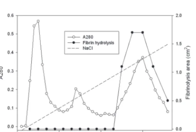
(3) Purification and Characterization of a Fibrinolytic Enzyme from Snake Venom of Macrovipera lebetina turanica. The venom powder (2 mg) was dissolved and dialyzed in 50 mM Tris-HCl, pH 7.6. The venom solution was loaded on the column (5 × 5.8 cm) of Q-Sepharose (GE, USA) equilibrated with the buffer. The column was eluted with the buffer and then with a concentration gradient of sodium chloride of up to 0.3 M. Two fractions with fibrinolytic activity were obtained (Fig 1). The first fraction eluted with 50 mM Tris-HCl, pH 7.6 was dialyzed in 50 mM Tris-HCl, pH 8.6. The dialyzed solution was loaded on a column (2.5 × 10 cm) of Q-Sepharose equilibrated 50 mM Tris-HCl, pH 8.6. The column was washed with the buffer and eluted with a concentration gradient of sodium chloride up to 0.2 M (Fig 2). The fraction with fibrinolytic activity was dialyzed in 50 mM Tris-HCl, pH 7.6, 0.15 M NaCl and separated on the column (2.5 × 120 cm) of Sephadex G-75. The second fraction released with the concentration gradient of sodium chloride from the column of Q-Sepharose (Fig 1) was dialyzed in 50 mM Tris-HCl, pH 7.6, 0.15 M NaCl and were separated on the column (2.5 × 120 cm) of Sephadex G-75 (GE, USA) equilibrated with the buffer. All the chromatography was performed at the refrigeration temperature.. 3. Fibrin plate assay Fibrinolytic activity was determined on fibrin plates according to the procedure of Astrup and Mullertz.21) An aliquot (200 μl) of thrombin (SigmaAldrich, USA) solution (10 U/ml in 100 mM TrisHCl, pH 7.8) were added to 9 ml of 0.1% fibrinogen (Sigma-Aldrich, USA) dissolved in the buffer solution. The mixed solution was added into petri dish with the diameter of 9 cm and incubated at 37 ℃ for 1 hr, until it converted into fibrin gel. An aliquot (10 μl) of the venom fraction was placed on the fibrin gel and then incubated at 37℃ for 18 hr. Diameters (r1 and r2) at a right angle of hydrolysis. 7. zone were measured. The fibrinolysis area was calculated following the formula of 0.785 r1 × r2.. 4. SDS-PAGE Sodium dodecyl sulfate - polyacrylamide gel electrophoresis (SDS-PAGE) was performed according to the procedure described by Laemmli.22) The molecular weight markers (Bio-Rad, USA) for the SDS-PAGE consisted of myosin (200,000), -galactosidase (116,250), phosphorylase b (97,400) bovine serum albumin (66,200), ovalbumin (45,000), carbonic anhydrase (31,000), trypsin inhibitor (21,500), lysozyme (14,000), and aprotinin (6,500).. 5. Fibrinogen hydrolysis The fibrinogen (4 mg) in 200 μl of 50 mM TrisHCl, pH 7.5, 0.15 M NaCl was added with 50 μl of the purified fibrinolytic enzyme and then the mixture was incubated at 37℃ for 6 hr. An aliquot (10 μl) of the mixture was taken at 0, 10, 30, 60, 120, and 240 min during the incubation and added with equal amount of the sample buffers for SDS-PAGE each time. The mixtures were added with a couple of drops of mineral oil, heated at 100℃ for 5 min, and analyzed in SDS-PAGE.. 6. Protein determination Protein concentration was determined using BCA Protein Assay Reagent (Pierce, USA). An aliquot (100 μl) of samples was mixed with 2.0 ml of BCA Reagent which is prepared following the producer s manure. The mixture was incubated at 37℃ for 30 min and then cooled at room termperature for 30 min. The absorbance at 562nm of the mixture was measured. Bovine serum albumin was used for calibration curve..
(4) 8. Journal of Pharmacopuncture 제 14권 제 2호( 2011년 6월). III. Results 1. Purification of fibrinolytic enzyme from the snake venom The snake venom was separated into two major fractions by using ion exchange chromatography of Q-Sepharose which was eluted using 50 mM TrisHCl, pH 7.6 and then using the buffer with a concentration gradient up to 0.1 M sodium chloride. The first fraction from 4 to 23 and the second fraction from 44 to 53 showed strong fibrinolytic activity in fibrin plate assay (Fig 1). The first fraction was dialyzed in 50mM Tri-HCl, pH 8.6. The dialyzed sample was separated on a column of Q-Sepharose equilibrated with the buffer (Fig 2). The fraction release from the column with the buffer did not show fibinolytic activity (results not shown). The column was then eluted using the buffer with a concentration gradient of sodium chloride up to 0.3 M sodium chloride. The chromatogram in Fig 2 showed three fractions released from the column. The last fraction from 25 to 36 showed fibrinolytic activity in fibrin plate assay. The second fraction in Fig 1 and the last protein fraction in Fig 2 were dialyzed in 50 mM Tris-HCl, pH 7.6, 1.5 M NaCl, and then subjected to gel filtration chromatography using the column of Sephadex G-75, respectively (results not shown). The fractions with fibrinolytic activity were concentrated in dialysis bag using polyethylene glycol, and frozen stored. The volume and protein concentration of the fibrinolytic enzyme preparations isolated originally from the first fraction and the second fraction of ion exchange chromatography of Q-Sepharose in Fig 1 were 6.7ml and 3.29 mg/ml and 15.4ml and 3.92 mg/ml, respectively.. 2. SDS-PAGE analysis. SDS-PAGE of venom of M.l. turanica in SDSPAGE showed that the venom contained several major polypeptides with the molecular weights of 66,000, 35,000, 27,500, and 15,000 (Fig 3). The fibrinolytic enzyme preparation from the first fraction in Fig 1 showed a single polypeptide with the molecular weight of 27,500 (Fig 4). The fibrinolytic enzyme preparation from the second fraction in Fig 1 showed a major polypeptide with the molecular weight of 27,500 and minor polypeptides with the molecular weights of 42,000 and 15,000. The results suggested that the two preparations might contain the fibrinolytic enzymes with a same molecular weight. The fibrinolytic enzyme from the first fraction seemed to be purified and thus its enzymic characteristics were determined further.. 3. Fibrinogen hydrolysis Two hundred μl of 2% fibrinogen was added with 50 μl of the purified fibrinolytic enzyme and then the mixture was incubated at 37℃ for 4 hr. Ten μl of the mixture was taken at 0, 10, 30, 60, 120, and 240 min during the incubation and subjected to SDS-PAGE. The SDS-PAGE in Fig 5 showed that -chain of fibrinogen was hydrolyzed preferentially and no intact -chain was left after incubation for 240 min. -Chain of fibrinogen was hydrolyzed more slowly than -chain but -chain of fibrinogen was not hydrolyzed.. 4. Inhibition of the fibrinolysis by protease inhibitors and salts Thrombin was added into fibrinogen solution and then the mixture was incubated at 37℃ for 1 hr and then fibrinogen formed a translucent fibrin gel in the fibrin plate assay (Fig 6). The fibrinolytic enzyme which was placed on the gel hydrolyzed the fibrin gel and made a transparent circular zone.
(5) Purification and Characterization of a Fibrinolytic Enzyme from Snake Venom of Macrovipera lebetina turanica. filled with liquid as shown in Fig 6. The relationship between the area of the zone which was calculated from the measured diameter and the concentration of the fibrinolytic enzyme was shown in Fig 7. The results showed that the fibrinolysis area increased steeply at the lower concentrations from 0.01 to 0.02 mg/ml and then the slope became lower at the higher concentrations. Protease inhibitors were added to the fibrinolytic enzyme of 0.02 mg/ml and 10μl of the mixed solutions were placed on the gel in fibrin plate assay to determine their inhibitory effects on the fibrinolysis (Table 1.). The metalloprotease inhibitors, EDTA, EGTA, and 1,10-phenanthroline, inhibited the fibrinolysis completely. The dithiol-reducing compounds, cysteine and dithiothreitol, showed partial and complete inhibition on the fibrinolysis, respectively. Serine protease inhibitors, PMSF and TLCK, an acid protease inhibitor, pepstatin A, and a cysteine protease inhibitor, E-64, showed few inhibition on the fibrinolysis. Salts of 10 mM were added to the fibrinolytic enzyme of 0.02 mg/ml and 10 μl of the mixed solutions were placed on the gel in fibrin plate assay to determine their inhibitory effects on the fibrinolysis. Calcium chloride, iron(III) chloride, mercuric chloride, and cobalt (II) chloride inhibited the finolysis completely. Cupric sulfate inhibited the fibrinolysis strongly. Manganese chloride, zinc sulfate, and cesium chloride showed partial inhibition on the fibrinolysis. Strontium chloride and magnesium chloride did not inhibit the fibrinolysis. The fibrinolysis zone which was formed after addition of zinc chloride was clearer than the control.. IV. Discussion The venom of M. l. lebetina has various proteases, which act on blood coagulation through both pro- and anticoagulant mechanisms and contains (1) proteases that degrade fibrinogen, but not fibrin;. 9. (2) fibrinolytic enzyme (lebetase); (3) factor X activator, (4) factor V activator. The fibrin(ogen)olytic enzymes cleave of fragments from the C-terminals of - and -chains of fibrinogen, rendering it not clottable by thrombin.16) Lebetase, is an ( )-fibrin(ogen)olytic metalloprotease, possesses direct-acting fibrinolytic activity which involves direct cleavage of fibrin and hydrolyze -chain of fibrinogen faster than -chain of fibrinogen. Its fibrinolytic activity is inhibited by EDTA and dithiothreitol. The protease consists of a single polypeptide chain with a molecular weight of 23,700 and is a metalloenzyme that contains one mole of Zn2+ and one mole of Ca2+ per mole of protein.18),19) Two major fractions with fibrinolytic activity were separated from the venom of M. l. turanica by using ion exchange chromatography. The two preparations with fibrinolytic activity from the two major fractions isolated after additional ion exchange chromatography and gel filtration chromatography contained the major polypeptide with a molecular weight of 27,500, which suggested the polypeptides were the fibrinolytic enzyme. The fibrinolytic enzyme purified from the venom of M. l. turanica in this study seemed to be similar with lebatase. The fibrinolytic enzyme hydrolyzed -chain of fibrinogen faster than -chain of fibrinogen. Its fibrinolytic activity was inhibited by metalloprotease inhibitors, EDTA, EGTA, and 1,10phenanthroline and also by dithiothreitol, indicating that the fibrinolytic enzyme is metalloprotease containing dithiol groups. However, the molecular weight of the fibrinolytic enzyme, 27,000, was a little higher than that of labetase. Its fibrinolytic acitivity was inhibited completely by calcium chloride completely. The hydrolysis zone formed after addition of zinc chloride was smaller but clearer than the control. These results suggested that the fibrinolytic enzyme preparation might have lost some cationic salts during the purification, if the fibrinolytic enzyme were lebetase..
(6) 10. Journal of Pharmacopuncture 제 14권 제 2호( 2011년 6월). Further studies are necessary to determine the effects of salts on the fibrinolysis. In the present study a fibrinolytic enzyme was purified from the venom of M. l. turanica and its enzymatic characteristics were investigated. The purified enzyme preparation may be applied for a pharmacopuncturological therapy, which may enable treatment of acute isochaemic stroke by removing fibrinogen from the circulation and treatment of occlusive thrombi by dissolving blood clot. The fibrinolytic enzyme should dissolve plasma fibrinogen specifically without side effects for clinical application.. V. References 1. Markland, F. S. : Snake venoms and the hemostatic system. Toxicon 36:1749-1800, 1998. 2. Russel, F. E. : Toxic effects of animal toxins. In Klassen, C.D., Amdur, M.O., and Doull, J. (Ed.) Casarett and Doull s toxicology-the basic science of poisons, 3rd ed. MacMillan, New York, pp. 706-756, 1986. 3. Ramos, O. H. P. and Selistre-de-Araujo, H. S. : Snake venome metalloproteases-structure and function of catalytic and disintegrin domains. Comp. Biochem. Physiol. Part C. 142:328-346, 2006. 4. Marsh, N. and Willams, V. : Practical applications of snake venom toxins in haemostasis. Toxicon 45:1171-1181, 2005. 5. Kamiguti, A. S., Hay, C. R. M., Theakston, R. D. G., and Zuzel, M. : Insights into the mechanism of haemorrhage caused by snake venom metalloproteinases. Toxicon 34:627-642, 1996. 6. Faiz, M. A., Falkous, G., Harris, J. B., and Mantle, D. : Comparison of protease and related enzyme activities in snake venoms. Comp. Biochem. Physiol. B. 113:199-204, 1996. 7. Marsh, N. A. : Snake venoms affecting the haemostatic mechanism - a consideration of. their mechanisms, practical applications and biological significance. Blood Coagul. Fibrinolysis. 5:399-410, 1994. 8. Samsa, G. P., Matchar, D. B., Williams, G. R., Levy, D. E. : Cost-effectiveness of ancrod treatment of acute ischaemic stroke: results from the Stroke Treatment with Ancrod Trial (STAT). J. Eval. Clin. Pract. 8:61-70, 2002. 9. Markland, F. S., Reddy, K. N. N., and Guan, L. F. : Purification and characterization of a directacting fibrinolytic enzyme from Southern copperhead venom. In: Pirkle, H., Markland, F. S. (Eds.), Hemostasis and Animal Venoms. Marcel Dekker, New York, pp. 173-189, 1988. 10. Swenson, S. and Markland Jr. F. S. : Snake venom fibrin(ogen)olytic enzymes. Toxicon 45:1021-1039, 2005. 11. Toombs, C. F. : Alfimplrase: pharmacology of a novel fibrinolytic metalloproteinase for thrombolysis. Haemostasis 31:141-147, 2001. 12. Baker, B. J., Tu, A. T. : Atroxase: a fibrinolytic enzyme isolated from the venom of western diamondback rattlesnake: isolation, characterization and cloning. Adv. Exp. Med. Biol. 391:203-211, 1996. 13. Gasmi, A., Chabchoub, A., Guermazi, S., Karoui, H., Elayeb, M., and Dellagi, K. : Further characterization and thrombolytic activity in a rat model of a fibrinogenase from Vipera lebetina venom. Thromb. Haemostas. 86:233-242, 1997. 14. McDiarmid, R. W., Campbell, J. A., and Toure, T. : Snake species of the world: Taxonomic and geographic reference, vol. 1. Herpetologists League pp. 511, 1999. 15. Integrated Taxonomic Information System : Macrovipera labetina.(http://www.itis.gov/ servlet/SingleRpt/SingleRpt?search_topic=TS N&search_value=634977), 2009. 16. Siigur, J., Aaspollu, A., Tonismagi, K., Trummal, K., Samel, M., Vija, H., Subbi, J., Siigur, E., : Proteases from Vipera lebetina.
(7) Purification and Characterization of a Fibrinolytic Enzyme from Snake Venom of Macrovipera lebetina turanica. 11. venom affecting coagulation and fibrinolysis. Haemostasis 31:123-132, 2001. 17. Siigur, E. and Siigur, J. Purification and characterization of lebetase, a fibrinolytic enzyme from Vipera lebetina (snake) venom. Biochim. Biophys. Acta. 1074:223-229, 1991. 18. Siigur, J., Samel, M., Tonismagi, K., Subbi, J., and Siigur, E. : Biochemical characterization of lebetase, a direct-acting fibrinolytic enzyme from Vipera lebetina snake venom. Thromb. Res. 90:39-49, 1998.. 19. Siigur, E., Aaspollu, A., Tu, A. T., and Siigur, J. : cDNA cloning and deduced amino acid sequence of fibrinolytic enzyme (lebetase) from Vipera lebetina snake venom. Biochem. Biophy. Res. Comm. 224:297-2236, 1996. 20. Astrup, T., and Mullertz, S. : The fibrin plate method for estimating of fibrinolytic activity. Archs. Biochem. Biophys. 40:346-351, 1952. 21. Laemmli, U. K. : Cleavage of structural proteins during the assembly of the head of bacteriophage T-4. Nature (London) 227: 680-685, 1970.. Fig 1. Ion exchange chromatography of the venom of M. l. turanica on a column (5 5.8 cm) of Q-Sepharose equilibrated with 50 mM Tris-HCl, pH 7.6.. Fig 2. Ion exchange chromatography of the first fraction in Fig 1 on a column (2.5 8 cm) of Q-Sepharose equilibrated with 50 mM Tris-HCl, pH 8.6..
(8) 12. Journal of Pharmacopuncture 제 14권 제 2호( 2011년 6월). 205,000 116,200 97,400 66,000 45,000. 31,000. 21,500. 14,400 1. 2. 3. 4. Fig 3. SDS-PAGE of the venom of M. l. turanica and its fibrinolytic enzyme preparations.1, standard molecular weight marker; 2, venom; 3, the fibrinolytic enzyme preparation from the first fraction in Fig 1; 4, the fibrinolytic enzyme preparation from the second fraction in Fig 1.. Fig 4. Molecular weight determination in SDS-PAGE of the fibrinolytic enzyme in the fibrinolytic enzyme preparation isolated from the first fractions in Fig 1. A, galactosidase (116,250); B, phosphorylase b (97,400); C, bovine serum albumin (66,200); D, ovalbumin (45,000); E, carbonic anhydrase (31,000); F, trypsin inhibitor (21,500); G, the fibrinolytic enzyme.. 205,00 0 116,20 0 97,400 66,000. B. 45,000. A C 31,000. F E. D. 21,500 14,400 1. 2. 3. 4. 5. 6. 7. 8. Fig 5. Hydrolysis of fibrinogen by the purified fibrinolytic enzyme from the venom of M. l. turanica. 1, Standard molecular weight marker; 2, fibrinogen without the fibrinolytic enzyme; 3, 4, 5, 6, 7, and 8, fibrinogen taken at 0, 10, 30, 60, 120, and 240 min after incubating with the fibrinolytic enzyme at 37 .. Fig 6. Fibrin plate assay showing transparent fibrinolysis zones. The fibrinolytic enzyme (10 l) at the concentration of 0.01(A), 0.02(B), 0.04(C), 0.06(D), and 0.08(E) mg/ml was placed on the fibrin gel..
(9) Purification and Characterization of a Fibrinolytic Enzyme from Snake Venom of Macrovipera lebetina turanica. 13. Fig 7. The relationship between the concentration of the fibrinolytic enzyme and the fibrinolysis area in fibrin plate assay.. Table 1. Effects of protease inhibitors on the fibrinolysis by the fibrinolytic enzyme purified from the venom of M. l. turanica. Protease inhibitors. Concentration(mM). Relative fibrinolysis(%). Control. 0. 100. Pepstatin A. 0.01. 100. Iodoactate. 1. 107. PMSF. 1. 72. TLCK. 1. 101. E-64. 0.1. 115. Dithiothreitol. 10. 0. Cysteine. 10. 66. EDTA. 10. 0. EGTA. 10. 0. 1,10-phenanthroline. 10. 0.
(10) 14. Journal of Pharmacopuncture 제 14권 제 2호( 2011년 6월). Table 2. Effects of salts on the fibrinolysis by the fibrinolytic enzyme purified from snake venom of M. l. turanica Salts. Concentration(mM). Relative fibrinolysis(%). Control. 0. 100. MgCl2. 10. 100. CaCl2. 10. 0. ZnCl2. 10. 41. MnCl2. 10. 51. FeCl2. 10. 0. CuSO2. 10. 5. CoCl2. 10. 0. CsCl2. 10. 86. SrCl2. 10. 100. HgCl2. 10. 0.
(11)
수치


관련 문서
1 John Owen, Justification by Faith Alone, in The Works of John Owen, ed. John Bolt, trans. Scott Clark, "Do This and Live: Christ's Active Obedience as the
The sublimation purification method is similar to the vapor deposition process ,in which crystallization occurs after evaporation from a solid state to a
The fibrinolytic enzyme was purified by using DEAE sepharose CL-6B fast flow chromatography followed by Sephadex G-75 gel filtration and POROS 20
This report make a description of criminal special code for purification and improvement of terminology and sentence.. All the while, criminal special code make a
The 5-day-old seedlings overexpressing the AtTX12 -like genes originated from crops of Brassicaceae were used for the extraction of the total RNA from which cDNA was
First, computational fluid dynamics(CFD) analysis were used to predict the heat transfer and pressure drop characteristics of each plate heat exchanger with snake
This report make a description of criminal special code for purification and improvement of terminology and sentence.. All the while, criminal special code make
The breakthrough curve of ion exchange fiber(KC31) toward As(Ⅴ) at regenerated with 0.5 M NaCl... H., 2001, Removal of Chromium from Water and Wastewater by Ion Exchange
