Available at http://www.formulastudy.com
HFS
Original Article / 원저
EJ 인간 방광암 세포에서 bufalin 의 TRAIL 저항성 극복 효과
홍수현
*동의대학교 한의과대학 생화학교실
The Effect of overcoming the TRAIL resistance through bufalin in EJ human bladder cancer cell
Su Hyun Hong
*Department of Biochemistry, College of Korean Medicine, Dong-Eui University
ABSTRACT
Objectives : Bufalin is one of the bioactive component of 'Sum Su (蟾酥)', which is obtained from the skin and parotid venom gland of toad. Bufalin has been known to possess the inhibitory effects on cell proliferation and inducing apoptosis in various cancer cells. The tumor necrosis factor (TNF)-related apoptosis-inducing ligand (TRAIL) has concerned, because it can selectively induce apoptotic cell death in many types of malignant cells, while it is relatively non-toxic to normal cells. Here, we investigated whether bufalin can trigger TRAIL-induced apoptotic cell death in EJ human bladder cancer cells.
Methods : Effects on the cell viability and apoptotic activity were quantified using MTT assay and flow cytometry analysis, respectively. To investigate the morphological change of nucleus, DAPI staining was performed. Protein expressions were measured by immunoblotting.
Results : A combined treatment with bufalin (10 nM) and TRAIL (50 ng/ml) significantly promoted TRAIL-mediated growth inhibition and apoptosis in EJ cells. The apoptotic effects were associated with the up-regulation of death receptor proteins, and the down-regulation of cFLIP and XIAP. Moreover, our data showed that bufalin and TRAIL combination activated caspases and subsequently increased degradation of poly(ADP-ribose) polymerase.
Conclusions : Taken altogether, the nontoxic doses of bufalin sensitized TRAIL-mediated apoptosis in EJ cells.
Therefore, bufalin might be an effective therapeutic strategy for the safe treatment of TRAIL-resistant bladder cancers.
Key words : Bufalin, TRAIL, apoptosis, caspase, death receptor 5, bladder cancer
ⓒ 2017 The Korean Medicine Society For The Herbal Formula Study
This paper is available at http://www.formulastudy.com which permits unrestricted non-commercial use, distribution, and reproduction in any medium, provided the original work is properly cited.
Ⅰ. 서론
3)방광암은 흔한 비뇨 생식기 악성 신생물 중 하나로 나이가 많을수록 발병률이 증가하는 것으로 알려져 있다1,2). 전 세계적으로 노인 인구의 증가에 따라 방 광암의 발생률이 증가하고 있는 추세이지만, 진행성 방광암의 경우에는 평균 생존률이 14-15개월 정도로 일반적으로 치료가 잘 되지 않는 상황이다. 최근 다 양한 연구 성과로 전립선암과 신세포암과 같은 비뇨 생식기 암의 치료법에는 괄목한 말한 성과가 있는 반 면에, 방광암에 대한 치료 성과는 상대적으로 미미한 편이다3). 특히 사망률이 높은 진행성 방광암의 치료 율을 높이기 위한 방법의 하나로 Tumor necrosis factor (TNF)-related apoptosis-inducing ligand (TRAIL, 또한 Apo2L 및 TNFSF10)에 의해 유도되는 세포 사 멸에 관심이 높아지고 있다4,5).
TRAIL은 세포 내의 death domain을 가지고 있는 death receptors 4 (DR4, 또한 TRAIL-R1 및 TNFRSF10a) 또는 death receptors 5 (DR5, 또한 TRAIL-R2와 TNFRSF10b) 와 결합하여 apoptosis 관련 단백질들의 신호 전달 기전을 통해 apotposis를 유도하는 것으로 알려져 있다
6,7). TRAIL은 많은 종류의 형질 전환 또는 악성 세포 에서 세포 사멸 세포 사멸을 선택적으로 유도할 수 있는 반면, 정상 세포에는 상대적으로 독성이 없기 때문에 새로운 항암치료의 대안으로 관심이 증대되었 다4,8). 그러나 일부 악성 세포는 TRAIL에 내성이 있 음이 밝혀져 임상 적용에 한계를 가지고 있다9,10). 따 라서 TRAIL에 저항성을 가지는 암세포에 TRAIL과 병용 치료하여 TRAIL 유도 apoptosis를 촉진할 수 있는 새로운 약물을 발견하려는 노력이 계속되고 있
다8,11). 현재까지 여러 가지 화학 요법제 또는 천연
물질과 TRAIL의 조합으로 DR의 상향 조절, caspases 활성화, cellular Fas-associated death domain-like interleukin-1β-converting enzyme inhibitory protein (cFLIP) 및 X-linked inhibitor of apoptosis protein (XIAP)와 같은 생존 단백질의 하향 조절과 Bcl-2 계 열 단백질의 조절에 의해 apoptosis가 유도되는 것으 로 보고되었다12-15). 본 연구에서는 이전의 연구에서 TRAIL에 저항성을 가지고 있는 것으로 나타난 EJ
인간 방광암 세포의 저항성을 bufalin으로 극복할 수 있는가에 대해 연구하였다.
Bufalin은 두꺼비의 이후선(耳後腺)에서 분비되는 흰색의 장액인 '섬수(蟾酥, Bufonis Venenum)'의 주 요 생리 활성물질 중 하나로
마취, 호흡 억제, 혈압 상승 뿐 아니라 및 항종양 효과 등 다양한 생리적 능력을 가지고 있는 것으로 알려져 있다16-18). 그 중 항종양 작용으로 bufalin은 폐암, 전립선암, 난소암, 위암, 간암, 유방암, 백혈병 및 방광암과 같이 광범위한 암세포에서 세포 증식을 억제하고 세포 사멸을 일으키는 것으로 입증되었다
16,17). 하지만 현재까지 EJ 인간 방광암 세포에서
bufalin과 TRAIL의 병용 투여에 대한 연구는 미비한 실정이다. 따라서 본 연구에서는 저용량의 bufalin이 EJ 세포의 TRAIL에 대한 내성을 극복하여 세포 증 식을 억제하고 apoptosis를 촉진시킬 수 있는지를 조 사하고, 그 근본적인 분자 기전을 이해하는데 초점을 두었으며, 의미 있는 결과를 도출하였기에 보고하는 바이다.
Ⅱ. 재료 및 방법
1. 시약 및 항체
재조합 TRAIL은 Koma Biotech Inc. (Seoul, Korea) 에서 구입하여 phosphate buffered saline (PBS)에 녹여 사용하였다. Bufalin은 Sigma-Aldrich Chemical Co. (St. Louis, MO, USA)에서 구입하여 dimethyl sulfoxide (DMSO)에 녹여 사용하였다. 3-(4,5-dimethylthiazol- 2-yl)- 2,5-diphenyltetrazolium bromide (MTT), Propidium iodide (PI), and 4, 6-diamidino-2-phenylindole (DAPI) 는 Sigma-Aldrich Chemical Co.에서 구입하였다.
Caspase activity assay kit는 R&D Systems (Minneapolis, MN, USA) 제품을 사용하였으며, 본 연구에 사용된 일차 항체 및 peroxidase-labeled donkey anti-rabbit and sheep anti-mouse immunoglobulin은 Santa Cruz Biotechnology (Santa Cruz, CA, USA) 제품을 이용 하였으며, 나머지 언급되지 않은 시약들은 Sigma- Aldrich Chemical Co.에서 구입하였다.
* Corresponding author:Su Hyun Hong. College of Korean Medicine, Dong-Eui University, 52-57, Yangjeong-ro, Busanjin-gu, Busan, 47227, Republic of Korea.
Tel:+82-51-850-8664, Fax : +82-51-850-4036, E-mail:[email protected]
∙ Received : May 10, 2017 / Accepted : May 24, 2017
2. 세포배양
실험에 사용한 EJ 인간 방광암 세포는 American Type Culture Collection (Rockville, MD, USA)에서 분양 받았으며, 암세포의 배양을 위해 Dulbecco’s modified Eagle’s medium (DMEM) 배지와 10%의 FBS 에 100 U/㎖ penicillin 및 100 ㎍/㎖ streptomycin (Gobco-BRL)이 포함된 배지를 사용하여 37℃ 및 5%
CO2조건에서 배양하였다. 세포수의 증식에 따른 과밀 도 현상을 방지하고 적정수의 세포를 유지하기 위하 여 성장배지의 교환을 48시간 주기로 실시하였다.
3. MTT assay를 이용한 세포 생존율의 측정 세포 배양용 6 well plate에 1.5 X 10⁵ 개/㎖로 세 포를 분주하고 24시간 동안 안정화시킨 후, TRAIL (25, 50 ng/ml) 또는 bufalin (5, 10 nM) 또는 같이 처리하여 각 well 당 처리하였다. 약물 처리 24시간 후 MTT 시약을 0.5 ㎎/㎖ 농도로 희석하여 200 ㎕씩 분주하고 빛을 차단하여 37℃에서 2시간 동안 다시 배양하였다. 배양이 끝난 다음 MTT 시약을 제거하고 DMSO를 2 ㎖씩 각 well에 분주하여 생성된 formazan을 모두 녹인 후 96 well plate에 200 ㎕씩을 microplate로 옮겨서 ELISA reader (Molecular Devices, Sunnyvale, CA, USA)로 540 nm에서 흡광도를 측정하였다.
4. 세포의 형태 관찰
세포 배양용 6 well plate에 1.5 X 10⁵ 개/㎖로 EJ 세포를 분주한 후, 50 ng/ml 농도의 TRAIL 또는 10 nM 농도의 bufalin 또는 같이 처리하여 37℃, 5% CO2조건 하에서 24시간 동안 배양시켰다. 도립 광학 현미경 (Carl Zeiss AG, Oberkochen, Germany)을 이용하여 50배의 배율로 약물 처리에 따른 암세포의 형태 변화 를 관찰하여 가장 대표적인 사진을 촬영하였다.
5. DAPI staining에 의한 세포핵의 형태 관찰
Apoptosis가 유발되었을 경우 특이적으로 나타나는 핵의 형태적 변화를 관찰하기 위하여 TRAIL (50 ng/ml) 또는 bufalin (10 nM) 또는 같이 처리된 EJ 세포를 모은 다음 2,000 rpm으로 5분간 원심 분리하여 상층 액을 제거하고 37% formaldehyde 용액과 PBS를 1:9의 비율로 섞어 만든 fixing solution을 모아진 세포에 500 ㎕ 첨가하여 충분히 섞은 후, 상온에서 10분 동안 고정하였다. 고정된 세포를 2,000 rpm으
로 5분간 원심 분리하여 fixing solution을 제거하고 PBS 200 ㎕에 부유시킨 후 세포가 포함되어 있는 PBS 80 ㎕를 slide glass 위에 떨어뜨리고 1,000 rpm에서 5분간 cytospin하여 세포를 slide glass에 부착하였다. 세포가 부착된 slide glass를 PBS로 2∼
3회 정도 세척하고 PBS가 마르기 전에 0.2%의 Triton X-100 (Amresco, Solon, OH, USA)을 첨가하여 상 온에서 10분간 고정한 후 2.5 ㎍/㎖ 농도의 DAPI 용 액을 처리하여 상온에서 15분간 염색하였다. 염색이 끝난 후 DAPI 용액을 충분하게 세척하고 mounting solution을 처리한 후 형광 현미경(Carl Zeiss AG)을 이용하여 200배의 배율로 시약 처리에 따른 암세포 의 핵의 형태 변화를 관찰한 다음 Axio Vision 프로 그램을 이용하여 사진 촬영을 하였다.
6. Apoptosis 유발의 정량적 분석
시약에 유도된 apoptosis를 정량적으로 분석하기 위하여 TRAIL (50 ng/ml) 또는 bufalin (10 nM) 또 는 같이 24시간 동안 처리하여 세포들을 모은 다음 2,000 rpm으로 5분간 원심분리하여 상층액을 제거한 후 PBS를 이용하여 2∼3회 정도 세척하였다. 준비된 세포는 Cycle TEST PLUS DNA REAGENT Kit (Becton Dickinson, San Jose, CA, USA)를 이용하 여 고정 및 염색을 하여 4℃, 암실에서 30분 동안 반 응을 시켰다. 반응시킨 세포를 35-mm mesh를 이용 하여 단일세포로 분리한 후 FACSCalibur (Becton Dickinson, San Jose, CA, USA)를 적용시켜 형광반 응에 따른 Cellular DNA content 및 histogram을 CellQuest software 및 ModiFit LT (Becton Dickinson) 프로그램을 이용하여 분석하였다.
7. Western blot analysis
TRAIL 및 bufalin 처리에 따른 단백질 발현 변화 관찰을 위하여 준비된 세포들을 모은 다음, 적당량의 lysis buffer [25 mM Tris-Cl (pH 7.5), 250 mM NaCl, 5 mM EDTA, 1% NP-40, 1 mM pheny- methylsulfonyl fluoride (PMSF), 5 mM dithiothreitol (DTT)]를 첨가 하여 용해시켰다. 상층액의 단백질 농도는 Bio-Rad 단백질 정량 시약(Bio-Rad, Herculs, CA, USA)과 그 사용방법에 따라 정량한 다음 동량의 Laemilni sample buffer (Bio-Rad)를 섞어서 protein sample을 만들었다. 정량한 후, 동일한 양의 단백질을 sodium
dodecyl sulphate (SDS)-polyacrylamide gel을 이용하 여 전기영동으로 분리하여 nitrocellulose membrane (Schleicher and Schuell, Keene, NH, USA)으로 전이 시켰다. 단백질이 전이된 nitrocellulose membrane 을 5% skim milk를 1시간 처리하여 비특이적인 단 백질들에 대한 blocking을 실시하고 1차 antibody를 처리하여 상온에서 2시간 이상 또는 4℃에서 over night 시킨 다음 PBS-T로 세척하였다. 그 후 1차 antibody에 맞는 2차 antibody (PBS-T로 1:1500으 로 희석하여 사용)를 사용하여 상온에서 1시간 정도 반응시킨 후, enhanced chemiluminoesence solution (Amersham Life Science Corp., Arlington Heights, IL, USA)을 이용하여 특정 단백질의 발현 정도를 분 석하였다.
8. In vitro caspase activity 측정
Caspase-3,-8 및 –9의 in vitro 활성 측정을 위하 여 세포 배양용 6 well plate에 1.5 X 10⁵개/㎖로 세 포를 분주하고 안정화 시킨 후, TRAIL (50 ng/ml) 또는 bufalin (10 nM) 또는 같이 각 well 당 24시간 동안 처리하였다. 배양된 세포를 모은 뒤 단백질을 추출하고 정량하여 각각 20 μg의 단백질을 fluorogenic peptide 기질 100 μM이 함유된 50 μl의 extraction buffer [40 mM 4-(2-hydroxyethyl)-1-piperazinee- thanesulfonic acid (pH 7.4), 20% glycerol (v/v), 1 mM EDTA, 0.2% NP-40 및 10 mM DTT]에 혼합하였으 며, microplate에 각 시료당 총 volume이 100 μl가 되게 extraction buffer로 희석하였다. 준비된 microplate를 37℃에서 2시간 동안 반응시킨 후 ELISA reader를 이용하여 405 nm의 흡광도에서 반응의 정도를 측정 하였다. 실험에 사용된 기질은 각각 caspase-3은 Asp-Clu-Val-Asp (DEVD)-p-nitroaniline (pNA), caspase-8은 Ile-Glu-Thr-Asp (IETD)- pNA, caspase-9 은 Leu-Glu-His-Asp (LEHD)-pNA이였다.
9. 통계 처리
모든 실험결과는 평균 ± 표준편차로 표시하였고, SigmaPlot (systat Software Inc., San Joes, CA, UA)을 이용하여 Student t-test를 이용하여 통계적 유의성을 얻었다.
Ⅲ. 결과
1. Bufalin과 TRAIL의 병용 처리에 의한 EJ 방광암 세포 증식 억제 효과
TRAIL에 저항성을 보이는 EJ 인체 방광암 세포에 서 bufalin의 병용 처리가 세포 성장 억제를 향상시 킬 수 있는지 여부를 MTT 분석을 통해 관찰해보았 다. Bufalin (5 또는 10 nM) 또는 TRAIL (25 또는 50 ng/ml)을 단독으로 처리하거나 두 약제를 동시에 처리한 후 24 시간 동안 세포 배양기에서 배양하였 다. Fig. 1에서 보여 지는 바와 같이, bufalin 및 TRAIL 단독 처리는 세포 생존 능력에 유의한 영향을 미치지 않는 조건에서 bufalin과 TRAIL의 병용 처리 는 EJ 세포의 성장률을 뚜렷하게 감소시켰다. 이러한 결과는 bufalin과 TRAIL의 병용 처리가 TRAIL에 저 항성을 보이는 방광암 세포의 성장 억제에 효과적이 라는 것을 보여준다.
2. Bufalin의 TRAIL 매개 apoptosis 증강 효과 Bufalin과 TRAIL의 병용 처리로 억제된 세포 증식이 apoptosis와 관련이 있는지 조사하였다. 이를 위해 EJ 세포를 bufalin (10 nM) 또는 TRAIL (50 ng/ml) 또 는 이들의 조합으로 24시간 동안 처리 한 후, 현미경 관찰과 유세포 분석기 검사를 통해 apoptosis의 특징 을 관찰하였다. 도립 광학 현미경과(Fig. 2A) 형광 현미경 관찰에서(Fig. 2B) Bufalin 및 TRAIL의 단독 처리에서는 약간의 형태학적 변화만을 보였던 세포들 이 bufalin과 TRAIL 병용 처리에 의해서는 염색질의 응축 및 세포 밀도 감소가 확연하게 나타났다. 이러 한 염색질의 응축은 apoptosis가 일어난 세포에서 전 형적으로 관찰되는 apoptotic body와 관련이 되어 있을 것으로 추측되어 apoptosis 유발의 정량적인 조 사를 위하여 동일한 조건에서 배양된 세포의 세포주 기를 측정하였다. Apoptosis가 유발된 세포에서는 DNA의 단편화에 의하여 세포주기 중 sub-G1 주기 에 해당하는 세포의 비율이 증가하는 것으로 알려져 있으므로 bufalin과 TRAIL 처리에 의한 세포주기를 유세포 분석기를 이용하여 측정하여 보았다19). Fig.
2C의 결과에서 알 수 있듯이, EJ 세포에 bufalin 및 TRAIL을 단독 처리한 경우에는 sub-G1기에 해당하 는 세포의 비율이 각각 9.27% 및 8.77% 정도인 것에 반해, bufalin과 TRAIL을 병용 처리하였을 경우에는
65.78%로 그 비율이 크게 증가되는 것을 알 수 있었 다. 이러한 결과들은 bufalin과 TRAIL의 병용 투여 에 의한 EJ세포의 세포 증식 억제 및 생존율 저하는 apoptosis 유발에 의한 현상임을 시사한다.
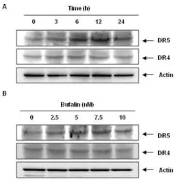
3. Bufalin의 EJ 방광암 세포의 DR 발현 조절에 미 치는 영향
DR4 및 DR5는 많은 암 세포들의 막에서 발현되어 TRAIL과의 결합을 통해 apoptosis의 extrinsic pathway 의 개시를 허용하는 것으로 알려져 있다20). 따라서 EJ 세포에서 bufalin 단독 처리가 DR4와 DR5의 발 현에 영향을 미치는 여부를 조사해 보았다. 이를 위 하여 EJ 세포를 0, 3, 6, 12, 24시간 동안 bufalin (10 nM)에 노출시키고 Western blot 분석을 수행하 였다. 또한, 24시간 동안 bufalin을 0, 2.5, 5, 7.5, 10 nM 농도로 처리하여 DR4와 DR5의 발현 변화를 조사하였다. Fig. 3A에서 보여 지는 바와 같이, bufalin의 자극은 EJ 세포에서 DR4 발현은 크게 변 화를 미치지 않았으나 DR5의 발현은 뚜렷하게 증가 시켰다. 이상의 결과는 bufalin에 의한 세포 사멸 유 도 증가는 EJ 세포의 DR5 단백질 수준의 상향 조절 에 중요하게 작용함을 시사한다.
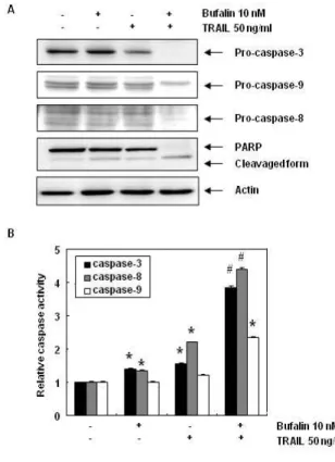
4. EJ 방광암 세포에서 bufalin 및 TRAIL 병용 처 리가 caspase의 활성화에 미치는 영향
Caspases protease는 정상적으로 생존 및 증식하 는 세포에서는 핵과 mitochondria의 외막에 불활성 상태(proenzyme)으로 존재하지만, 여러 가지 자극에 의하여 활성화되면 기질 단백질들을 선택적으로 분해 함으로써 apoptosis 신호 전달 경로의 중요한 조절자 로 작용한다는 것이 잘 알려져 있다(24). 따라서 Western blot 분석과 in vitro caspase activity test를 이용하여 bufalin의 TRAIL 매개 apoptosis에 다양한 caspase의 관련성을 조사하였다. Fig. 4A의 결과가 나타내듯이, EJ 세포에서 bufalin 또는 TRAIL 단일 처리는 pro-form capase의 단백질 분 해를 유도하지 않았지만, bufalin 및 TRAIL의 병용 투여는 caspase-3, -8 및 -9를 효과적으로 감소시 켰다. 비록 본 실험에서 활성화된 caspase를 직접 조 사하지는 못 했지만, pro-form caspase의 뚜렷한 발현 감소는 caspase의 활성이 나타났음으로 유추할 수 있다. Poly (ADP-ribose)-polymerase (PARP)는
활성화된 caspase에 의해서 분해되는 대표적인 기질 단백질로서 caspase에 의해 분해되면 정상적인 DNA 수복 및 유전자 안정성 유지의 기능을 상실하게 되 며, 이는 caspase 활성 의존적 apoptosis 유도의 생 화학적 지표로 사용되고 있다19,22). Bufalin과 TRAIL 의 병용 처리에 의해서 caspase-3의 기질 단백질인 PARP는 명백하게 분해되는 것을 알 수 있었다. 다음 으로, bufalin 및 TRAIL으로 처리된 EJ 세포로부터 동등한 양의 총 단백질을 정량하여 caspase 활성을 평가하였다. Fig. 4B의 결과에서 볼 수 있듯이, bufalin 과 TRAIL을 병용 처리는 EJ 세포의 caspase-3, -8 및 -9 활성이 뚜렷하게 촉진되었다. 이는 bufalin 및 TRAIL의 단독 처리에 의해서 caspase 활성이 증가 되지 않거나 다소 증가되는 것과는 대조적인 결과이 다. 이상의 결과는 bufalin이 TRAIL 매개 apoptosis 에 촉진에 caspase 의존적인 기전이 작용한다는 것 을 나타낸다.
5. Bufalin과 TRAIL의 병용 처리가 세포 자멸 관련 단백질 발현에 미치는 영향
Bufalin의 TRAIL 매개 apoptosis 유도 분자 기전 을 확인하기 위해 Western blot analysis를 이용하 여 apoptosis 관련 단백질 발현 조절을 평가하였다.
먼저 bufalin과 TRAIL의 병용 투여로 인한 DR의 발 현 변화를 알아본 결과, TRAIL의 단독 처리로는 증 가하지 않던 DR5의 발현이 bufalin과 TRAIL의 병용 처리에 의해서는 확연하게 증가하는 것을 알 수 있었 다. 또한, DR4의 발현은 단독 처리에 의해서는 변화 가 없다가 병용 처리에 의해서는 다소 증가하는 양상 을 나타내었다(Fig. 4A). 이러한 결과는 Fig. 3과도 일치하는 것으로 bufalin의 투여는 DR5의 발현을 향 상시켜 TRAIL 유도 apoptosis를 촉진하는 것으로 생 각되어진다. 다음으로 apoptosis의 mitochondria 연 관 intrinsic pathway의 조절에 핵심적으로 작용하 는 Bcl-2 family 유전자들의 발현을 조사하였다.
Bcl-2 family는 Bax 및 Bid와 같이 apoptosis를 유 도하는 pro-apoptotic proteins과 Bcl-2 및 Bcl-xL 과 같은 apoptosis 유도를 억제하는 anti-apoptotic proteins으로 구성되어 있다19, 23). Bufalin과 TRAIL 의 단독 혹은 병용 처리는 Bax 및 Bcl-2의 발현에는 영향을 미치지 않았으나, Bid의 발현 감소는 뚜렷하 게 나타났다. Bid는 BH3 interacting-domain death
agonist로 apoptosis 유도에 의하여 단편화되어 (truncated Bid, tBid) apoptosis의 intrinsic 및 extrinsic pathway를 연결하는 것으로 알려져 있다
24). 비록 본 연구에서는 tBid를 발현을 직접적으로 확인할 수는 없었지만, Bid의 발현 감소는 상대적으 로 tBid의 발현 증가를 의미하는 것으로 bufalin과 TRAIL에 의한 apoptosis 유도는 extrinsic pathway 뿐 아니라 intrinsic pathway의 활성이 동시에 관련 되어 있을 것임을 시사한다. XIAP과 cFLIP은 caspase의 활성을 억제하는 것으로 알려져 있는데25,26), bufalin 과 TRAIL의 병용 투여는 두 단백질의 발현을 현저하 게 감소시킴으로써 caspase의 활성을 증가시킴을 알 수 있었다.
이상의 결과를 종합해 보면, EJ 인간 방광암 세포 에서 bufalin의 처리로 촉진된 TRAIL 매개 apoptosis에 는 DR5의 발현 증가를 통한 caspase-8의 활성 증가 로 이어지는 extrinsic pathway가 주요 기전으로 작 용하며, Bid의 단편화, caspase-3의 활성 및 PARP 의 기능상실로 이어지는 intrinsic pathway도 관련 되어 있음을 시사한다.
Ⅳ. 고찰
본 연구에서는 EJ 인간 방광암 세포에서 TRAIL 저항성을 극복 할 수 있는 새로운 약물로써 bufalin 의 가능성을 증명하고자 하였다. Bufalin은 두꺼비의 이후선(耳後腺)에서 분비되는 흰색의 장액인 '섬수(蟾 酥, Bufonis Venenum)'의 주요 생리 활성물질 중 하 나로 여러 암종에서 apoptosis를 유도하는 것으로 밝
혀졌다16,17). 섬수는 해독소종(解毒消腫), 지통(止痛)의
효능을 가지고 있어, 나력(瘰癧), 소아감적(小兒疳積), 심 쇠(心衰), 옹저(癰疽), 정창(疔瘡), 풍충아통(風蟲牙 痛)의 치료에 사용된다18). 나력, 옹저, 정창 등은 현 재의 염증 및 종양 질환과 유사성이 있어 섬수의 생 리활성물질인 bufalin이 종양의 치료에 높은 효과가 있음을 유추해볼 수 있다.
본 실험에서 사용한 EJ 세포는 방광암의 TRAIL의 저항성을 조사한 선행 연구에서 J82 세포와 더불어 TRAIL에 저항성을 가지고 있는 것으로 나타난 반면, 5637 세포는 TRAIL에 민감성을 보였다27). 이번 실험 에서 사용한 bufalin 10 nM과 TRAIL 50 ng/ml은 각각 단독으로 처리하였을 경우 세포 증식 및 생존율
에 유의한 영향을 주지 않았지만, 같은 용량으로 병 용 처리하였을 경우 bufalin은 EJ 세포의 TRAIL 매 개 apoptosis를 유의하게 촉진한다는 것을 보여주었 다(Fig. 1 & 2).
TRAIL에 저항성을 가지는 방광암 세포가 TRAIL 매개 apoptosis를 극복하는 여러 기전들이 보고 되어 있다. 이 중 DR의 발현 조절이 중요하게 작용하는 데, 이는 실험적으로 DR 발현의 향상이 TRAIL의 저 항성을 극복하게 한다는 연구 뿐 아니라, 임상 연구 에서도 DR4 또는 DR5 발현이 높은 방광암 환자 경 우에 수술 후 재발률이 낮은 것으로 보고되었다
7,27,28). 본 연구에서는 bufalin의 단독 및 TRAIL과의 병용 투여는 DR4의 발현에는 큰 영향이 없었지만, DR5의 발현은 뚜렷하게 증가시키는 것으로 나타났다 (Fig. 3 & 5). 이러한 양상은 DR5의 발현을 증가시 키는 것이 TRAIL에 암세포를 민감하게 하는데 있어 서 DR4보다 더 매력적인 표적이 될 수 있음을 보여 주는 다른 연구와도 일맥상통하였다11,28).
DR에 결합된 TRAIL은 DISC를 형성하여 caspase-8을 비롯한 initiator caspases가 활성화되고 이는 caspase-3와 같은 effector caspases를 순차적으로 활성화시켜 death receptor 연관 extrinsic pathway를 통해 apoptosis 를 유발한다29). Bufalin과 TRAIL을 병용 투여한 경 우 단독으로 투여한 경우에 비해 pro-form caspase-3, -8 및 -9의 단편화 및 활성화를 유도하였는데, 특히 caspase-3와 caspase-8의 활성이 caspase-9의 활 성보다 뚜렷하게 나타나는 것으로 보아 bufalin의 촉 진하는 TRAIL 매개 apoptosis는 extrinsic pathway 가 주요 경로로 작용함을 뒷받침해준다. 또한, caspase를 억제하는 XIAP 및 cFLIP가 감소는 caspase의 활성 화를 뚜렷하게 유도하며, 이는 PARP의 절단으로 이 어지는 caspase 의존적인 경로가 작용함을 보여주었 다(Fig. 4 & 5). 그리고 Bid의 활성화를 통해 mitochondria 연관 intrinsic pathway도 일부 관련되어 있을 것으 로 추측된다.
이상의 결과를 종합하여 보면, bufalin과 TRAIL의 병용 투여는 DR5의 수준을 상향 조절하여 extrinsic pathway를 통한 apoptosis 신호를 증폭시키고 Bid 의 단편화를 통해 intrinsic pathway 신호를 자극하 여 저용량의 bufalin 투여가 EJ 방광암 세포에서 TRAIL 매개 apoptosis를 촉진함을 알 수 있었다. 이 는 TRAIL 내성 방광암 환자의 내성을 극복하기 위한
새로운 치료제로써의 bufalin의 높은 가능성을 제시 하는 바이다.
Ⅴ. 결론
본 연구에서는 TRAIL 내성을 가진 EJ 인체 방광 암 세포의 TRAIL 매개 apoptosis를 촉진하는 bufalin의 효과를 조사하여 다음과 같은 결과를 얻었다.
1. Bufalin과 TRAIL의 병용 처리는 각각의 약물이 독성이 없는 조건에서 EJ 방광암 세포의 세포 성 장 억제 및 생존율을 유의하게 감소시켰다.
2. Bufalin과 TRAIL의 병용 처리는 TRAIL 매개 apoptosis를 촉진함으로써 세포 성장을 억제하였다.
3. Bufalin의 단독 처리는 시간 및 농도 의존적으로 DR5의 발현을 상향 조절하였다.
4. Bufalin과 TRAIL의 병용 처리는 caspase 의존적 인 apoptosis를 유도하였다.
5. Bufalin과 TRAIL의 병용 처리는 DR5의 수준을 향상시켰으며, Bid의 단편화 및 XIAP, cFLIP의 수 준을 저하시켰다.
이상의 결과로부터 bufalin은 TRAIL에 저항성을 가진 EJ 세포의 저항성 극복을 위해 DR5의 발현을 상향 조절하여 caspase 의존적인 apoptosis 유도를 촉진함을 알 수 있었다.
References
1. Guancial EA, Roussel B, Bergsma DP, Bylund KC, Sahasrabudhe D, Messing E, Mohile SG, Fung C.
Bladder cancer in the elderly patient: challenges and solutions. Clin Interv Aging. 2015 ; 10 : 939-49.
2. Shamseddine A, Saleh A, Charafeddine M, Seoud M, Mukherji D, Temraz S, Sibai AM. Cancer trends in Lebanon: a review of incidence rates for the period of 2003-2008 and projections until 2018. Popul Health Metr. 2014 ; 12(1) : 4.
3. Syn SJ. Systemic Treatment for Metastatic Bladder Cancer. Kor J Med. 2015 ; 29(5) : 515-21.
4. Lim B, Allen JE, Prabhu VV, Talekar MK, Finnberg NK, El-Deiry WS. Targeting TRAIL in the
treatment of cancer: new developments. Expert Opin Ther Targets. 2015 ; 19(9) : 1171-85.
5. Ploeg M, Aben KK and Kiemeney LA: The present and future burden of urinary bladder cancer in the world. World J Urol. 2009 ; 27(3) : 289-93.
6. Dai X, Zhang J, Arfuso F, Chinnathambi A, Zayed ME, Alharbi SA, Kumar AP, Ahn KS, Sethi G.
Targeting TNF-related apoptosis-inducing ligand (TRAIL) receptor by natural products as a potential therapeutic approach for cancer therapy. Exp Biol Med (Maywood). 2015 ; 240(6) : 760-73.
7. Szliszka E, Mazur B, Zydowicz G, Czuba ZP, Król W. TRAIL-induced apoptosis and expression of death receptor TRAIL-R1 and TRAIL-R2 in bladder cancer cells. Folia Histochem Cytobiol.
2009; 47(4) : 579-85.
8. Trivedi R, Mishra DP. Trailing TRAIL Resistance:
Novel Targets for TRAIL Sensitization in Cancer Cells. Front Oncol. 2015; 5 : 69.
9. Shuzhen C, Lei F, Shruti MR, Ping Y, Fadlo RK and Sun SY. Dissecting the roles of DR4, DR5 and c-FLIP in the regulation of Geranylgeranyltransferase I inhibition-mediated augmentation of TRAIL- induced apoptosis. Mol Cancer. 2010 ; 9 : 23.
10. Wang S, El-Deiry WS. TRAIL and apoptosis induction by TNF-family death receptors. Oncogene. 2003
; 22(53) : 8628-33.
11. Thorburn A, Behbakht K, Ford H. TRAIL receptor- targeted therapeutics: resistance mechanisms and strategies to avoid them. Drug Resist Updat.
2008 ; 11(1-2) : 17-24.
12. Yan S, Qu X, Xu L, Che X, Ma Y, Zhang L, Teng Y, Zou H, Liu Y. Bufalin enhances TRAIL-induced apoptosis by redistributing death receptors in lipid rafts in breast cancer cells. Anticancer Drugs. 2014; 25(6) : 683-9.
13. White-Gilbertson SJ, Kasman L, McKillop J, Tirodkar T, Lu P, Voelkel-Johnson C. Oxidative stress sensitizes bladder cancer cells to TRAIL mediated apoptosis by down-regulating anti- apoptotic proteins. J Urol. 2009 ; 182(3) : 1178-85.
14. LeBlanc H, Lawrence D, Varfolomeev E, Totpal K, Morlan J, Schow P, Fong S, Schwall R, Sinicropi D, Ashkenazi A. Tumor-cell resistance to death receptor-induced apoptosis through mutational inactivation of the proapoptotic Bcl-2 homolog Bax. Nat Med. 2002 ; 8(3) : 274–81.
15. Ahmed SM, Wu X, Jin X, Zhang X, Togo Y, Suzuki T, Li Y, Kanematsu A, Nojima M, Yamamoto S, Sugimoto M, Kakehi Y. Synergistic induction of apoptosis by mapatumumab and anthracyclines in human bladder cancer cells. Oncol Rep. 2015
; 33(2) : 566-72.
16. Yin PH, Liu X, Qiu YY, Cai JF, Qin JM, Zhu HR, Li Q. Anti-tumor activity and apoptosis- regulation mechanisms of bufalin in various cancers: new hope for cancer patients. Asian Pac J Cancer Prev. 2012 ; 13(11) : 5339-43.
17. Qi F, Li A, Inagaki Y, Kokudo N, Tamura S, Nakata M, Tang W. Antitumor activity of extracts and compounds from the skin of the toad Bufo bufo gargarizans Cantor. Int Immunopharmacol 2011 ; 11(3) : 342-9.
18. http://www.koreantk.com/
19. Park C, Hong SH, Choi SH, Lee SR, Lean SH, Choi YH. Sagantang-induced Apoptotic Cell Death is Associated with the Activation of Caspases in AGS Human Gastric Carcinoma Cells. J Life Sicence. 2015 ; 25(12) : 1384-1392.
20. Kelley RF, Totpal K, Lindstrom SH, Mathieu M, Billeci K, Deforge L, Pai R, Hymowitz SG, Ashkenazi A. Receptor-selective mutants of apoptosis-inducing ligand 2/tumor necrosis factor- related apoptosis-inducing ligand reveal a greater contribution of death receptor (DR) 5 than DR4 to apoptosis signaling. J Biol Chem.
2005 ; 280(3) : 2205-12.
21. Shalini S, Dorstyn L, Dawar S, Kumar S. Old,
new and emerging functions of caspases. Cell Death Differ. 2015 ; 22(4) : 526-39.
22. Duriez PJ, Shah GM. Cleavage of poly(ADP-ribose) polymerase: a sensitive parameter to study cell death. Biochem Cell Biol. 1997 ; 75(4) : 337-49.
23. Yin XM. Signal transduction mediated by Bid, a pro-death Bcl-2 family proteins, connects the death receptor and mitochondria apoptosis pathways. Cell Res. 2010 ; 10(3) : 161-7.
24. Kim JH, Kim MJ, Choi KC, Son J. Quercetin sensitizes pancreatic cancer cells to TRAIL- induced apoptosis through JNK-mediated cFLIP turnover. Int J Biochem Cell Biol. 2016 ; 78 : 327-34.
25. Safa AR. c-FLIP, a master anti-apoptotic regulator. Exp Oncol. 2012 ; 34(3) : 176-84.
26. Ewald F, Ueffing N, Brockmann L, Hader C, Telieps T, Schuster M, Schulz WA, Schmitz I.
The role of c-FLIP splice variants in urothelial tumours. Cell Death Dis. 2011; 2 : e245.
27. Jin CY, Park C, Hwang HJ, Kim GY, Choi BT, Kim BW, Choi YH. Naringenin up-regulates the expression of death receptor 5 and enhances TRAIL-induced apoptosis in human lung cancer A549 cells. Mol Nutr Food Res. 2011
; 55(2) : 300-9.
28. Li Y, Jin X, Li J, Jin X, Yu J, Sun X, Chu Y, Xu C, Li X, Wang X, Kakehi Y, Wu X.
Expression of TRAIL, DR4, and DR5 in bladder cancer: correlation with response to adjuvant therapy and implications of prognosis. Urology.
2012 ; 79(4) : 968.e7-15.
29. Chen Z, Sangwan V, Banerjee S, Chugh R, Dudeja V, Vickers SM, Saluja AK. Triptolide sensitizes pancreatic cancer cells to TRAIL- induced activation of the death receptor pathway.
Cancer Lett. 2014 ; 348(1-2) : 156-66.
Figures and figure legends
Fig. 1. Inhibition of cell proliferation by co- treatment with bufalin and TRAIL in human bladder carcinoma cells. EJ cells were seeded at 1 x 10⁵ cells in 6-well plates. On the second day, the cells were treated with bufalin (5 or 10 nM), TRAIL (25 or 50 ng/ml), or a combination of both for 24 h. Then, cell viability was measured using the metabolic-dye based MTT assay. The data are shown as mean ± SD of three independent experiments. *, p < 0.05, # , p<0.01, as compared with untreated cells.
Fig. 2. Morphological changes and apoptotic formation by co-treatment with bufalin and TRAIL in human bladder cancer cells. The indicated cell lines were seeded in 6-well
plates and incubated for 24 h. EJ cells were treated with bufalin (10 nM), TRAIL (50 ng/ml), or both for 24 h. (A) The morphological changes of the cells were observed using an inverted microscope (magnification, × 50). (B) The cells were harvested, centrifuged and fixed. The cells were stained with a DAPI solution for 10 min, and the stained nuclei were observed under a fluorescent microscope using a blue filter (magnification, × 400).
(C) To quantify the degree of apoptosis induced by the combination stimulation with bufalin and TRAIL, a flow cytometric analysis was carried out for the sub-G1 DNA contents. Each experiment was carried out at least in triplicate.
Fig. 3. Involvement of death receptor (DR) in the apoptosis-enhancing property in human bladder cancer cells. EJ cells were incubated with various times of bufalin 10 nM (A) or various concentrations of bufalin for the indicated times (B). Equa
l amounts of protein (30 μl) was subjected to electrophoresis on SDS-polyacrylamide gels and transferred onto nitrocellulose membranes, and probed with DR5 and DR4 antibodies. Proteins were visualized using an ECL detection system. Actin was used as an internal control. The blot is representative of independent two experiments. (C, D) The relative protein levels of DR5 and DR4 were shown.
Fig. 4. Effects on the activation of caspase and the cleavage of PARP by combination treatment with bufalin and TRAIL. (A) EJ cells were incubated with bufalin (10 nM), TRAIL (50 ng/ml), or a combination of both for 24 h. Equal amounts of protein (30 μl) were subjected to electrophoresis on SDS-polyacrylamid gel and transferred to nitrocellulose membranes, and probed with specific antibodies. Actin was used as an internal control. (B) In EJ cells, the activities of caspase -3, -8 and -9 were determined using a caspase assay kit obtained from R&D, and used according to the protocol of the manufacturer.
The data are expressed as mean ± SD of the three independent experiments. The significance was determined using the Student’s t-test (*p < 0.05 or # , p<0.01 vs. untreated control).
Fig. 5. Effects of bufalin and TRAIL on the expression of apoptosis related proteins.
EJ cells were incubated with bufalin (10 nM), TRAIL (50 ng/ml), or both in combination for 24 h. Equal amounts of protein (30 μl) were subjected to electrophoresis on SDS-polyacrylamid gel and transferred to nitrocellulose membranes, and probed with specific antibodies.
Actin expression was used as a loading control. The blot is representative of three experiments.