ISSN 1225-6552, eISSN 2287-7630 http://dx.doi.org/10.7853/kjvs.2014.37.4.247
< Original Article >
Veterinary Service
Available online at http://kjves.org
*Corresponding author: Okjin Kim, Tel. +82-63-850-6668, Fax. +82-63-850-7308, E-mail. [email protected]
Mycoplasma hyopneumoniae 와 Mycoplasma hyorhinis 동시 감별진단을 위한 다중진단 중합효소반응
홍선화
1ㆍ이현아
1ㆍ김동우
1ㆍ김태완
2ㆍ김옥진
1*
원광대학교 동물자원개발연구센터1, 경북대학교 수의과대학2
Simultaneous diagnosis and differentiation of Mycoplasma hyopneumoniae and Mycoplasma hyorhinis
infections by multiplex PCR
Sunhwa Hong
1, Hyun-A Lee
1, Dong-Woo Kim
1, Tae-Wan Kim
2, Okjin Kim
1*
1Center for Animal Resource Development, Wonkwang University, Iksan 570-749, Korea
2College of Veterinary Medicine, Kyungpook National University, Daegu 702-701, Korea (Received 11 August 2014; revised 21 October 2014; accepted 5 November 2014)
Abstract
The economic impact of swine mycoplasma infection is high. An accurate diagnosis is often difficult and time consuming. We report the development and validation of an effective multiplex polymerase chain reaction (PCR) assay that detects Mycoplasma (M.) hyopneumoniae and M. hyorhinis. The multi detection of M. hyopneumoniae and M. hyorhinis primer set were employed to detect mycoplasma spe- cies and typing of the species was performed on the basis of sequence analysis of the PCR product.
The target nucleic acid fragments were specifically amplified by M. hyopneumoniae and M. hyorhinis PCR with 16S ribosomal DNA primers. Single and mixed Mycoplasma species DNA templates were used to evaluate the specificity of the multiplex assay. The corresponding specific DNA products were amplified for each pathogen. The multiplex PCR assay provides a novel tool for simultaneous detection and differentiation of M. hyopneumoniae and M. hyorhinis.
Key words : Mycoplasma, M. hyopneumoniae, M. hyorhinis, Multiplex, PCR
서 론
Mycoplasma 세균과 같이 인공배양이 가능한 미생 물중 세포벽을 가지지 않는 세균들을 Molicutes로 분 류하는데(Razin 등, 1998), 직경이 0.2∼2 m로 작아 세포배양용 배지 여과에 사용되는 0.22∼0.45 m의 필터를 통과할 수 있는 매우 작은 크기로, Molicutes 중 가장 잘 알려진 것이
Mycoplasma이다(Hay 등,
1989). 돼지 Mycoplasma성 폐렴은 돼지에서 발생하는 세균성 호흡기 질병 중 가장 흔하게 관찰되는 질병으로서 발병률이 매우 높고 경제적 손실도 큰 것으로 보고되었다(Lee 등, 2003). 특히 국내는 계절별 온도, 습도 등의 기후변화가 심하고 농장의 다두 집약사육 으로 인해 호흡기의 만성 혼합감염이 발생하고 있으 며 이에 따른 사료효율의 저하와 발육 불량 등 양돈 경영에 경제적 큰 손실을 입히고 있다(Hayflick, 1965). 양돈 농가에 막대한 경제적 손실을 입히는 돼 지유행성폐렴의 원인체로 알려진
M. hyopneumoniae
는 돼지에서 만성 기관지폐렴을 일으키는 원인체로 전세게적으로 발생되고 있으며 돼지의 생산성을 저 하시키는 중요한 병원체로 알려져 있다. M. hyopneu-moniae는 폐 상피세포의 섬모부분에 부착하여 섬모
Table 1.Components of culture medium used at the Mycoplasma hyopneumoniae and Mycoplasma hyorhinis cultivation
Components Source Dose
PPLO broth BD Difco 8.7 g
Brain heart infusion broth Oxoid 8.2 g
HBSS (X10) Sigma 50 mL
2% Thallium acetate Sigma 5.5 mL
Phenol red Difco 4.5 mL
Yeast extract Duchefa 60 mL
Horse serum Sigma 100 mL
Porcine serum Sigma 100 mL
Glucose solution Gibco 60 g
Ampicillin Sigma 6 g
Distilled water - 750 mL
정체와 섬모소실을 일으켜 상피세포 손상의 원인이 되며 이러한 손상으로 인하여 다른 병원체에 의한 2 차 감염을 유발할 수 있다(Taylor-Robinson, 1996). M.
hyorhinis는 돼지의 호흡기 점막에 흔하게 존재하는
원인체로 발병율은 매우 높지만 치사율은 낮은 질병 이며, 카타르성 기관지폐렴과 간질성 폐렴에 관련이 있는 2차적인 빌병요인으로 알려져 있다(Schilman 등, 1970). 또한 porcine reproductive and respiratory syn- drom (PRRS) 진단 시에도 함께 분리가 되기도 한다 (Kobayashi 등, 1996). M. hyorhinis는 돼지의 상부 호 흡기 기관과 편도에서 분리가 잘 되며, 늑막염, 복막 염, 심막염, 다발성 장막염, 다발성 관절염을 유발하 며, 성장 지연, 사료 효율 저하, 염증 반응 유발, 면역 반응 억제와 다른 전염성 질병의 감수성을 높여 경제 적인 손실을 일으키는 질병이다(Friis 등, 1994). 수의 학적 측면에서 돼지 mycoplasma 폐렴(mycoplasma pneumonia of swine; MPS)은 전세계적으로 발생되고 있으며 돼지를 생산하는 거의 모든 나라에서 생산성 을 저하시키는 중요한 인자로서 그원인체인M. hy- opneumoniae는 세균 감염 여부를 신속히 진단하는 것
은 경제적 측면이나 질병 관리측면에서 매우 중요하 다(Lee 등, 2003). 현재 Mycoplasma 감염 여부를 확인 하기 위한 방법 중 국가검정 동물용의약품 검정기준 에 세균시험의 일부로 직접배양법을 이용한 myco- plasma부정시험법을 적용하고 있다(Hopert 등, 1993).직접배양법은 시간이 많이 소요될 뿐만 아니라, 분리 배지의 조성, 시료의 특성 및 배양조건 등 많은 요인 에 의해 영향을 받을 수 있다는 단점이 있다(Hopert 등, 1993; Tang 등, 2000). 따라서 최근에는 신속한 Mycoplasma 진단 방법으로 특이 병원체 염기서열 부 위를 검출하는 polymerase chain reaction (PCR) 방법 이 보고되고 있다(Harasawa 등, 1993; Tang 등, 2000).
그러나 기존의 Mycoplasma 세균을 검출하기 위한 PCR 방법들은 대부분 한 종을 검출하는데 그치고 있 어 돼지유행성폐렴과 같이 여러 종의 Mycoplasma 세 균들이 감염될 확률이 있는 경우에 다른 primer 세트 를 이용하여 여러 번의 PCR 분석을 수행해야 하는 불편이 있다. Mycoplasma 속 세균의 감염이나 오염 여부를 신속하게 한 번의 작업으로 검출할 수 있는 기술이 필요하다(Spaepen 등, 1992).
최근에는 Mycoplasma species의 여러 유전자들의 염기 배열이 밝혀지고, 목적하는 부분의 DNA만을 선 택적으로 시험관내에서 대량으로 증폭시킬 수 있는 polymerase chain reaction (PCR) 방법이 개발되었다.
하지만 돼지유행성폐렴의 병원체를 동시에 여러 질 병 원인체를 검출할 수 있는 multiplex PCR에 관한 보고는 없다. 따라서 본 연구는 돼지유행성폐렴의 병 원체인 M. hyopneumoniae와 M. hyorhinis에 특이적인 영역을 검출할 수 있고, 신속하게 동시에 감별진단할 수 있는 multiplex PCR을 개발하고자 계획되었다.
재료 및 방법
사용균주 및 배양
본 연구에 사용된 균주는 American Type Cell Culture (ATCC)로부터 M. hyopneumoniae (ATCC, 25934), M. hyorhinis (ATCC, 27717), Helicobacter py-
lori (ATCC, 43504), Salmonella gallinarum (ATCC,
9184)을 구입하여 사용하였다. Helicobacter pylori는 10% calf serum이 첨가된 브루셀라 한천배지에 접종 후, 10% CO2 및 100% 습도가 유지되는 37oC in- cubator에서 3일간 배양 하여 사용하였고, Salmonellagallinarum는 LB 배지에 접종 후, 100% 습도가 유지
되는 37oC incubator에서 3일간 배양하여 사용하였으 며, M. hyopneumoniae와 M. hyorhinis는 Friis 배지 (Table 1)를 이용하여 37oC에서 5% CO2 함유 incuba- tor에서 14일 동안 배양하여 사용하였다(Friis, 1975).Genomic DNA의 분리
배양한 각각의 Mycoplasma 균주들과 Helicobacter
pylori 및 Salmonella gallinarum의 DNA 추출은
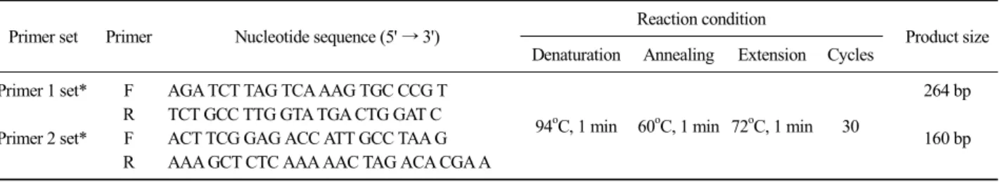
Table 2. Primer sets used in this study
Primer set Primer Nucleotide sequence (5' → 3') Reaction condition
Product size Denaturation Annealing Extension Cycles
Primer 1 set* F AGA TCT TAG TCA AAG TGC CCG T
94oC, 1 min 60oC, 1 min 72oC, 1 min 30
264 bp R TCT GCC TTG GTA TGA CTG GAT C
Primer 2 set* F ACT TCG GAG ACC ATT GCC TAA G 160 bp
R AAA GCT CTC AAA AAC TAG ACA CGA A
*Primer 1 set: Mycoplasma hyopneumoniae, Primer 2 set: Mycoplasma hyorhinis.
AccuPrepⓇ genomic DNA extraction kit (Bioneer Co., Daejeon, Korea)를 사용하였다 (Kim 등, 2010). 배양된 균주들을 13,000 g에서 30분 동안 원심분리하여 얻은 펠렛 검체를 1 mL 멸균 증류수가 들어있는 Mini- Bead Beater (Biospec product) 전용 2 mL 튜브에 무균 채취하여 세절한 후 멸균 3차 증류수에 부유시킨 glass bead (0.1 mm size, Biospec product) 200 L와 phenol-chlorform-isoamyl alcohol 용액(50:49:1(v/v/v)) 200 L를 넣어 Mini-Bead Beater (Biospec product)로 30초간 5,000 rpm으로 진탕하였다. 진탕 후 4oC에서 12,000 rpm으로 15분간 원심분리한 후 상층액을 멸균 2 mL 튜브에 옮겼다. 3 M 초산나트륨(sodium acetate) 10 L와 ice-cold 에탄올 250 L를 넣어 -20oC에서 10 분간 정체시킨 후, 15,000 rpm으로 15분간 원심분리 하였다. 침전물은 70% 알코올로 세척하여 실온에서 건조시키고 Tris EDTA (pH 8.0) 60 L에 용해시켜 실 험에 사용하였다.
종 특이적 프라이머의 개발
Mycoplasma universal primers는 유전자 변이가 거 의 없는 것으로 알려진 16S rRNA 유전자의 특이적 부분을 증폭시킬 수 있는 유전자로 본 연구에서 신규 디자인하였으며 사용되어진 primer는 Bioneer (Daejeon, Korea)에 합성을 의뢰하여 consensus primer 세트와
M. hyopneumoniae 및 M. hyorhinis primer 세트를 제조
하였다(Table 2). 또한 분석한 결과를 NCBI의 blast search (http://ncbi.nlm.nih.gov/)의 표적 유전자 염기서 열과의 비교하여 확인하였다.PCR 반응은 DNA thermal cycler (PTC-100 Thermo- cycler; MJ Research, USA)를 이용하여 반응을 실시하 였다. PCR을 위한 반응시약의 조성은 1 L forward 및 reverse primer (10 pmol/L), 2.5 L 10× reaction buffer, 500 M deoxy nucleotide, 1.5 U Taq polymer- ase를 각각 넣고, 총 반응량이 25 L이 되도록 멸균
된 증류수를 첨가하여 Table 2와 같은 조건으로 PCR 반응을 실시하였다.
PCR 프라이머의 특이도
개발한 프라이머의 특이도(specificity)를 확인하기 위해, M. hyopneumoniae (ATCC, 25934), M. hyorhinis (ATCC, 27717)를 제작한 최적화 배양 배지를 이용하 여 37oC에서 5% CO2 함유 incubator에서 14일간 배양 하여 사용하였고 음성 대조 균주로는 consensus prime 에 반응하지 않는
Helicobacter pylori (ATCC, 43504), Salmonella gallinarum (ATCC, 9184)를 사용하였으며,
앞에서 언급한 PCR 반응조건으로 PCR을 실시하여 각각의 PCR 반응의 특이성을 확인하였다.PCR 프라이머의 민감도
개발한 프라이머의 민감도(sensitivity)를 확인하기 위해
M. hyopneumoniae (ATCC, 25934), M. hyorhinis
(ATCC, 27717), M. pulmonis (ATCC, 19612)를 제작한 최적화 배양 배지를 이용하여 37oC에서 5% CO2 함유 incubator에서 14일간 배양하여 사용하였다. Mycopla- sma 균주를 DNA추출하여 Spectrophotometer를 이용 하여 DNA 정량을 한 후, 단계 희석하여 순수분리된M. hyopneumoniae DNA와 M. hyorhinis DNA을 10
4, 103, 102, 10 및 1 pg의 농도로 M. hyopneumoniae DNA 와 M. hyorhinis DNA을 정량하여 각각의 PCR을 수행 하였다.결 과
마이코플라즈마 배양 확인
본 연구에 사용된 Mycoplasma는 제작한 최적화 배
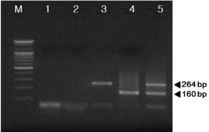
Fig. 1. Specificity of the developed multiplex polymerase chain reaction. Lane M: DNA marker (100 bp DNA ladder), 1:Helicobacter pylori, 2: M. hyopneumoniae, 3: M. hyorhinis, 4: M. hyopneumoniae + M. hyorhinis.
Table 3. Detection limit of M. hyopneumoniae and M. hyorhinis by the multiplex polymerase chain reaction
Strain Template DNA (pg)
104 103 102 10 1
M. hyopneumoniae +++ ++ + + -
M. hyorhinis +++ +++ ++ + -
+++: Strong reaction, ++: moderate, +: mild, -: No reaction.
Fig. 2.Detection limit of Mycoplasma hyopneumoniae by the de- veloped mutiplex PCR. Lane M: DNA marker (100 bp DNA ladder), 1; 104 pg DNA of M. hyopneumoniae, 2; 103 pg DNA of M. hyopneu- moniae, 3; 102 pg DNA of M. hyopneumoniae, 4; 10 pg DNA of M.
hyopneumoniae, 5; 1 pg DNA of M. hyopneumoniae.
Fig. 3.Detection limit of Mycoplasma hyorhinis by the developed mutiplex PCR. Lane M: DNA marker (100 bp DNA ladder), 1; 104 pg DNA of M. hyorhinis, 2; 103 pg DNA of M. hyorhinis, 3; 102 pg DNA of M. hyorhinis, 4; 10 pg DNA of M. hyorhinis, 5; 1 pg DNA of M.
hyorhinis.
지를 이용하여 37oC에서 5% CO2 함유 incubator에서 14일 동안 배양하고, 배지의 색깔이 붉은색에서 노란 색으로 변하면 Mycoplasma가 자란 것으로 추정하고, 색깔이 변한 최대희석 배수의 역지수를 색깔변화단 위(Color Change Unit; CCU)를 관찰하였다.
종 특이적 프라이머 개발 확인
개발한 Mycoplasma의 다중 검출 프라이머 세트를 확인하기 위해 PCR Primer 1 set (Mycoplasma hy-
opneumoniae)와 Primer 2 set (Mycoplasma hyorhinis)를
95oC에서 1분간 pre-denaturation을 실시 한 후 95oC에 서 1 분, 60oC에서 1분, 72oC에서 1분을 30 주기로 시 행하고 마지막으로 72oC에서 1분간 post-polymer- ization을 실시하여 나온 PCR 증폭 산물은 1.5% agar- ose에 넣고 100 volt에서 20분간 전기영동하여 ethi- dium bromide로 염색한 후 Gel-Documentation system (Uvitec cambridge, EEC)을 이용하여 Mycoplasma 특이 밴드의 유무를 확인하였다. Primer 1 set (Mycoplasmahyopneumoniae)를 이용하여 DNA를 증폭 하였을 때
264 bp에서 밴드를 확인하였고, Primer 2 set (Myco-plasma hyorhinis)를 이용하여 DNA를 PCR 증폭한 결
과 160 bp에서 밴드를 확인하였다(Fig. 1).PCR 프라이머의 특이도
2개의 primer set의 DNA를 PCR 증폭하여 특이도 (specificity)를 확인한 결과, M. hyopneumoniae, M. hy-
orhinis DNA 샘플에만 특이적으로 반응하였고 음성
대조균주인 Helicobacter pylori, Salmonella gallinarum DNA 샘플에서는 반응하지 않았음을 확인하였다(Fig. 1).PCR 프라이머의 민감도
특이도와 같이 2개의 primer set의 DNA를 PCR 증
폭하여 민감도(sensitivity)를 확인한 결과, M. hyopneu-
moniae DNA의 농도 1, 10, 10
2, 103, 104 pg으로 높을 수록 민감도가 높아짐을 확인할 수 있었다(Table 3)(Fig. 2). M. hyorhinis DNA의 농도 1, 10, 102, 103, 104 pg으로 높을수록 민감도가 높아짐을 확인할 수 있었다(Table 3)(Fig. 3).고 찰
Mycoplasma 세균은 사람과 동물의 호흡기나 생식 기 등의 각종 장기에 상주하며 심각한 질병을 일으키 는데, 특히 돼지에서는 폐렴에 의한 사망을 유발하여 농축산 산업에 막대한 손실을 초래할 수 있는 병원체 이다(Razin 등, 1998). 이러한 Mycoplasma 세균은 항 생물질에 내성이 있고 크기가 0.15∼0.8 m 정도이기 때문에 여과멸균에 의하여 제거되지 않으며, 형태학 적으로도 다형적(pleomorphic)특성이 있어 광학현미 경을 이용한 관찰이 어렵다(Hay 등, 1989). 현재 각종 세포배양과 동물에 오염된 Mycoplasma 세균을 검출 하기 위한 많은 PCR 기법 및 시판키트가 개발되어 실용화 되고 있다(Harasawa 등, 1993; Hu 등, 1995).
본 연구에서는
M. hyopneumoniae 및 M. hyorhinis에
각각 특이적인 primer 세트를 개발한 후 이들 primer 세트가 같은 PCR 반응 조건에서 동시에 반응하는지 를 확인하였다. PCR 진단은 병원체를 민감하게 검출 할 수 있는 방법으로 많이 이용되고 있으나 병원체 종류별로 각각의 검사가 이루어져야 한다(Wong-Lee 와 Lovett, 1993). 본 연구에서는 돼지유행성폐렴의 원 인체로 알려진 M. hyopneumoniae와 M. hyorhinis를 동 시에 감별진단할 수 있는 multiplex PCR을 개발하여 각각에 특이적인 증폭산물을 확인하였다. 일반적으로 세포배양에 오염된 Mycoplasma 검출 PCR기법의 민 감도는 104 cfu/mL의 검출한계를 보인다고 알려져 왔 으나(Spaepen 등, 1992; Tang 등, 2000; Toji 등, 1998), 최근에는 DNA 추출방법, nested PCR법, hot-start Taq DNA polymerase 사용으로 검출한계가 1∼100 cfu/mL 과 유사한 민감도를 보인다고 보고하고 있다(Sasaki 등, 1996; Tang 등, 2000; Eldering 등, 2004). 본 연구 에서는 M. hyopneumoniae와 M. hyorhinis의 민감도는 DNA의 농도 1, 10, 102, 103, 104 pg으로, 기존의 Mycoplasma 검출 연구결과들과 비교해 볼 때 높은 민감도를 확인하였다.본 연구를 통하여 개발된 multiplex PCR은 양돈 농
가에 큰 피해를 입히는 돼지유행성폐렴의 원인체 M.
hyopneumoniae와 M. hyorhinis를 한 번의 검사를 통하
여 동시에 감별진단하는 데 이용될 수 있다.감사의 글
본 연구는 2014년도 원광대학교 교비 연구비 지원 을 지원받아 수행되었으며, 이에 감사드립니다.
참 고 문 헌
Barile MF, Hopps HE, Grabowski MW, Riggs DB, DelGiudice RA. 1974. The identification and sources of myco- plasmas isolated from contaminated cell culture. Ann NY Acad Sci 225: 251-264.
Eldering JA, Felten C, Veilleux CA, Potts BJ. 2004. Development of a PCR method for mycoplasma testing of Chinese hamster ovary cell cultures used in the manufacture of recombinant therapeutic proteins. Biologicals 32:
183-193.
Friis NF. 1975. Some recommendations concerning primary iso- lation of Mycoplasma suipneumoniae and Mycoplasma flocculare a survey. Nord Vet Med 27: 337-339.
Friis NF, Feenstra AA. 1994. Mycoplasma hyorhinis in the etiol- ogy of serositis among piglets, Acta Vet Scand 35:
93-98.
Harasawa R, Mizusawa H, Nozawa K, Nakagawa T, Asada K, Kato I. 1993. Detection and tentative identification of dominant Mycoplasma species in cell cultures by re- striction analysis of the 16S-23S rRNA intergenic spacer regions. Res Microbiol 144: 489-493.
Hay RJ, Macy ML, Chen TR. 1989. Mycoplasma infection of cultured cells. Nature 339: 487-488.
Hayflick L. 1965. Tissue cultures and mycoplasmas. Tex Rep Biol Med 23: 285-303.
Hopert AC, Uphoff M, Wirth H, Hauser, DrexleH GR. 1993.
Specificity and sensitivity of polymerase chain reaction (PCR) in comparison with other methods for the de- tection of mycoplasma contamination in cell lines. J Immunol Methods 164: 91-100.
Hu M1, Buck C, Jacobs D, Paulino G, Khouri H. 1995.
Application of PCR for detection and identification of mycoplasma contamination in virus stocks. In Vitro Cell Dev Biol Anim 31: 710-715.
Kim HW, Kim JH, Rhim SR, Lee KA, Kim CJ, Paik HD. 2010.
A Multiplex PCR assay for the dection of food-borne pathogens in meat products. Korean J. Food Sci. Ani.
Resour. 30(4): 590-596.
Kobayashi H, Morozumi T, Munthall G, Mitani K, Ito N, Yamamoto K. 1996. Macrolide susceptibility of Myco-
plasma hyorhinis isolated from piglets. Antimicrob Agent Chemother 40: 1030-1032.
Lee YJ, Kim KS, Kwon YK, Tak RB. 2003. Biochemical charac- teristics and antimicrobials susceptibility of Salmonella gallinarum isolated in Korea. J Vet Sci 4: 161-166.
Razin S, Yogev D, Naot Y. 1998. Molecular biology and patho- genicity of mycoplasmas. Microbiol Mol Biol Rev 62:
1094-156.
Sasaki TR, Harasawa M, Shintani H, Fujiwara Y, Sasaki A, Horino T, Kenri K, Asada I, Kato, Chino F. 1996.
Application of PCR for detection of mycoplasma DNA and pestivirus RNA in human live viral vaccines.
Biologicals 24: 371-375.
Schilman A, Estola T, Garry-Anderson AS. 1970. On the occur- rence of Mycolasma hyorhinis in the respiratory organs of pigs, with special reference to enzootic pneumonia.
Zenrealbl Veterinarmed B 17: 549-553.
Spaepen M, Angulo AF, Marynen P, Cassiman JJ. 1992.
Detection of bacterial and mycoplasma contamination in cell cultures by polymerase chain reaction. FEMS
Microbiol Lett. 99: 89-94.
Stipkovits L, Bodon L, Romvary J, Varge L. 1975. Direct iso- lation of mycoplasmas and acholeplasmas from sera and kidneys of calves. Acta Microbiol Acad Sci Hung 22:
45-51.
Tang J, Hu M, Lee S, Roblin R. 2000. A polymerase chain re- action based method for detecting Mycoplasma/Achole- plasma contaminants in cell culture. J Microbiol Method 39: 121-126.
Taylor-Robinson D. 1996. Infections due to species of Myco- plasma and Ureaplasma. Clin Infect Dis 23: 671-682.
Toji LH, Lenchitz TC, Kwiatkowski VA, Sarama JA, Mulivor RA. 1998. Validation of routine mycoplasma testing by PCR. In Vitro Cell Dev Biol Anim 34: 356-358.
Wong Lee JG, Lovett M. 1993. Rapid and sensitive PCR method for identification of Mycoplasma species in tissue culture. pp. 257-260. In: (Persing DH, Smith TF, Tenover FC, and White TJ(ed.). Diagnostic molecular microbiology principles and applications. American Society for Microbiology. Washington DC.