3T3-L1 세포의 지방세포형성과정에서 Baicalin에 의한 유전자 발현 프로파일 분석
이해용1․강련화1․정상인1․조수현2․윤유식1†
1
중앙대학교 의과대학 미생물학교실
2
중앙대학교 용산병원 가정의학과
Effects of Baicalin on Gene Expression Profiles during Adipogenesis of 3T3-L1 Cells
Haeyong Lee1, Ryunhwa Kang1, Sang-In Chung1, Soo Hyun Cho2, and Yoosik Yoon1†
1Dept. of Microbiology, Chung-Ang University College of Medicine, Seoul 156-756, Korea
2Dept. of Family Medicine, Yongsan Hospital, Chung-Ang University, Seoul 140-757, Korea
Abstract
Baicalin, a flavonoid, was shown to have diverse effects such as anti-inflammatory, anti-cancer, anti-viral, anti-bacterial and others. Recently, we found that the baicalin inhibits adipogenesis through the modulations of anti-adipogenic and pro-adipogenic factors of the adipogenesis pathway. In the present study, we further characterized the molecular mechanism of the anti-adipogenic effect of baicalin using microarray technology.
Microarray analyses were conducted to analyze the gene expression profiles during the differentiation time course (0 day, 2 day, 4 day and 7 day) in 3T3-L1 cells with or without baicalin treatment. We identified a total of 3972 genes of which expressions were changed more than 2 fold. These 3972 genes were further analyzed using hierarchical clustering analysis, resulting in 20 clusters. Four clusters among 20 showed clearly up-regulated expression patterns (cluster 8 and cluster 10) or clearly down-regulated expression patterns (cluster 12 and cluster 14) by baicalin treatment for over-all differentiation period. The cluster 8 and cluster 10 included many genes which enhance cell proliferation or inhibit adipogenesis. On the other hand, the cluster 12 and cluster 14 included many genes which are related with proliferation inhibition, cell cycle arrest, cell growth suppression or adipogenesis induction. In conclusion, these data provide detailed information on the molecular mechanism of baicalin-induced inhibition of adipogenesis.
Key words: baicalin, adipogenesis, microarray, gene expression profile
†
Corresponding author. E-mail: thanks@cau.ac.kr
†
Phone: 82-2-820-5767, Fax: 82-2-820-5767
서 론
Adipogenesis(지방세포 형성)는 세포 형태의 변화, 호르 몬 민감성의 변화, 유전자 발현의 변화 및 단백질 발현의 변화가 일어나는 복합적인 과정이다. 비만, 당뇨 등의 여러 질병과 연계되어 있는 adipogenesis는 CCAAT/enhancer- binding proteins(C/EBPs)와 peroxidase proliferator-acti- vated receptor-γ(PPARγ)에 의해 조절된다(1,2). 분화가 시작되면 전사인자인 C/EBPβ와 C/EBPδ가 초기에 발현되 어 aidpogenesis의 핵심적인 기능을 하는 C/EBPα와 PPAR γ 의 발현을 증가시킨다(3). 핵심 조절자인 PPARγ와 C/EBPα는 서로 상호작용하여 상승효과를 일으키고 그 결과 로 ADIPOQ(adiponectin), LPL(lipoprotein lipase)과 같은 terminal marker(최종 마커)들의 발현이 유도된다(4).
최근, 발현되는 유전자의 양을 cDNA/oligonucleotide microarray 기술을 이용하여 그 차이를 비교하고 분석할 수
있게 되면서 생명과학은 급속도로 발전하였다(5). 특정 세포 나 조직에서 전체 유전자 발현의 변화를 확인할 수 있는 mi- croarray 기술은 3T3-L1 세포를 이용한
in vitro와 mouse를 이용한
in vivo의 adipogenesis 및 비만 연구에도 적용되고 있다(6). Microarray 기술을 이용한 adipogenesis 보고들은 수백 혹은 수천 개의 유전자들의 발현이 증가하거나 감소하 면서 지방세포(adipocytes)의 분화에 관여한다고 보고하고 있다. 또한, microarray는 유의 유전자(DEG, Differentially Expressed Gene) 분석, 유전자 정보(GO, Gene Ontology) 분석, 신호전달(pathway) 분석 그리고 군집(clustering) 분 석 등 결과 분석법이 다양해지면서 결과 해석과 이후 실험 진행에 크게 영향을 주고 있다.
Flavonoid는 과일이나 채소 등의 다양한 식물에 일반적으
로 함유되어 있는 polyphenolic compounds의 한 종류이다
(7). 식물성인 flavonoid는 항암, 항염증, 항산화 및 심장 질환
등의 많은 질병과 기작에 매우 유의적으로 연관되어 있다
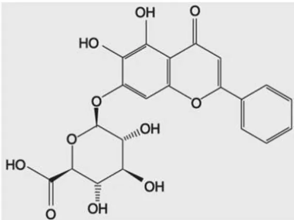
Fig. 1. Chemical structure of baicalin.
(8,9). Flavonoid 계열의 baicalin은 황금(
Scutellaria baica- lensisGeorgi)의 주요 성분으로써, 항염증, 항세균, 항바이 러스, 항암 등의 다양한 질병에 중요한 기능을 하는 것으로 보고되어 왔다(10-12). 바이칼린의 구조는 Fig. 1과 같다.
최근 본 연구진에 의해 baicalin의 adipogenesis 억제 및 adipogenesis 관련 유전자 발현 조절 효과가 규명되었다 (13). 이번 연구에서는 microarray analysis를 이용하여 adi- pogenesis에서 분화 시간에 따라 baicalin에 의해 변화되는 다양한 유전자들의 발현 변화를 전반적으로 살펴보고 군집 분석을 통해 유전자 발현 변화의 양상을 확인하였다.
재료 및 방법
세포 배양 및 시약
실험에 사용된 3T3-L1 전지방세포(preadipocyte)는 한국 세포주은행(Seoul, Korea)에서 구입하여 Dulbecco’s modi- fied Eagle’s medium(DMEM)에 10% calf serum, 100 μg/
mL streptomycin, 100 units/mL penicillin과 함께 37
oC, 5%
CO
2세포 배양기에서 배양하였다. Baicalin과 다른 시약들은 Sigma Chemical Co.(St. Louis, MO, USA)에서 구입하였다.
Adipogenesis 유도
3T3-L1 세포는 phosphate buffered saline(PBS) 용액으 로 씻어준 후, 1~2×10
5cells/mL을 6 well-plate에 세포밀도 가 100%가 될 때까지 배양하였다. 2일 후(day 0), 10% fetal bovine serum(FBS)이 첨가된 DMEM에 1 mg/mL insulin, 0.25 mM dexamethasone 그리고 0.5 mM 3-isobutyl-1- methylxanthine이 처리된 분화유도배지(differentiation- induction medium)로 교체하여 48시간 처리하였다. 그 후 2일 간격으로 10% FBS가 첨가된 새 배지에 1 mg/mL in- sulin이 처리된 분화유지배지(differentiation-maintenance medium)로 교체되었다. Adipogenesis가 진행될 때, baica- lin이 주는 영향을 확인하기 위해, 분화유도배지와 분화유지 배지를 첨가할 때 baicalin을 함께 처리하였다.
Total RNA 추출
Total RNA는 acid-phenol 추출법을 이용한 RNeasy kit(Qiagen, Hilden, Germany)를 사용하여 분리 정제되었다.
추출된 total RNA는 분광광도계(spectrophotometer)를 이 용하여 optical density(OD)값을 측정하고 -80
oC에서 cDNA microarray에 보합결합(hybridization)까지 냉동보관 하였다.
Microarray 분석
300 ng의 total RNA는 cDNA로 역전사하고 GeneChip IVT laveling kit(Affymetrix, Santa Clara, CA, USA)를 사 용하여 표기되었다. 표기된 cDNA는 35,557개의 유전자를 인지할 수 있는 probe들이 포함된 Mouse Gene 1.0 ST array 와 45
oC에서 17시간 동안 보합결합 된 후, GeneChip Fluidics Station 450(Affymetrix)을 사용하여 25
oC에서 non-strin- gent wash buffer로 세척되었다. 세척 후, Streptavidin- phycoerythrin reagents로 염색과정을 거친 microarray는 GeneChip scanner 3000(Affymetrix)을 이용하여 스캐닝 하 였다. GeneChip Operating Software(Affymetrix)를 이용하 여 측정되고 수치화된 형광 발현 정도는 RMA-sketch 알고 리즘을 통해 M-A plot을 측정하여 표준화하였다. 표준화 작업에는 Expression Console 1.0(Affymetrix) 프로그램이 사 용되었다. 표준화된 값들은 GenPlex software(Affymetrix) 을 이용하여 fold change와 t-test 그리고 군집 분석 등을 진행하였고, 유전자들의 기능 분석은 GO Miner를 이용하여 진행되었다(14).
결 과
Baicalin의 adipogenesis 억제 효과
Baicalin이 adipogenesis에 주는 효과를 살펴보기 위해 3T3-L1 지방세포를 이용하여 중성지방의 축적을 검사하였 다. IBMX(isobutylmethylxanthine), dexamethasone, in- sulin 및 FBS(fetal bovine serum)가 포함된 배지에 200 μM baicalin을 첨가하여 3T3-L1 전지방세포에서 지방세포로 7 일간 분화시킨 후 축적된 중성지방을 Oil Red O 염색법으로 확인하였다. 그 결과, baicalin을 첨가한 경우가 첨가하지 않 은 경우에 비해 중성지방의 축적이 현저하게 줄어든 것을 확인하였다(Fig. 2). 이러한 Baicalin에 의한 중성지방 축적 감소 결과는 baicalin이 adipogenesis를 억제함을 보여준다.
Baicalin에 의한 adipogenesis 억제효과는 본 연구진의 선행 연구에 의해 보다 상세하게 규명된 바 있다(13).
Microarray 결과의 전체 유전자 초기 분석
Baicalin이 유도하는 anti-adipogenesis 효과에 대한 분자
적 기작(molecular mechanism)을 좀 더 상세히 살펴보기
위해 microarray 분석을 진행하였다. 세포가 plate의 표면에
100% 확장된 후부터 2일이 지난 시점을 0일(0 day)로 하여,
0 day에 전지방 세포의 total RNA가 정제되었다. 분화 시간
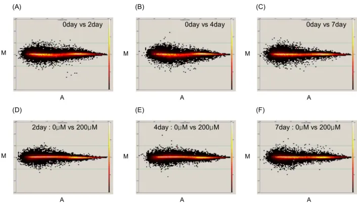
A
0day vs 2day 0day vs 4day 0day vs 7day
2day : 0μM vs 200μM 4day : 0μM vs 200μM 7day : 0μM vs 200μM
A A
A A A
M M M
M M M
(A) (B) (C)
(D) (E) (F)
Fig. 3. M-A-plot after normalization. Black dots represent the spot before standardization and red spots after standardization. Upper panels show comparison between preadipocytes (0 day) and differentiated adipocytes (2 day (A), 4 day (B), and 7 day (C)). Lower panels show comparison between 0 μM baicalin treatment and 200 μM baicalin treatment in differentiated adipocytes 2 day (D), 4 day (E), and 7 day (F)).
0 0.2 0.4 0.6 0.8 1.0 1.2
0 200
baicalin (μM) T ri g ly c e ri d e a c c u m u la ti o n (r a ti o t o 0 μ M v a lu e )
0 0.2 0.4 0.6 0.8 1.0 1.2
0 200
baicalin (μM) T ri g ly c e ri d e a c c u m u la ti o n (r a ti o t o 0 μ M v a lu e )
0 μM baicalin 200 μM baicalin
0 μM baicalin 200 μM baicalin
Fig. 2. Inhibitory effects of baicalin on triglyceride accu- mulation. 3T3-L1 cells were differentiated during 7 days with or without 200 μM baicalin treatment, and lipid droplet in differ- entiated cells were stained with Oil Red O.
별 시료들은 분화 유도 물질 및 baicalin을 2일 간격으로 첨 가하여 각 시간에 맞게 준비되었다. 이러한 과정으로 정제된 total RNA 시료들은 총 7개의 군으로 구성되었다: 전지방 세포(preadipocyte, 0 day), 2일 분화(2 day), 4일 분화(4 day), 7일 분화(7 day), 2일 분화+baicalin(2 day+baicalin), 4일 분화+baicalin(4 day+baicalin), 7일 분화+baicalin(7 day+baicalin).
7개의 total RNA 시료들은 cDNA로 역전사하여 표식화 작업을 거친 뒤, 35,557개의 probe로 구성된 Affymetrix Mouse Gene 1.0 ST array에 적용되었다. Microarray 결과 는 실험 재료와 첨가된 시약의 특성 및 실험에서의 변이 등 을 보정하고 결과의 신뢰성을 보장하기 위해 normalization 을 진행하였다(Fig. 3). 유전자 발현의 상호비교는 먼저 전지 방 세포와 각 2일, 4일 및 7일 분화세포의 비교 그리고 분화 된 2일, 4일 및 7일 동안 baicalin을 첨가한 세포와 첨가하지 않은 세포 간의 비교를 구분하여 수행하였다. 그 결과, 전지 방 세포와 비교하여 2배 이상 유의하게 증가 혹은 감소한 유전자의 수는 2일 분화세포에서 1,636개(증가: 690개, 감소:
946개), 4일 분화세포에서 1,792개(증가: 1,113개, 감소: 679
개), 7일 분화세포에서 1,006개(증가: 408개, 감소: 598개)였
다(Table 1). 또한, baicalin을 처리하지 않은 세포와 처리한
세포의 비교에서, 2배 이상 증가 혹은 감소한 유전자 수는
2일 분화 후 739개(증가: 401개, 감소: 338개), 4일 분화 후
1,296개(증가: 440개, 감소: 856개), 7일 분화 후 977개(증가:
Table 1. Number of genes of which expression levels were changed more than 2 fold at each time points of differ- entiation
up down total
preadipocytes vs 2 day preadipocytes vs 4 day preadipocytes vs 7 day
690 1113 408
946 679 598
1,636 1,792 1,006 The genes are selected if their expression levels in differ- entiated adipocytes were changes more than 2-fold compared with undifferentiated preadipocyte.
Table 2. Number of genes of which expression levels were changed more than 2 fold by baicalin treatment
up down total
2 day: 0 μM vs 200 μM 4 day: 0 μM vs 200 μM 7 day: 0 μM vs 200 μM
401 440 472
338 856 505
739 1,296
977 The genes are selected if their expression levels in baica- lin-treated adipocytes were changes more than 2-fold com- pared with untreated adipocyte at each time points of differentiation.
472개, 감소: 505개)였다(Table 2). 이러한 결과는 지방세포 가 분화되는 과정에 수많은 유전자들이 작용하며 baicalin의 첨가가 수많은 유전자들의 발현에 영향을 미침을 제시한다.
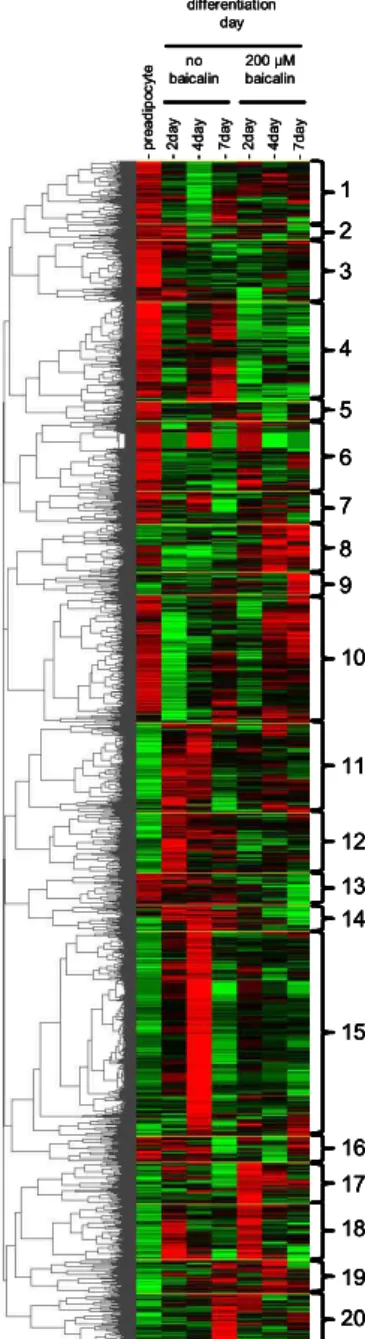
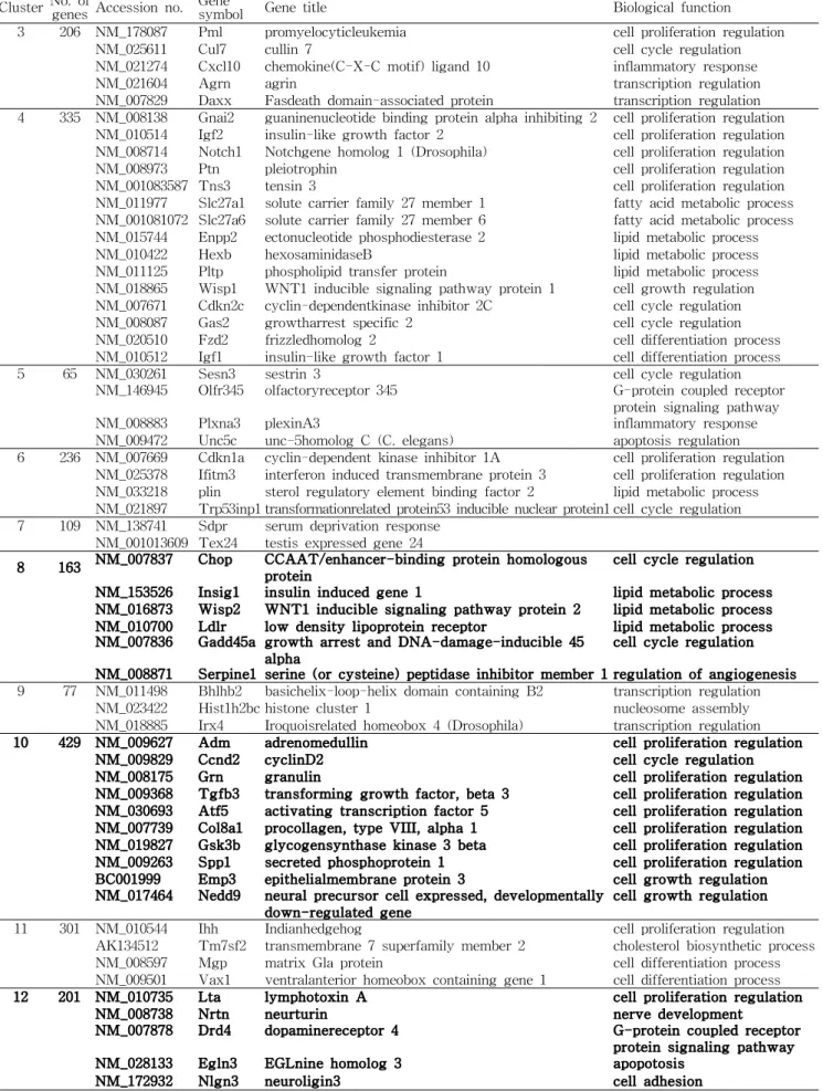
Hierarchical clustering을 통한 유전자 발현 양상의 분석 Baicalin에 의한 유전자 발현 양상의 전반적 분석을 위하 여 2배 이상 변화하는 유의 유전자들에 대해 그룹화를 진행 한 후, 특정 유전자들을 선별하는 작업을 진행하였다. Table 1과 Table 2에 정리된 2배 이상 증가 혹은 감소된 유전자들 을 합집합 하여 3,972개의 유전자를 얻었다. 이 유전자들을 hierarchical clustering으로 유전자 발현 양상을 살펴본 결 과, 분화 시간의 흐름과 baicalin의 처리에 따라 총 20개의 cluster를 얻을 수 있었다(Fig. 4). Fig. 5는 각 cluster들의 발현 양상을 0일, 2일, 4일 그리고 7일간의 분화 기간에 따라 baicalin을 첨가한 샘플과 첨가하지 않은 샘플을 구분하여 비교해서 보여준다. 각 cluster에 포함된 특정 유전자의 명 칭, 기능 및 수는 Table 3에 정리하였다. Baicalin의 효과에 가장 크게 작용하는 유전자 양상을 살펴보기 위해, 20개의 cluster 중 adipogenesis가 유도될 때 전반적인 분화 기간에 서 억제되었다가 baicalin의 첨가에 의해 크게 상승되는 양 상을 보이는 cluster들(cluster 8, cluster 10)과 반대로 전반 적으로 증가되었다가 baicalin의 첨가에 의해 크게 억제되는 양상을 보이는 cluster들(cluster 12, cluster 14)을 선별하였 다. Baicalin의 첨가에 의해 상승되어 분화를 억제하는 양상 을 보이는 cluster 8과 cluster 10에는 각각 163개(CHOP, INSIG1, WISP 등)와 429개(ADM, CCND2 등)의 유전자가 포함되었다. 또한 지방세포의 분화 시 증가되었다가 baica- lin의 첨가에 의해 감소되는 양상을 보이는 cluster 12에는 LTA 등의 201개 유전자가 포함되었다. Cluster 12와 같은 발현 양상을 보이는 cluster 14(89개)에는 ACADSB,
-preadipocyte -2day -2day
-4day -4day
-7day -7day
no baicalin
200 µM baicalin differentiation
day
1 2
5 3
4
6 7 8 9
10
11
12 13 14
15
16 17 18 19 20
-preadipocyte -2day -2day
-4day -4day
-7day -7day
no baicalin
200 µM baicalin differentiation
day
-preadipocyte -2day -2day
-4day -4day
-7day -7day
no baicalin
200 µM baicalin differentiation
day
1 2
5 3
4
6 7 8 9
10
11
12 13 14
15
16 17 18 19 20 1 2
5 3
4
6 7 8 9
10
11
12 13 14
15
16 17 18 19 20 1 2
5 3
4
6 7 8 9
10
11
12 13 14
15
16 17 18 19 20
Fig. 4. Hierarchical clustering analyses of 3972 selected genes. The red color refers to the up-regulation of the gene ex- pression and the green color indicates the down-regulation of the gene expression compared to the preadipocyte. Cluster numbers were marked in the right side of the figure.
HMGCS2 등이 포함되었다.
고 찰
Adipocytes의 기능 장애는 비만의 발생과 인슐린 저항성 을 포함한 다양한 질병과 밀접한 관련성을 가진다(15).
Adipocytes의 주요 작용인 adipogenesis는 C/EBPs와
PPAR
γ를 중심으로 수백 개의 유전자의 발현이 조절되어
일어나는 복잡한 과정이다. 최근 여러 보고에서 genistein,
sakuranetin, isorhamnetin 등의 flabonoid 물질들이 adipo-
0μM baicalin 200μM baicalin
Cluster 1 (212)
0 day 2 day 4 day 7 day
0μM baicalin 200μM baicalin
Cluster 1 (212)
0 day 2 day 4 day 7 day
0μM baicalin 200μM baicalin
Cluster 2 (53)
0 day 2 day 4 day 7 day
0μM baicalin 200μM baicalin
Cluster 2 (53)
0 day 2 day 4 day 7 day
0μM baicalin 200μM baicalin
Cluster 3 (208)
0 day 2 day 4 day 7 day
0μM baicalin 200μM baicalin
Cluster 3 (208)
0 day 2 day 4 day 7 day
0μM baicalin 200μM baicalin
Cluster 4 (335)
Relative expression
0 day 2 day 4 day 7 day
0μM baicalin 200μM baicalin
Cluster 4 (335)
Relative expression
0 day 2 day 4 day 7 day
0μM baicalin 200μM baicalin
Cluster 5 (65)
0 day 2 day 4 day 7 day
0μM baicalin 200μM baicalin
Cluster 5 (65)
0 day 2 day 4 day 7 day
0μM baicalin 200μM baicalin
Cluster 6 (236)
0 day 2 day 4 day 7 day
0μM baicalin 200μM baicalin
Cluster 6 (236)
0 day 2 day 4 day 7 day
0μM baicalin 200μM baicalin
Cluster 7 (109)
0 day 2 day 4 day 7 day
0μM baicalin 200μM baicalin
Cluster 7 (109)
0 day 2 day 4 day 7 day
0μM baicalin 200μM baicalin
Cluster 8 (163)
0 day 2 day 4 day 7 day
0μM baicalin 200μM baicalin
Cluster 8 (163)
0 day 2 day 4 day 7 day
0μM baicalin 200μM baicalin
Cluster 9 (77)
0 day 2 day 4 day 7 day
0μM baicalin 200μM baicalin
Cluster 9 (77)
0 day 2 day 4 day 7 day
0μM baicalin 200μM baicalin
Cluster 10 (429)
0 day 2 day 4 day 7 day
0μM baicalin 200μM baicalin
Cluster 10 (429)
0 day 2 day 4 day 7 day
0μM baicalin 200μM baicalin
Cluster 11 (301)
0 day 2 day 4 day 7 day
0μM baicalin 200μM baicalin
Cluster 11 (301)
0 day 2 day 4 day 7 day
0μM baicalin 200μM baicalin
Cluster 12 (201)
0 day 2 day 4 day 7 day
0μM baicalin 200μM baicalin
Cluster 12 (201)
0 day 2 day 4 day 7 day
0μM baicalin 200μM baicalin
Cluster 13 (107)
0 day 2 day 4 day 7 day
0μM baicalin 200μM baicalin
Cluster 13 (107)
0 day 2 day 4 day 7 day
0μM baicalin 200μM baicalin
Cluster 14 (89)
0 day 2 day 4 day 7 day
0μM baicalin 200μM baicalin
Cluster 14 (89)
0 day 2 day 4 day 7 day
0μM baicalin 200μM baicalin
Cluster 15 (688)
0 day 2 day 4 day 7 day
0μM baicalin 200μM baicalin
Cluster 15 (688)
0 day 2 day 4 day 7 day
0μM baicalin 200μM baicalin
Cluster 16 (86)
0 day 2 day 4 day 7 day
0μM baicalin 200μM baicalin
Cluster 16 (86)
0 day 2 day 4 day 7 day
0μM baicalin 200μM baicalin
Cluster 17 (141)
0 day 2 day 4 day 7 day
0μM baicalin 200μM baicalin
Cluster 17 (141)
0 day 2 day 4 day 7 day
0μM baicalin 200μM baicalin
Cluster 18 (187)
0 day 2 day 4 day 7 day
0μM baicalin 200μM baicalin
Cluster 18 (187)
0 day 2 day 4 day 7 day
0μM baicalin 200μM baicalin
Cluster 19 (115)
0 day 2 day 4 day 7 day
0μM baicalin 200μM baicalin
Cluster 19 (115)
0 day 2 day 4 day 7 day
R e la ti v e e x p re s s io n
0μM baicalin 200μM baicalin
Cluster 20 (170)
0 day 2 day 4 day 7 day
0μM baicalin 200μM baicalin
Cluster 20 (170)
0 day 2 day 4 day 7 day
R e la ti v e e x p re s s io n R e la ti v e e x p re s s io n R e la ti v e e x p re s s io n
R e la ti v e e x p re s s io n R e la ti v e e x p re s s io n R e la ti v e e x p re s s io n R e la ti v e e x p re s s io n
R e la ti v e e x p re s s io n R e la ti v e e x p re s s io n R e la ti v e e x p re s s io n R e la ti v e e x p re s s io n
R e la ti v e e x p re s s io n R e la ti v e e x p re s s io n R e la ti v e e x p re s s io n R e la ti v e e x p re s s io n
R e la ti v e e x p re s s io n R e la ti v e e x p re s s io n R e la ti v e e x p re s s io n R e la ti v e e x p re s s io n
Fig. 5. Average expression profiles of each cluster (cluster 1~20) of the hierarchical clustering analysis of differentiated adipo- cytes treated with or without baicalin. The y-axis represents relative expression levels and the x-axis represents the time course.
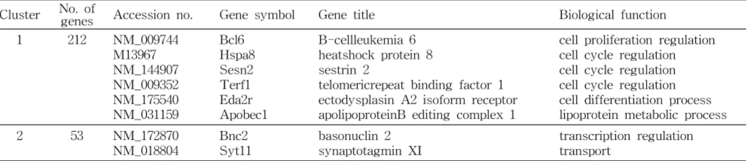
Table 3. List of genes in each cluster. The number of the genes, GenBank accession number, gene symbol, gene title and gene function are included for each cluster
Cluster No. of
genes Accession no. Gene symbol Gene title Biological function
1 212 NM_009744
M13967 NM_144907 NM_009352 NM_175540 NM_031159
Bcl6 Hspa8 Sesn2 Terf1 Eda2r Apobec1
B-cellleukemia 6 heatshock protein 8 sestrin 2
telomericrepeat binding factor 1 ectodysplasin A2 isoform receptor apolipoproteinB editing complex 1
cell proliferation regulation cell cycle regulation cell cycle regulation cell cycle regulation cell differentiation process lipoprotein metabolic process
2 53
NM_172870
NM_018804 Bnc2
Syt11 basonuclin 2
synaptotagmin XI transcription regulation
transport
Table 3. Continued
Cluster No. of genes Accession no. Gene
symbol Gene title Biological function
3 206 NM_178087 Pml promyelocyticleukemia cell proliferation regulation
NM_025611 Cul7 cullin 7 cell cycle regulation
NM_021274 Cxcl10 chemokine(C-X-C motif) ligand 10 inflammatory response
NM_021604 Agrn agrin transcription regulation
NM_007829 Daxx Fasdeath domain-associated protein transcription regulation 4 335 NM_008138 Gnai2 guaninenucleotide binding protein alpha inhibiting 2 cell proliferation regulation
NM_010514 Igf2 insulin-like growth factor 2 cell proliferation regulation NM_008714 Notch1 Notchgene homolog 1 (Drosophila) cell proliferation regulation
NM_008973 Ptn pleiotrophin cell proliferation regulation
NM_001083587 Tns3 tensin 3 cell proliferation regulation
NM_011977 Slc27a1 solute carrier family 27 member 1 fatty acid metabolic process NM_001081072 Slc27a6 solute carrier family 27 member 6 fatty acid metabolic process NM_015744 Enpp2 ectonucleotide phosphodiesterase 2 lipid metabolic process
NM_010422 Hexb hexosaminidaseB lipid metabolic process
NM_011125 Pltp phospholipid transfer protein lipid metabolic process NM_018865 Wisp1 WNT1 inducible signaling pathway protein 1 cell growth regulation NM_007671 Cdkn2c cyclin-dependentkinase inhibitor 2C cell cycle regulation
NM_008087 Gas2 growtharrest specific 2 cell cycle regulation
NM_020510 Fzd2 frizzledhomolog 2 cell differentiation process
NM_010512 Igf1 insulin-like growth factor 1 cell differentiation process
5 65 NM_030261 Sesn3 sestrin 3 cell cycle regulation
NM_146945 Olfr345 olfactoryreceptor 345 G-protein coupled receptor protein signaling pathway
NM_008883 Plxna3 plexinA3 inflammatory response
NM_009472 Unc5c unc-5homolog C (C. elegans) apoptosis regulation 6 236 NM_007669 Cdkn1a cyclin-dependent kinase inhibitor 1A cell proliferation regulation
NM_025378 Ifitm3 interferon induced transmembrane protein 3 cell proliferation regulation NM_033218 plin sterol regulatory element binding factor 2 lipid metabolic process NM_021897 Trp53inp1 transformationrelated protein53 inducible nuclear protein1 cell cycle regulation 7 109 NM_138741 Sdpr serum deprivation response
NM_001013609 Tex24 testis expressed gene 24
8 163 NM_007837 Chop CCAAT/enhancer-binding protein homologous
protein cell cycle regulation
NM_153526 Insig1 insulin induced gene 1 lipid metabolic process NM_016873 Wisp2 WNT1 inducible signaling pathway protein 2 lipid metabolic process NM_010700 Ldlr low density lipoprotein receptor lipid metabolic process NM_007836 Gadd45a growth arrest and DNA-damage-inducible 45
alpha cell cycle regulation
NM_008871 Serpine1 serine (or cysteine) peptidase inhibitor member 1 regulation of angiogenesis 9 77 NM_011498 Bhlhb2 basichelix-loop-helix domain containing B2 transcription regulation
NM_023422 Hist1h2bc histone cluster 1 nucleosome assembly
NM_018885 Irx4 Iroquoisrelated homeobox 4 (Drosophila) transcription regulation
10 429 NM_009627 Adm adrenomedullin cell proliferation regulation
NM_009829 Ccnd2 cyclinD2 cell cycle regulation
NM_008175 Grn granulin cell proliferation regulation
NM_009368 Tgfb3 transforming growth factor, beta 3 cell proliferation regulation NM_030693 Atf5 activating transcription factor 5 cell proliferation regulation NM_007739 Col8a1 procollagen, type VIII, alpha 1 cell proliferation regulation NM_019827 Gsk3b glycogensynthase kinase 3 beta cell proliferation regulation NM_009263 Spp1 secreted phosphoprotein 1 cell proliferation regulation BC001999 Emp3 epithelialmembrane protein 3 cell growth regulation NM_017464 Nedd9 neural precursor cell expressed, developmentally
down-regulated gene cell growth regulation
11 301 NM_010544 Ihh Indianhedgehog cell proliferation regulation
AK134512 Tm7sf2 transmembrane 7 superfamily member 2 cholesterol biosynthetic process
NM_008597 Mgp matrix Gla protein cell differentiation process
NM_009501 Vax1 ventralanterior homeobox containing gene 1 cell differentiation process
12 201 NM_010735 Lta lymphotoxin A cell proliferation regulation
NM_008738 Nrtn neurturin nerve development
NM_007878 Drd4 dopaminereceptor 4 G-protein coupled receptor
protein signaling pathway
NM_028133 Egln3 EGLnine homolog 3 apopotosis
NM_172932 Nlgn3 neuroligin3 cell adhesion
Table 3. Continued
Cluster No. of genes Accession no. Gene
symbol Gene title Biological function
13 107 NM_010216 Figf c-fos induced growth factor cell proliferation regulation NM_013584 Lifr leukemia inhibitory factor receptor cell proliferation regulation NM_010788 Mecp2 methyl CpG binding protein 2 cell proliferation regulation NM_026058 Lass4 longevity assurance homolog 4 lipid metabolic process NM_007922 Elk1 member of ETS oncogene family cell cycle regulation
NM_021457 Fzd1 frizzledhomolog 1 cell differentiation process
NM_007470 Apod apolipoprotein D transport
14 89 NM_025826 Acadsb acyl-CoenzymeA dehydrogenase, short, branched
chain fatty acid metabolic process
NM_008256 Hmgcs2 3-hydroxy-3-methylglutaryl-CoenzymeA
synthase 2 lipid metabolic process
NM_008048 Igfbp7 insulin-like growth factor binding protein 7 cell growth regulation NM_008587 Mertk c-merproto-oncogene tyrosine kinase cell cycle regulation NM_175445 Rassf2 Ras association (RalGDS/AF-6) domain family 2 cell cycle regulation NM_133955 Rhou ras homolog gene family, member U cell cycle regulation
NM_001013370 Sesn1 sestrin1 cell cycle regulation
15 688 NM_133670 Sult1a1 sulfotransferase family 1A, phenol-preferring,
member 1 lipid metabolic process
NM_011335 Ccl21a chemokine (C-C motif) ligand 21a cell differentiation process NM_023052 Ccl21c chemokine (C-C motif) ligand 21c cell differentiation process NM_011124 Ccl21b chemokine (C-C motif) ligand 21b inflammatory response NM_207273 Tdpoz5 TD and POZ domain containing 5 biological process
NM_019917 V2r1b vomeronasal 2 receptor 1b G-protein coupled receptor protein signaling pathway 16 86 NM_020260 Cdgap Cdc42 GTPase-activating protein small GTPase mediated signal
transduction
17 141 NM_007428 Agt angiotensinogen cell proliferation regulation
NM_007950 Ereg epiregulin cell proliferation regulation
NM_013598 Kitl kitligand cell proliferation regulation
NM_172671 Lgr4 leucine-richrepeat-containing G protein-coupled
receptor4 cell proliferation regulation
NM_010849 Myc myelocytomatosisoncogene cell proliferation regulation NM_011198 Ptgs2 prostaglandin-endoperoxidesynthase 2 fatty acid metabolic process NM_031167 Il1rn interleukin1 receptor antagonist lipid metabolic process NM_013642 Dusp1 dualspecificity phosphatase 1 cell cycle regulation NM_009061 Rgs2 regulatorof G-protein signaling 2 cell cycle regulation NM_009883 Cebpb CCAAT/enhancerbinding protein beta cell differentiation process NM_013562 Ifrd1 interferon-relateddevelopmental regulator 1 cell differentiation process 18 187 NM_008489 Lbp lipopolysaccharidebinding protein lipid metabolic process
NM_019521 Gas6 growtharrest specific 6 cell growth regulation
NM_008343 Igfbp3 insulin-likegrowth factor binding protein 3 cell growth regulation
NM_010591 Jun Junoncogene cell cycle regulation
NM_007483 Rhob rashomolog gene family member B cell cycle regulation
NM_001039554 Angptl7 angiopoietin-like7 signal transduction
NM_007679 Cebpd CCAAT/enhancerbinding protein delta transcription regulation NM_018866 Cxcl13 chemokine (C-X-C motif) ligand 13 inflammatory response
NM_009769 Klf5 Kruppel-likefactor 5 transcription regulation
NM_010638 Klf9 Kruppel-likefactor 9 transcription regulation
19 115 NM_020581 Angptl4 angiopoietin-like4 lipid metabolic process
NM_024406 Fabp4 fattyacid binding protein 4 cholesterol biosynthetic process NM_011146 Pparg peroxisomeproliferator activated receptor gamma cell differentiation process NM_009760 Bnip3 BCL2/adenovirusE1B interacting protein 1; NIP3 apoptosis regulation regulation NM_178373 Cidec celldeath-inducing DFFA-like effector c apoptosis regulation regulation NM_010930 Nov nephroblastomaoverexpressed gene cell growth regulation 20 170 NM_007981 Acsl1 acyl-CoAsynthetase long-chain family member 1 fatty acid metabolic process
NM_009605 Adipoq adiponectin fatty acid metabolic process
NM_026384 Dgat2 diacylglycerol O-acyltransferase 2 lipid metabolic process
NM_008509 Lpl lipoprotein lipase lipid metabolic process
NM_175640 Plin perilipin lipid metabolic process
NM_022984 Retn resistin lipid metabolic process
AK087727 Prlr prolactin receptor cell differentiation process
NM_027852 Rarres2 retinoicacid receptor responder 2 defense response
genesis 및 관련 유전자들을 조절한다고 보고하고 있다 (16-18). 황금(
Scutellaria baicalensisGeorgi)의 주요 성분 인 baicalin은 flavonoid 계열의 한 물질로서 항염증, 항암, 항바이러스 등 다양한 분야에서 그 기능을 보이고 있고 최근 본 연구진에 의해 adipogenesis 억제효능이 발견되었다(13).
본 연구에서는 microarray 분석을 이용하여 baicalin에 의한 adipogenesis 억제 시, 세포내의 다양한 pathway에 위치한 전반적인 유전자들의 발현 양상을 살펴보았다. 전지방 세포 에서 지방세포로 분화되는 동안 cell cycle regulator(세포주 기 조절자)와 cell growth regulator(세포성장 조절자) 및 adipogeneic factor(지방세포 형성 인자)들의 상호작용이 이 루어진다. 이 과정에서 전지방 세포의 cell proliferation(세포 분열)이 중지되고 cell cycle에서 G1기의 세포들이 증가되면 서 지방세포의 분화가 시작된다(19). Hierarchical cluster- ing에서 분화기간 전반에 걸쳐 baicalin에 의해 크게 증가 혹은 감소된 cluster(cluster 8, cluster 10, cluster 12, cluster 14)들에 포함된 유전자들의 기능을 살펴보면 adipogenesis 와 연관된 metabolism을 비롯하여 cell proliferation, cell growth 및 cell cycle regulation과 관련된 유전자가 많았다.
Cluster 8과 Cluster 10의 경우 adipogenesis를 억제하는 유 전자이거나 cell proliferation과 cell growth을 유도하는 기 능을 가진 유전자들이 다수 포함되었다. 그 중 C/EBP fam- ily의 한 멤버인 CHOP(CCAAT/enhancer-binding protein homologous protein)은 C/EBPβ와 heterodimer를 형성하여 C/EBPβ의 활성을 억제함으로써 anti-adipogenic factor(지 방세포 형성 억제 인자)로 작용하며, 지방세포 유도물질인 FBS(fetal bovine serum)가 첨가되어 분화가 유도되면 억제 된다고 알려져 있다(20). 또한 INSIG1(insulin induced gene 1)은 adipogenesis에서 PPARγ를 조절하는 pro-adipo- genic factor(지방세포 형성 유도 인자)로 알려진 SREBP (sterol-regulatory element binding protein)를 억제하였다.
Adams의 보고에 따르면, INSIG의 두 유전자인 INSIG1과 INSIG2를 설치류에서 비활성화 시키면 SREBP가 크게 상 승한다(21). 따라서 INSIG1은 adipogenesis에서 anti-adi- pogenic factor로 작용할 가능성이 높다. WISP2(WNT1 in- ducible signaling pathway protein 2)는 adipogenesis을 저 해하는 canonical WNT pathway에 포함되어 anti-adipo- genesis로 작용될 것으로 추정되고 있다. 또한 WISP2는 cell growth에도 관여한다고 알려져 있는데, WISP2의 과다 발 현은 종양 세포의 proliferation을 증가시킨다(22). ADM (adrenomedullin) 역시 피부, 구강, 혈관 등의 다양한 세포에 서 proliferogenic effect(세포증식 유도 효과)를 가진다고 알 려져 있다. 최근, Ouafik의 보고에서 ADM은 cyclin D1을 증가시키고 cell cycle을 변화시킨다고 보고되었다(23). Cell cycle의 positive regulation의 기능을 가지며, 다양한 기관과 세포에서 cell proliferation을 증가시킨다고 널리 알려져 있 는 CCND2(cyclin D2) 역시 baiclain에 의해 그 발현이 크게
상승되었다(24,25). 이 밖에도 GRN(granulin)이나 TGFB3 (transforming growth factor, beta 3)을 포함한 cell pro- liferation이나 cell growth에 관련된 유전자들의 발현이 지 방세포 형성에서 baicalin에 의해 상승되었다(26,27). 이와 같이 adipogenesis를 억제하는 기능과 cell proliferation을 유도하는 기능을 가지는 CHOP, INSIG1, WISP2, ADM, CCND2, GRN, TGFB3 등의 많은 유전자들은 지방세포가 분화될 때, 감소하였다가 baicalin의 첨가에 의해 크게 상승 되는 cluster 8과 cluster 10의 발현 양상을 가졌다. 따라서 baicalin은 anti-adipogenesis와 cell proliferation의 기능을 가지는 유전자들의 발현을 조절하여 adipogenesis를 억제하 는 것으로 사료된다.
이와는 반대로, cluster 12와 cluster 14에 포함된 유전자 들은 adipogenesis의 전반적인 기간에서 증가하였다가 bai- calin에 의해 크게 감소하는 양상을 보였다. 이러한 발현 양 상을 보이는 LTA(lympotoxin A), ACADSB(acyl-Coenzyme A dehydrogenase, short/branched chain), HMGCS2 (3-hydroxy-3-methylglutaryl-CoenzymeA synthase 2), IGFBP7(insulin-like growth factor b faing protein 7), MERTK(c-merproto-oncogene tyrosine kinase), RASSF2 (ras assotoation(RalGDS/AF-6) domain family, 2), RHOU (ras homolog gene family, member U), SESN1(sestrin1) 등의 유전자들은 cell proliferation이나 cell growth를 억제 하고 adipogenesis를 유도하는 기능을 가질 것으로 예상된 다. 그 중 ACADSB는 acyl-CoA esters의 α,β-dehydroge- nation를 촉매 하는 효소 중 하나로써 energy metabolic process(에너지 대사과정)에 포함되어진다(28). 이러한 ACADSB는 지방세포의 분화에서 그 유전자의 발현이 증가 되었다가 baicalin에 의해 감소되는 양상을 보였다.
HMGCS2는 에너지 대사와 지방생성 등에 관련된다고 보고
되고 있는데 역시 baicalin에 의해 발현이 크게 감소되었다
(29,30). IGFBP-rP1, mrc25, TAF, angiomodulin으로 불리
는 IGFBP7는 분비형 단백질로써 설치류의 간암에서 유전자
의 발현이 낮다고 보고되고 있으며 TGF-beta family의 조
절을 통해 cell growth를 억제한다고 알려져 있다(31,32). 따
라서 baicalin에 의한 IGFBP7의 감소는 baicalin이 유도하는
지방세포 형성 억제의 결과와 부합된다. Axl/Mer/Tyro3
receptor tyrosine kinase family에 속하는 MERTK 역시
IL3(interleukin 3)과 함께 처리하면 cell growth를 억제한다
고 보고하고 있다(33). 이 밖에도 LTA, RASSF2, RHOU,
SESN1 등과 같이 아직 정확한 기작을 밝히는 보고는 거의
없지만 cell proliferation이나 cell growth에 관련된 유전자
들의 발현이 지방세포 형성에서 증가하였다가 baicalin에 의
해 크게 감소되었다. 이와 같이 baicalin은 에너지 대사와
세포 증식을 유도하는 유전자들의 발현을 조절함으로써
adipogenesis를 저해하는 것으로 사료된다. Differentiation
과정의 일종인 adipogenesis는 cell growth 및 proliferation
과 상대되는 개념으로 실제로 adipogenesis 시에는 cell cy- cle arrest가 일어난다(19). 또한 cell proliferation에 관여하 는 많은 유전자들이 differentiation 과정에서 억제되며, 반대 로 differentiation 관련 유전자들은 cell proliferation 과정에 서 억제되는 양상을 보인다. 본 연구에서 baicalin은 cell proliferation을 촉진시키는 유전자의 발현을 증가시키고, 이 를 억제하는 유전자의 발현을 억제와 관련 differentiation (adipogenesis)과 proliferation의 균형을 proliferation 쪽으 로 이동시킨다고 사료된다.
우리는 이번 연구를 통해 microarray를 이용하여 baicalin 이 유도하는 adipogenesis 억제의 유전자 발현 양상을 확인 하고자 하였다. 여러 cluster 중 baicalin의 첨가에 의해 크게 증가되거나 감소되는 양상의 cluster들을 선별하였고 그 cluster들에서 adipogenesis와 cell proliferation 및 cell growth에 관련된 유전자들을 발견하였다. 이러한 유전자들 은 baicalin의 사용에 대한 중요한 생물학적 원리를 밝히고, 향후 adipogenesis와 관련이 깊은 비만 및 각종 성인병과 관련된 연구들에 적용될 중요하고 잠재성 있는 유전자 재료 들이 될 것으로 예상된다. 또한 일상에서 쉽게 접할 수 있는 식물성 식품들의 주요 성분인 baicalin의 이러한 adipo- genesis 및 이에 따른 지방축적 기능은 점점 더 증가하는 비만과 비만에 의한 각종 성인병에 대한 중요한 단서가 될 수 있으리라 사료된다.
요 약
Flavonoid 계열의 한 종류인 baicalin은 항염증, 항암, 항 바이러스, 항세균 등의 효능을 가진다. 본 연구진은 선행연 구를 통한 이전의 보고에서 baiclain이 adipogenesis path- way(지방세포 형성 경로)의 anti-adipogenic(지방세포 형성 억제)과 pro-adipogenic(지방세포 형성 유도) factor들을 조 절함으로써 비만 및 adipogenesis를 억제함을 밝혔다. 본 연 구에서는, microarray 기술을 이용하여 3T3-L1 세포에서 baiclain이 유도하는 지방세포 형성 억제 효과에 대한 분자 적 기작을 보다 상세하게 연구하고자 하였다. 지방세포의 분화 시간(0일, 2일, 4일 및 7일)과 분화 시 baicalin의 처리 유무에 따라 유전자 발현 양상을 분석하기 위해 해당 시료들 을 microarray에 적용하였다. Microarray 결과로부터 2배 이상의 변화가 있는 3972개의 유전자를 확보하였다. 그 유전 자들의 발현 양상을 좀 더 자세히 살펴보기 위해 hier- archical clustering 분석을 진행하였고 그 결과로 20개의 cluster를 분류할 수 있었다. 그들 중 4개의 cluster는 분화의 전반적인 기간에서 baicalin의 첨가에 의해 뚜렷하게 상승 (cluster 8과 cluster 10)하거나 반대로 뚜렷하게 감소 (cluster 12와 cluster 14)하는 양상을 보였다. Cluster 8과 cluster 10에는 CHOP(CCAAT/enhancer-binding protein homologous protein), INSIG1(insulin induced gene 1),
WISP2(WNT1 inducible signaling pathway protein 2), ADM(adrenomedullin), CCND2(cyclin D2), GRN(gran- ulin) 및 TGFB3(transforming growth factor, beta 3)과 같 은 세포 증식과 지방세포 형성 억제를 상승시키는 유전자들 이 다수 포함되었다. 반대로 cluster 12와 cluster 14에는 세 포 증식 억제, 세포 주기 억제 및 세포 성장 억제와 연관되거 나 지방세포를 유도하는 유전자인 LTA(lympotoxin A), ACADSB(acyl-Coenzyme A dehydrogenase, short/
branched chain), HMGCS2(3-hydroxy-3-methylglutaryl- Coenzyme A synthase 2), IGFBP7(insulin-like growth factor binding protein 7), MERTK(c-merproto-oncogene tyrosine kinase), RASSF2(ras association(RalGDS/AF-6) domain family 2), RHOU(ras homolog gene family, mem- ber U) 및 SESN1(sestrin1) 등이 포함되었다. 결론적으로 baicalin은 세포 증식 및 지방세포 형성과 연관된 유전자들 을 조절함으로써 지방세포의 분화를 억제하는 것으로 사료 된다. 이러한 결과는 baicalin이 유도하는 지방세포 형성 억 제 및 비만 억제 효과의 분자적 기작에 대한 중요한 정보를 제시한다.
감사의 글
본 연구는 보건복지가족부 한의약선도기술개발사업 (B080020) 및 농촌진흥청의 바이오그린 21사업(20070301- 034-031)의 지원으로 이루어진 결과이며 이에 감사드립니다.
문 헌