R E S E A R C H Open Access
Radiographic study of the distribution of maxillary intraosseous vascular canal in Koreans
JuHyon Lee
1, Nara Kang
2, Young-Mi Moon
3and Eun-Kyoung Pang
4*Abstract
Background: This study aimed to investigate the distribution and prevalence of intraosseous loop (anastomosis between posterior superior alveolar artery and infraorbital artery) in Koreans detected on computed tomography (CT) images taken prior to sinus augmentation surgery.
Methods: From the 177 patients who underwent sinus augmentation with lateral approach at Ewha Womans University Department of Implant Dentistry, 284 CT scans were evaluated. The canal height (CH), ridge height (RH), and canal height from the sinus floor (CHS) were measured on para-axial views at the first premolar, first molar, and second molar. The horizontal positions of the bony canals in the lateral wall were also classified. One-way analysis of variance (ANOVA) and t test were used to estimate the statistical differences (p < 0.05).
Results: The intraosseous loops were detected in 92 CT scans (32 %). The mean vertical height of the bony canals from the alveolar crest (CH) was 23.45 ± 2.81, 15.92 ± 2.65, and 16.61 ± 2.92 mm at the second premolar, first molar, and second molar, respectively. In the horizontal positions of the bony canals, intraosseous type was the most predominant. The canal heights more than 15 mm and less than 17 mm were most prevalent (33.7 %) and those under 13 mm were 12.0 %.
Conclusions: The radiographic findings in this study could be used to decide the lateral osteotomy line avoiding potential vascular complication. However, only one third of the canals could be detected in CT scans; a precaution should be taken for the possibility of severe bleeding during lateral osteotomy.
Keywords: Maxillary artery, Posterior superior alveolar artery, Sinus augmentation
Background
Sinus augmentation with lateral osteotomy is a predict- able surgical technique that allows successful placement of dental implants to patients with extremely atrophic posterior maxilla. Despite the high level of safety and predictability [1 –3], severe vascular complications may occur during lateral osteotomy as a result of arterial injury [4, 5]. Therefore, knowledge of the arterial supply of the maxillary sinus region is crucial to avoid untoward complications [6, 7].
The arterial supply of the maxilla originates from the posterior superior alveolar artery (PSAA) and infraorbital
artery (IOA). PSAA is the first branch of the third portion of the maxillary artery (MA) and usually arises just before the MA enters the pterygopalatine fossa [8]. PSAA divides into intraosseous branch (IObr) and extraosseous branch (EObr) before entering the posterior superior alveolar foramen. Each branch forms an anastomosis with IOA and creates intraosseous loop and extraosseous loop [7, 9].
The IOA frequently arises from a common trunk with the PSAA and runs anteriorly along the roof of the max- illary sinus (orbital floor). Within the orbit, it gives rise to muscular branches as well as the anterior superior alveolar arteries, which forms an anastomosis with the PSAA [7 –11].
The intraosseous loop, an anastomosis between the IObr of the PSAA and IOA, was found in 100 % of ana- tomic specimens at the lateral antral wall, 18.9 ± 2.82 mm
* Correspondence:[email protected]
4Department of Periodontology, Graduate School of Medicine, Ewha Womans University, 1071, Anyangcheon-ro, Yangcheon-gu, Seoul 158-710, South Korea
Full list of author information is available at the end of the article
© 2015 Lee et al. Open Access This article is distributed under the terms of the Creative Commons Attribution 4.0 International License (http://creativecommons.org/licenses/by/4.0/), which permits unrestricted use, distribution, and reproduction in any medium, provided you give appropriate credit to the original author(s) and the source, provide a link to the Creative Commons license, and indicate if changes were made.
Lee et al. Maxillofacial Plastic and Reconstructive Surgery (2016) 38:1 DOI 10.1186/s40902-015-0045-x
from the crestal margin [7, 9]. Elian et al. examined 50 computed tomography (CT) scans of the maxillary sinus from 625 patients and detected the bony canal (intraoss- eous loop) in 53 % of cases [12].
Owing to its location, the intraosseous loop has the po- tential to cause bleeding during lateral window osteotomies [7, 9, 11, 12]. Thus, it is clinically important to detect the intrabony course of the vessels for planning the proper osteotomy line to avoid damage to the vessels and to main- tain perfusion of the entire bone segment [9]. However, few studies of the distribution of the intraosseous loop have been done, with none involving Koreans. The purpose of
this study was to investigate the distribution and the preva- lence of the intraosseous loop in Koreans detected on CT images taken prior to sinus augmentation surgery.
Methods
This study was approved by the Ewha Womans University
** Hospital IRB (EUMC. 2014-08-009), and all participants signed an informed consent agreement.
Materials
CT images from 177 patients (284 scans) who under- went sinus augmentation with a lateral approach at the Department of Implant Dentistry, Ewha Womans University from January 2002 to December 2008 were evaluated. In case of bilateral sinus augmentations, each sinus was counted separately. In CT images, only maxillary para-axial images were included (patients who did not have para-axial reconstruction were excluded).
The axial cuts at 1-mm intervals were reconstructed into 3-mm cross sections (para-axial). Of 177 patients and 284 sinus CT scans, 68 (109 scans) were women and 109 (175 scans) were men; age ranged from 33 to 78 years (mean 55.9 years). Sixty (21 %) of the cases were fully edentulous and 224 (79 %) were partially edentulous.
The right and left sinuses comprised 138 (51.4 %) and 146 (48.6 %) cases, respectively.
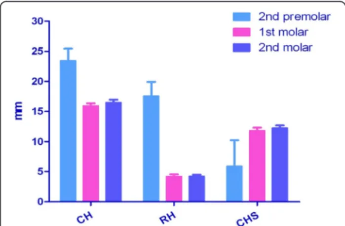
Fig. 1 The vertical position of the bony canals according to the tooth region on para-axial sinus CT scan. CH (canal height), distance between alveolar crest and inferior border of the canal; RH (ridge height), distance between alveolar crest and sinus floor; CHS (canal height from the sinus); CH-RH
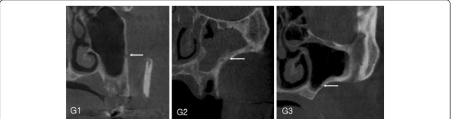
Fig. 2 The horizontal position of the bony canals. G1 extrasinusal, G2 intraosseous, G3 intrasinusal
Table 1 The vertical position of the bony canals according to the tooth region (mean ± SD, mm)
Tooth region 2nd premolar 1st molar 2nd molar Identified/investigated 2/152 (1.3 %) 44/264 (16.7 %) 86/247 (34.8 %)
CH 23.45 (±2.81) 15.92 (±2.65) 16.61 (±2.92)
RH 17.56 (±3.30) 4.19 (±2.15) 4.21 (±1.86)
CHS 5.89 (±6.11) 11.83 (±3.16) 12.21 (±2.87)
CH canal height (distance between alveolar crest and inferior border of the canal), RH ridge height (distance between alveolar crest and sinus floor), CHS canal height from sinus (CH-RH), G1 extrasinusal, G2 intraosseous, G3 intrasinusal
Lee et al. Maxillofacial Plastic and Reconstructive Surgery (2016) 38:1 Page 2 of 4
Methods
Para-axial CT images were evaluated for the presence of a bony canal of the intraosseous loop in the lateral sinus wall.
The canal height (CH) and ridge height (RH) were mea- sured on para-axial cut using a digital caliper. CH defined a vertical distance from the alveolar crest to the inferior border of the canal. CH from the sinus (CHS) was calcu- lated by subtraction of RH from CH. CH was measured at each tooth position from the first premolar to the second molar (Fig. 1). The horizontal position of the bony canals in the lateral wall mesiodistally from the sinus was classified into three categories: G1, in which the canal bulged towards outside of the wall (extrasinusal); G2, with the canal em- bedded in the sinus wall (intraossoeous); and G3, in which the canal bulged towards inside of the wall (intrasinusal) (Fig. 2).
Statistical analysis
The measured values according to the tooth region and age were analyzed by one-way analysis of variance (ANOVA), and those according to the gender, edentulous/partial eden- tulous, and right/left were analyzed with t test (p < 0.05).
Results
Two hundred eighty-four CT images were examined.
The bony canal of the intraosseous loop was identified in 92 images (32 %). Concerning the vertical canal position according to the tooth region, the canal was observed to run most inferiorly from the alveolar crest at the first molar. CH from the sinus floor increased as the canal
passed the posterior region (Table 1, Fig. 3). No signifi- cant differences were evident among the measured values according to the tooth region. The G2 (intraosseous) type of horizontal canal position was the most predominant in the lateral wall in the first molar and second molar re- gions (Table 2). The measured CH and RH values and calculated CHS values according to age, gender, edentu- lism, and right/left side are summarized in Tables 3 and 4. There were no statistical significances between the groups. CHs exceeding 15 mm and less than 17 mm were most prevalent (33.7 %), followed by CH over 19 mm (18.5 %) and CH under 13 mm (12.0 %) (Table 5).
Discussion
A bony canal of the intraosseous loop at the lateral wall was presently identified in 32 % of the examined CT im- ages. The mean canal height was 23.45 ± 2.81, 15.92 ± 2.65, and 16.61 ± 2.92 mm at the second premolar, first molar, and second molar, respectively. The determined mean heights between the canal and alveolar crest of the particular tooth area was shorter than 50 % of those previously reported in radiographic studies [12, 13]. The reconstruction of CT images in the 3-mm interval para- axial cut cannot be ruled out as the reason for the shorter prevalence because the CT images were reconstructed from 1-mm interval para-axial cuts in other studies.
In a cadaveric study, the intraosseous loop was always found at the lateral wall of the anatomic specimens while the extraosseous loop was found only in 44.4 % near the periosteum at the level of 22.75 ± 1.49 mm from the alveolar crest [7, 9]. In a Korean anatomic study, the intraosseous loop was also found in all examined cadavers;
the average external diameter was 0.9 ± 0.3 mm and the mean height from the CEJ was 24.1 ± 4.6, 21.1 ± 4.8, and 22.4 ± 3.7 mm in the second premolar, first molar, and second molar, respectively [10]. These results were much higher than those of the present study. The previous study had a reference point on the CEJ of the tooth, while the
Fig. 3 The vertical position of canals according to the tooth region
Table 2 The horizontal position of the bony canals according to the tooth region
Tooth region Number G1 G2 G3
2nd premolar 2 2 (100 %) – –
1st molar 44 8 (18.18 %) 34 (77.27 %) 2 (4.55 %)
2nd molar 86 – 75 (87.21 %) 11 (12.79 %)
Table 3 Canal height according to age (mean ± SD, mm)
Age 30s 40s 50s 60s 70s
Identified/
investigated
6/18 17/61 36/86 25/95 8/24
CH 18.06
(±4.11)
15.48 (±3.01)
16.49 (±2.87)
16.45 (±3.51)
16.75 (±2.82)
Table 4 Canal height according to gender, edentulism, and right/left (mean ± SD, mm)
Gender Edentulism Right/left
Female Male Full Partial Right Left
Identified/
investigated
31/109 61/175 18/60 74/224 45/138 47/146
CH 16.88
(±3.67) 16.18 (±2.85)
16.76 (±3.41)
16.36 (±3.10)
16.85 (±3.04)
16.25 (±3.49)
Lee et al. Maxillofacial Plastic and Reconstructive Surgery (2016) 38:1 Page 3 of 4
present study set a reference point on the alveolar crest of edentulous region. Alveolar bone resorption after extrac- tion could be an explanation of the presently higher re- sults. In the present study, as well as in anatomic studies [7, 9, 10, 14] and a radiographic study [13], the intraoss- eous loop formed a concave arch, with the first molar area being the lowest point of the bony canal arch course. Dur- ing surgical procedures including lateral window osteot- omy, more precautions should be taken at the first molar region than at the premolar region.
The most frequent horizontal position of the canal in the lateral wall was intraosseous or intrawall type (G2), 77.27 % in the first molar and 87.21 % in the second molar region. The results corresponded with another radiographic study [15], but in Korean cadavers, the canals were observed in G1 position most frequently [10].
Hur et al. [10] and Elian et al. [12] recommended de- signing the superior osteotomy line 13 to 15 mm from the alveolar crest for placing proper length of dental im- plant [10, 12]. In this study, a CH exceeding 13 mm was evident in 88 % of CT scans. This result indicates that 12 % of the cases could be followed by surgical vascular complications.
Even though the intraosseous loop was radiographic- ally evident in only 32 % in this study, the results could be used to prevent the arterial bleeding at the time of lateral window surgery, especially under local anesthesia.
Conclusions
In the horizontal positions of the bony canals, the intraosseous type was most predominant. CHs more than 15 mm and less than 17 mm were most prevalent (33.7 %) and those under 13 mm were 12.0 %. The radio- graphic findings in this study could be used to decide the lateral osteotomy line avoiding potential vascular compli- cation. However, since only one third of the canals could be detected in CT scans, precautions should be taken for the possibility of severe bleeding during osteotomy.
Competing interests
In consideration of publication of my contribution in Maxillofacial Plastic and Reconstructive Surgery, none of the authors have any competing interests in the manuscript.
Authors’ contributions
K and P designed and supervised this study, participated in the sequence alignment. L carries out radiographic measurements, statistic analysis and drafted the manuscript. M reviewed statistic analysis and data alignments. All authors read and approved the final manuscript.
Author details
1Department of Oral and Maxillofacial Surgery, College of Dentistry Jukjeon Dental Hospital, Dankook University, Yongin, South Korea.2Department of Oral and Maxillofacial Surgery, Kunkuk University Medical Center, Seoul, South Korea.3Department of Conservative Dentistry, College of Dentistry Daejeon Dental Hospital, Wonkwang University, Deajeon, South Korea.
4Department of Periodontology, Graduate School of Medicine, Ewha Womans University, 1071, Anyangcheon-ro, Yangcheon-gu, Seoul 158-710, South Korea.
Received: 18 August 2015 Accepted: 16 November 2015
References
1. Wallace SS, Froum SJ (2003) Effect of maxillary sinus augmentation on the survival of endosseous dental implants. A systematic review. Ann Periodontol 8:328–43
2. Del Fabbro M, Testori T, Francetti L, Weinstein R (2004) Systematic review of survival rates for implants placed in the grafted maxillary sinus. Int J Periodontics Restorative Dent 24:565–77
3. Schwartz-Arad D, Herzberg R, Dolev E (2004) The prevalence of surgical complications of the sinus graft procedure and their impact on implant survival. J Periodontol 75:511–6
4. Chanavaz M (1996) Sinus grafting related to implantology. Statistical analysis of 15 years of surgical experience (1979–1994). J Oral Implantol 22:119–30 5. Zijderveld SA, van den Bergh JP, Schulten EA, ten Bruggenkate CM (2008) Anatomical and surgical findings and complications in 100 consecutive maxillary sinus floor elevation procedures. J Oral Maxillofac Surg 66:1426–38 6. van den Bergh JP, ten Bruggenkate CM, Disch FJ, Tuinzing DB (2000)
Anatomical aspect of sinus floor elevations. Clin Oral Implants Res 11:256–65 7. Traxler H, Windisch A, Geyerhofer U, Surd R, Solar P, Firbas W (1999) Arterial
blood supply of the maxillary sinus. Clin Anat 12:417–21
8. Allen WE 3rd, Kier EL, Rothman SL (1973) The maxillary artery: normal arteriographic anatomy. Am J Roentgenol Radium Ther Nucl Med 118:517–27 9. Solar P, Geyerhofer U, Traxler H, Windisch A, Ulm C, Watzek G (1999) Bloody
supply to the maxillary sinus relevant to sinus floor elevation procedure.
Clin Oral Implants Res 10:34–44
10. Hur MS, Kim JK, Hu KS, Bae HE, Park HS, Kim HJ (2009) Clinical implications of the topography and distribution of the posterior superior alveolar artery.
J Craniofac Surg 20:551–4
11. Standring S (2005) Gray’s anatomy, 39th edn. Churchill Livingstone, London, pp 572–9
12. Elian N, Wallace S, Cho SC, Jalbout ZN, Froum S (2005) Distribution of the maxillary artery as it relates to sinus floor augmentation. Int J Oral Maxillofac Implants 20:784–7
13. Mardinger O, Abba M, Hirshberg A, Schwartz-Arad D (2007) Prevalence, diameter and course of the maxillary intraosseous vascular canal with relation to sinus augmentation procedure: a radiographic study. Int J Oral Maxillofac Surg 36:735–8
14. Rosano G, Taschieri S, Gaudy JF, Del Fabbro M (2009) Maxillary sinus vascularization: a cadaveric study. J Craniofac Surg 20:940–3
15. Ella B, Sédarat C, Noble Rda C, Normand E, Lauverjat Y, Siberchicot F et al (2008) Vascular connections of the lateral wall of the sinus: surgical effect in sinus augmentation. Int J Oral Maxillofac Implants 23:1047–52
Submit your manuscript to a journal and benefi t from:
7 Convenient online submission 7 Rigorous peer review
7 Immediate publication on acceptance 7 Open access: articles freely available online 7 High visibility within the fi eld
7 Retaining the copyright to your article
Submit your next manuscript at 7 springeropen.com Table 5 Distribution of canal height (mean ± SD, mm)
CH <11 <13 <15 <17 <19 <21 21≤ Sum
N (number) 2 9 17 31 18 9 6 92
N (%) 2.2 9.8 18.5 33.7 19.6 9.8 6.4 100
Lee et al. Maxillofacial Plastic and Reconstructive Surgery (2016) 38:1 Page 4 of 4