• 검색 결과가 없습니다.

Expression and Characterization of Human Immunodeficiency Virus-1 Oligomerized gp140 Protein in Mammalian Cells

N/A
N/A
Protected

Academic year: 2021

Share "Expression and Characterization of Human Immunodeficiency Virus-1 Oligomerized gp140 Protein in Mammalian Cells"

Copied!
10
0
0

로드 중.... (전체 텍스트 보기)

전체 글

(1)

INTRODUCTION

Acquired immune deficiency syndrome (AIDS) is caused by human immunodeficiency virus (HIV) and its clinical manifestations was originally observed from the failure of normal immune function. Moreover, in the absence of normal immunity, AIDS patients are sensitive to opportunistic infection by agents such as viruses, bacteria, fungi, and

protozoa against which a healthy individual would be resistant. The victims of AIDS also suffer from a high frequency of some types of cancers, particularly lymphomas and Kaposi's sarcoma

1

.

The HIV-1 envelope glycoproteins are initially synthesized as a polyprotein precursor, gp160, that undergoes the post- translational modifications including glycosylation, oligomerization, and proteolytic cleavage into the gp120 and gp41 subunits.

포유동물 세포에서 Human Immunodeficiency Virus-1의 Oligomeric gp140 단백의 발현 및 특성

김은옥, 김은, 김현수, 신광순, 김철중*

충남대학교 수의과대학

(게재승인 : 2002년 2월 1일)

Expression and Characterization of Human Immunodeficiency Virus-1 Oligomerized gp140 Protein in Mammalian Cells

Eun-Ok Kim, Eun Kim, Hyun-Soo Kim, Kwang-Soon Shin and Chul-Joong Kim

*

College of Veterinary Medicine, Chungnam National University, Daejon, 305-764, Korea

(Accepted : February 1, 2002)

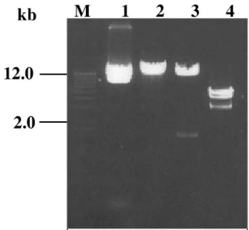
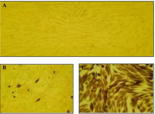
국문초록 : HIV-1의 envelope glycoprotein은 중화항체에 의한 체액성 면역반응의 중요한 target으로 surface glycoprotein인 gp120과 transmembrane glycoprotein인 gp41로 이루어져 있다. gp120과 gp41의 ectodomain으로 이루어진 gp140 유전자를 PCR의 방법으로 증폭하고 Semliki Forest virus(SFV) 유래 expression system을 이용하여 mammalian 세포에서 발현하였다. 발현된 gp140은 natural HIV-1에서와 같이 oligomer를 형성하였다. 발현된 gp140을 정제하여 BALB/c 마우스에 접종하여 항체가 형성되었 음을 확인하였다.

Abstract : The envelope glycoprotein of HIV-1 forms an oligomeric complex resulting in playing a role to induce neutralizing antibody and cell-mediate immune responses. The oligomer exists as a trimer of gp120-gp41 heterodimer which mediates HIV-1 attachment and fusion. We made a cDNA clone of gp140 consisting of gp120 and ectodomain of gp41 from the primary African isolate. To express the oligomeric gp140 in mammalian cells, we adopted the Semliki Forest virus (SFV) based expression system. The oligomeric gp140 in the secretory form was expressed and purified from the cell culture supernatant and characterized. The antibody inducing activity of the purified gp140 was also examined in mice inoculation.

*Corresponding author : +82-42-821-6783 (phone), [email protected] (email)

This work was supported by the National Research Laboratory (NRL) Program Grant (2000-N-NL-01-C-171), the Ministry of Science and Technology, Korea.

55

(2)

The mature envelope glycoproteins are transported to the cell surface, where they are incorporated into the virus as an oligomeric complex. The oligomer consists as a trimer of gp120-gp41 heterodimers

2

. The formation of hetero- oligomeric complexes of gp120/41 as the HIV-1 virion presumably causes the quaternary structural changes that could lead to new antigenic properties compared with those displayed by monomeric forms of these molecules

3

. The trimeric envelope glycoprotein complex mediates HIV-1 attachment and fusion

4

. So, the envelope glycoproteins are important targets for the humoral immune response in that neutralizing antibodies are known to interfere with virus- cell attachment and fusion

4,5

.

In the case of HIV, there are concerns that a killed whole virus might be an effective immunogen, and fears that a live, attenuated virus might be too dangerous. For these reasons, research has concentrated on developing vaccines based on viral protein subunits. To date, most recombinant HIV-1 glycoproteins tested as vaccine candidates have been gp120 monomers. The HIV gp120 has received the most attention because this molecule is located on the surface of the virus particle and is exposed to immune attack

10

. But, the antibody responses to gp120 are not usually effective in neutralizing primary HIV-1 isolates

2

.

Thus, identifying the factors that contribute to native env protein conformation will be important for a vaccine development and for characterizing the humoral response to infection and the mechanisms of neutralization. To attempt to mimic the native HIV-1 envelope glycoprotein, oligomer, soluble gp140 glycoproteins containing gp120 and the gp41 ectodomains have been created

2

. Recent studies have shown that the oligomeric nature of the HIV-1 envelope protein may strongly influence its antigenic structure. Like most other viral membrane proteins, HIV-1 env forms an oligomeric complex shortly after synthesis, with both dimers and higher-order structures have been reported

22

. Moreover, the proteins which faithfully represent the antigenic structure of the virion-associated envelope glycoprotein complex may be worth evaluating as vaccine immunogens

6,7,8,9

.

In this report, we described the production of secreted oligomeric HIV-1 envelope glycoproteins by using a novel expression system based on Semliki Forest Virus (SFV)

12,13,14

. Also, our data showed that gp140 was

adequately synthesized as the oligomer in SFV expression system. The purified gp140 protein was reacted with various antibodies against HIV-1 env protein and recognized as an immunogen candidate for immunization using laboratory animal.

MATERIALS AND METHODS

Cell Culture

To express a high-level of gp140 protein, Semliki Forest virus (SFV)-derived expression system and the mammalian cells (BHK-21 cell, COS-1 cell) were used. The BHK-21 cells were cultured in the minimum essential medium (α -MEM) with 10% tryptose phosphate broth, 5% fetal bovine serum (FBS), 20 mM HEPES, 2 mM glutamine (Gibco BRL, U.S.A.) and 0.1 U/ml penicillin and 0.1 ㎍/ml streptomycin. COS-1 cells were grown in Dulbecco's modified eagle medium (DMEM) with 10% FBS and 0.1 U/ml penicillin and 0.1 ㎍/ml streptomycin.

Cloning of gp140 Gene

The env gene from the primary isolate of HIV-1 clade E virus (CR4002)

40

derived from the Central African was used.

For the production of soluble, secreted HIV-1 envelope glycoprotein, gp140 portion of env gene was amplified by PCR using the forward primer (5'- CGCGCCTCGAGCG GGATCCCATGGCTCGCTCGGTGACCCTAGTCTTTCT GGTGCTTGTCTCACTGACCGGCTTGTATGCTATCCA GAAATCAGACAACTTGTGGGTTACAGTT - 3') and the reverse primer (5'- CGGCTAGCCGGATCCTTAGTGATG GTGATGGTGATGTATATACCACAGCCACCTTGTTAT -3') containing BamHⅠ restriction enzyme sites (underlined).

The forward primer contained β-2 microglobulin signal peptide to increase the secretory expression level. PCR conditions contained 30 cycles (composed of 10 min at 9 4℃ for denaturation, 2 min at 55℃ for annealing, and 3 min at 72℃ for extension) and l cycle (composed of 2 min at 55℃ for annealing and 10 min at 72℃ for extension.

The reaction was carried out in GeneAmp PCR system

2400 (Perkin Elmer, U.S.A.). The PCR product was cloned

into the pSFV-1 plasmid which was digested with BamHⅠ

and treated with calf intestine alkaline phosphatase (CIAP,

TaKaRa) for 1hr at 37℃. The cloned inserts were

identified by the several restriction enzyme digestion.

(3)

In vitro RNA Transcription and Transfection into Mammalian Cells by Electroporation.

Recombinant pSFV-gp140 plasmid was linearized by Sph

Ⅰ digestion and pSFV-Helper plasmid provided by Dr.

Marc Girard in Pasteur Institute was linearized by SpeⅠ digestion. These were used as templates for the in vitro transcription. Briefly, 50㎕ transcription reactions contained 40 mM Tris-HCl, pH7.5, 6 mM MgCl

2

, 2 mM spermidine, 5 mM DTT (TaKaRa), 1 mM each of ATP, CTP and UTP, 0.5 mM GTP (Roche), 1 mM Cap analogue m7G(5') PPP(5')G (Roche), 40 units RNase inhibitor (TaKaRa), and 50 units SP6 RNA polymerase (TaKaRa). This mixture was incubated at 37℃ for 1 hr and then added l ㎕ of SP6 RNA polymerase again and incubated for further 30 min.

The reaction was stopped by placing the tube on ice and an aliquot of RNA was analyzed on a 0.8% agarose gel.

The BHK-21 cells were washed twice and resuspended in PBS at 10

7

/ml. Cell suspension was mixed with 100 ㎕ (50

㎕ of pSFV-Helper RNA transcripts + 50 ㎕ of pSFV- gp140 RNA transcripts) of transcribed RNA. The mixture was electroporated with two consecutive pulses at 0.85 kv and 25 μF (BioRad Gene PulserⅡ, U.S.A.) and transferred to 100 mm tissue culture dishes and 24-well tissue plate (Nunc, Denmark).

Harvest of Recombinant Virus Particles

The transfected BHK-21 cells were incubated for 2 days, under the atmosphere of 5% CO

2

at 37℃. The culture fluid was harvested and clarified by centrifugation for 15 min at 3000 × g to remove cell debris. The supernatant was transferred to a fresh tube (Beckman) and ultracentrifugated at 35,000 × g, for 3 hr to collect virus particles (SW41 Ti rotor). The viral pellet was resuspended in TNE buffer (50 mM Tris-HCl [pH7.4], 100 mM NaCl and 0.5 mM EDTA) and stored quickly in aliquots at -70℃.

Infection with Recombinant SFV Particles The BHK-21 cells were seeded onto the 24-well tissue culture plate and tissue culture dishes. Prior to the infection, the cells were washed twice in PBS. The recombinant virus particles were thawed quickly at room temperature and treated with 500 ㎍/ml chymotrypsin (Roche) and 0.5 mM CaCl

2

and incubated on ice for 30 min. To inactivate chymotrypsin activity, 0.5 volume of aprotinin (2 ㎎/ml, Sigma) was added. The activated vrus

particles were diluted in MEM containing 1% FBS and inoculated into the BHK-21 cells and incubated for 90 min at 37℃, in atmosphere of 5% CO

2

. After 1 hr, the inoculum was removed and the new medium for BHK-21 cells was added. At 24 hr after infection, the cells were washed three times in PBS and lysed with cell lysis buffer (1% NP-40, 50 mM Tris-HCl [pH7.6], 150 mM NaCl, 2 mM EDTA, 1

㎍/ml PMSF) on ice for 1 hr. The lysates were centrifuged at 12,000× g for 5 min at 4℃ and used as samples in sodium dodecylsulfate-polyacrlyamide gel electrophoresis (SDS- PAGE). The culture supernatant was harvested, clarified by low-speed ultracentrifugation, and filtered using 0.45 ㎛ filter.

Sucrose Density Gradient ultracentrifugation The BHK-21 cells were infected with recombinant SFV-gp140 particles. After 24 hr, the supernatant was collected, clarified by low-speed ultracentrifugation and concentrated with a microconcentrator (M.W. cutoff = 30,000, Amicon). The proteins were separated by sucrose density gradient, 5-20% sucrose in 20 mM Tris-HCl, 150 mM NaCl (pH 7.4) at 35,000× g for 24 hr at 4℃ in an SW41 Ti rotor (Beckman) to separate monomeric and oligomeric forms of env. Following ultracentrifugation, 1ml fractions were obtained and each fraction was concentrated with a microconcentrator (M.W. cutoff = 100,000, Amicon). Each fraction was analyzed by immunoblotting with the various monoclonal and polyclonal antibodies.

Immunocytochemistry

The cells were rinsed twice PBS and fixed on the slide glass by ice-cold methanol at 4℃ for 15 min. The cells were washed three times in PBS. The cells were incubated at room temperature for 1 hr in PBS containing 1% gelatin to block the nonspecific binding. The anti-gp41 monoclonal antibody and human polyclonal antibody as primary antibodies for gp140 protein were added and incubated at room temperature for 3 hr. The cells were washed in PBS three times, followed by the reaction with a biotinylated anti-mouse, anti-human IgG (Vector, U.S.A.) at room temperature for 1 hr. The cells were washed in PBS and reacted with HRP-avidin-biotin reaction solution (Vector, U.S.A.) at room temperature for 30 min. The cells were finally washed in PBS and visualized by adding H

2

O

2

solution containing DAB (3,3'-diaminobenzidine, Vector,

U.S.A.).

(4)

SDS-PAGE and Western blot analyses

SDS-PAGE was performed by the method of Laemmli

26

. Protein samples were mixed with SDS-PAGE sample buffer (62.5 mM Tris-Cl [pH6.8], 10% glycerol, 2%

sodium dodecylsulfate [SDS], 5% 2-mercaptoet hanol, 0.05% bromophenol blue) and then boiled at 100℃ for 5 min and cooled on ice. The denatured protein samples were loaded into each well of prepared SDS- polyacrylamide gel (10%) and electrophoresis was performed. The proteins separated by SDS-PAGE were electrophoreticall y transferred to PVDF (polyvinylidine difluoride) membrane (Bio Rad, U.S.A.). The membrane was incubated in 5% skim milk with gentle shaking for 1 hr and reacted overnight with the primary antibody at room temperature in shaking camber.

After washing in washing buffer (20 mM Tris-HCl, 150 mM NaCl, 0.05% Tween 20), the membrane was reacted with a biotinylated anti-mouse, anti-human IgG (Vector, U.S.A.) at room temperature for 2 hr and then reacted with HRP-avidin-biotin reaction solution (Vector, U.S.A.) at room temperature for 30 min. After washing in washing buffer, the membrane incubated with chemiluminescence reagent (Supersignal

R

Substrate, PIERCE, U.S.A) and exposed to a Hyperfilm

TM

-MP (High performance autora- diography film, Amersham, Sweden) and also developed with DAB solution. The reaction was stopped by immersing the membrane in distilled water.

Mice Immunization

BALB/c mice were immunized with purified gp140 protein preparations with Freund's incomplete adjuvant (Sigma, U.S.A). Briefly, mice were inoculated intraperi- toneally three times at 3-week intervals with 20㎍ of purified HIV-1 gp140 protein per mouse. The serum samples were obtained 2 weeks after the final injection. The individual mouse sera were assayed by immunoblotting to ensure reactivity with the purified gp140 protein.

RESULTS

Cloning of gp140 Gene

To amplify gp140 portion of env gene of HIV-1 clade E virus, the PCR was carried out. The expected size, 1,600 bp was visualized in 0.8% agarose gel. The PCR product of gp140 was cloned into the BamHⅠ site of pSFV-1 (Fig.

1) Screening of recombinant DNA were performed by the restriction enzyme digestions with SmaⅠ, BglⅡ, and ScaⅠ (Fig. 2). The plasmid harboring the expected size and showing the expected restriction fragments was singled out and named as pSFV-gp140.

Expression of HIV-1 gp140 in Mammalian Cells We investigated the expression of gp140 protein after the trasnfection of BHK-21 cells with a pSFV-gp140 RNA transcript. The pSFV-gp140 plasmid was linearized by Sph Ⅰ

3.0 1.6

M 1 2

kb M 1 2 3 4

12.0 2.0 kb

Fig. 1. Amplified PCR products of HIV-1 gp140 gene (M : 1Kb DNA ladder, lanes 1-2 : gp140).

Fig. 2. The restriction fragment patterns of cloned

pSFV-gp140 by restriction enzymes (M: lkb DNA ladder,

lane 1: undigested plasmid, lane 2: SmaⅠ, lane 3: BglⅡ,

(5)

A B

C D

Fig. 4. Immunocytochemistry of HIV-1 gp140 protein expressed in BHK-21 cells (C and D) and COS-1 cells (A and B) infected with the pSFV-gp140 recombinant virus particles (A and C: control, B and D: infected).

A

B C

Fig. 3. Immunocytochemistry of HIV-1 gp140 protein expressed in BHK-21 cells posttransfection with the RNAs from control and pSFV-gp140 with pSFV-Helper (A: control, B: detected with mouse anti-gp41 monoclonal antibody, C:

detected with human polyclonal antibody).

(6)

and the pSFV-Helper was linearized by SpeⅠdigestion.

The linearized DNAs were used as the templates for in vitro RNA transcription under the control of SP6 promoter.

Expression of the HIV-1 gp140 protein was detected by immunocytochemistry using anti-gp41 monoclonal antibody

(Fig. 3-B) and human polyclonal antibody (Fig. 3-C).

Intracellular expression of gp140 protein was recognized in 60-80% of the electroporated cells and located in the cell cytoplasm.

Fig. 5. Western blot analysis of HIV-1 gp140 protein in BHK-21 cell lysates with mouse anti-gp41 monoclonal antibody (A) and human polyclonal antibody (B) (lane 1: control, lanes 2 and 3: infected cell lysate). The upper arrows indicate the aggregate, the middle arrows indicate gp140 oligomer, and the lower arrows indicate gp120.

A B

kDa 198

98 66 43

24

1 2 3 4 5 6

Fig. 6. Sucrose density gradient ultracentrifugation of HIV-1 gp140 protein. BHK-21 cells were infected with recombinant pSFV-gp140 defective virus particles. 24 hr after infection, the supernatant from infected cells was concentrated and separated on a 5-20% sucrose gradient (fractions were detected with polyclonal antibody directed to gp120-gp41 linkage site). The upper arrow indicates gp140 oligomer, the middle arrow indicates gp120, and the lower arrow indicates the

d

5 % 20%

(7)

Infection of Recombinant Virus Particles into Mammalian Cells

To investigate whether the SFV-derived system could be used in reconstituting assembly of the infectious viral particles carrying a target foreign gene, we infected the defective recombinant SFV particles into the BHK-21 and COS-1 cells. The gp140 protein expression was analyzed by immunocytochemistry (Fig. 4) and immunoblot (Fig. 5).

Immunocytochemistry with the human polyclonal antibody showed that the defective virus particles containing gp140 gene were successfully infected and gp140 protein was efficiently produced in the cytoplasm of the BHK-21 cell (Fig. 4-C, D) and COS-1 cell (Fig. 4-A, B). Also, the immunoblot analysis with anti-gp41 monoclonal antibody (Fig. 5-A) and human polyclonal antibody (Fig. 5-B) showed the expression of oligomeric gp140 and cleaved gp120 by the host cell proteases. The upper band was aggregates, the middle band was gp140 proteins and the lower band indicated the gp120 proteins (Fig. 5). In addition, these data indicated that SFV vectors could be used to produce the HIV-1 gp140 proteins to high levels and this protein was correctly processed, folded, and transported to the cell surface as the native conformation.

Sucrose Density Gradient Ultraultracentri- fugation Analysis

To investigate whether the gp140 was secreted in the mammalian cell as a native virion structure, the infected cell supernatant was harvested and concentrated to perform the sucrose density gradient ultraultracentrifugation. Each fraction of the gp140 protein was reacted with the specific antibody. Two different types of oligomers were identified (Fig.

6). Immunoblotting using polyclonal antibody directed to gp120-gp41 linkage site showed gp140 oligomer, gp120 cleavaged by host cell proteases, and gp41 ectodomain (Fig. 6). These data indicated that the expressed, secreted gp140 protein was adequately synthesized as the oligomer in the SFV expression system and reacted well with the antibody against HIV-1 gp140.

Mice Immunization

Five BALB/c mice were immunized with the purified gp140 protein. Mice were intraperitoneally inoculated three times at 3-week intervals with 20㎍ of the purified HIV-1 gp140 protein per mouse. The individual mouse serum was tested by immunoblot to ensure reactivity with the purified gp140 protein. Immunoblot analysis with the serum from mouse immunized with the purified gp140 protein showed that the monomeric gp140 and higher-order oligomeric form (Fig. 7). These data indicated that the purified gp140 Fig. 7. Western blot analysis of purified HIV-1 gp140 protein detected with mice serum (M : Protein marker, lane 1 : negative control, lanes 2-5 : fractions detected with individual mouse serum). The upper and lower arrows indicate the oligomeric and monomeric gp140, respectively.

kDa

98 66 43

24

M 1 2 3 4 5

(8)

protein induced the antibody production and was recognized as a possible immunogen.

DISCUSSION

Like most other viral membrane proteins, HIV-1 env forms an oligomeric complex that is essential for its structure and function. Trimers and tetramers of gp41 were detected in viral lysates of T cell line-adapted HIV-1 by immunoprecipitation, and recent biophysical analysis of recombinant protein fragment from the gp41 ectodomain confirmed their association into trimers

3

. Especially, the gp140 composed of gp120 and ectodomain of gp41 is organized into dimer or higher-order oligomer. In addition, the gp140 protein does not have a transmembrane domain of gp41, or secrets into host cell media.

Several lines of evidence suggests that secreted, oligomeric gp140 accurately reflected native env structure.

First, the protein was efficiently secreted. Since misfolded molecules are typically retained in the endoplasmic reticulum and degraded, it is unlikely that gp140 was grossly misfolded. Second, gp140 bounds sCD4 as well as the conformation-dependent MAb F105 efficiently. Because the CD4 and F105 binding sites are discontinous in nature

20

, the gp120 portion of the molecule appeared to be relatively intact. However, the strongest evidence that purified gp140 accurately reflects native env structure came from analysis of the antibodies raised by env on the surface of chronically infected cells. These results demonstrate that when oligomeric env is used for immunization, antibodies to conformation-dependent epitopes are readily generated and suggest that env tertiary and quarternary structure are important determinants for the humoral response

15

.

Oligomeric env closely reflects the antigenic characteristic of env protein on the surface of virions and infected cells, retains highly conserved epitopes that are recognized by antibodies raised against different clades, and makes it possible to detect a much greater fraction of total anti-HIV-1 env activity in sera than does native monomeric gp120.

The envelope glycoproteins of HIV-1, gp120 and gp41, are targets of neutralizing antibodies in infected individuals.

That env protein conformation is an important for the humoral response is indicated by the findings that antibodies to conformational epitopes are prevalent in human sera and that the most broadly cross-reactive

neutralizing antibodies characterized to recognize discontinuous epitopes in gp120. Thus, identifying the factors that contribute to native env protein conformation will be important for vaccine development and for characterizing the humoral response to infection and the mechanisms of neutralization

11

.

To investigate whether the oligomeric form of HIV-1 gp140 was expressed and secreted in mammalian cell, we used a SFV-derived expressed system. SFV has been adapted as an expression system by generating a plasmid containing the entire genome of the virus, without the genes for the four structural proteins (the capsid and three component of the envelope protein)

6,7,8,9,12

. Replacement of these structural protein genes with foreign DNA results in high-level expression of the heterologous proteins

12,13

. Thus, the SFV system may offer advantages in the expression of HIV-gp140 proteins. BHK cells were used for testing the transfected pSFV-1 gp140 vector and immunostaining revealed that this construct induced the expression of HIV-1 gp140 protein. Here we showed that HIV-1 gp140 was successfully expressed and was adequately synthesized as the oligomer. This gp140 was reacted well with polyclonal antibody directed to gp120-gp41 linkage site.

This oligomeric form of gp140 has a many implications for antigenic structures and is a target in the detection of antibodies and for studying correlations between binding and neutralizing activity. First, the formation of hetero- oligomeric complexes of gp120/gp41 causes quartenary structural changes that could lead to new antigenic properties compared with those displayed by monomeric forms of these molecules

3

. Oligomeric nature of the HIV-1 envelope protein may strongly influence its antigenic structure

5

.

Second, the envelope glycoproteins are important targets

for the humoral immune response. So this oligomerization

have implications for vaccine development and for the

study of the immune response against HIV-1

3

. Recent

studies showed that oligomeric structure of env-encoded

proteins may provided a more effective immune response

than monomeric structure of env-encoded proteins

3,16

. Finally,

most recombinant HIV-glycoproteins were tested as vaccine

candidates, especially a subunit vaccine. So many expression

vectors were used to express the recombinant HIV-1

envelope glycoproteins

17,18,19

, for example, a vaccinia virus

expression system. However, we used a SFV expression

(9)

system, and observed a high level expression of gp140.

Also, the gp140 was adequately synthesized as the oligomer and reacted well with antibodies against HIV-1. It may be used to be a important subunit vaccine candidates.

In conclusion, these results showed that oligomeric form of gp140 have significant effects on the antigenic structure of HIV-env and, as a consequence, the oligomeric state of gp140 should be taken into account in the design of potential subunit vaccine. So, further study is required to investigate whether the gp140 is similar to native, virion-associated env structure and antigenicity.

REFERENCES

1. Hung F, Ross F. Conner, Luis P. Villarreal, AIDS in brief. 1999. The biology of AIDS(4th Edition):2-5 2. XinZhen Yang, Michael Farzan, Richard Wyatt, and

Joseph Sodroski. 2000. Characterization of stable trimers containing complete ectodomains of Human immunodeficiency virus type 1 envelope glycoproteins.

2000. Journal of Virology. June. 5716-5725

3. Miroslaw K. Gorny, Thomas C. Vancott, Constance Williams, Kathy Revesz, and Susan Zolla-Pazner. 2000.

Effects of oligomerization on the epitopes of the Human immunodeficiency virus type 1 envelope glycoproteins.

Virology. 267:220-228

4. Rogier W. Sanders, Linnea Schiffner, Aditi Master, Francis Kajumo, Yong Guo, Tatjana Dragic, John P.

Moore and James M. Binley. 2000. Variable-loop- deleted variants of the Human immunodeficiency virus type 1 envelope glycoprotein can be stabilized by an intermolecular disulfide bond between the gp120 and gp41 subunits. Journal of Virology. June. 5091-5100 5. Trkola, A., T. Dragic, H. Arthos, J. M. Binley, W, C,

Olson, G. P. Allaway, C. Cheng-Mayer, J. Robinson, P.J. Maddon, and J. P. Moore. 1996. CD4-dependent, antibody-sensitive interaction between HIV-1 and its co-receptor CCR-5. Nature 384:184-187

6. James M. Binley, Rogier W. Sanders, Brian Class, Norbert Schuelke, Aditi Master, Yong guo, Francis Kajumo, Deborah. Anselma, Paul J, Maddon, Willim C. Olson, and John P, Moore. 2000. A recombinant Human immunodeficiency virus type 1 envelope glycoprotein complex stabilized by an intermolecular disulfide bond between the gp120 and gp41 subunits is

an antigenic mimic of the trimeric virion- associated structure. Journal of Virology. Jan. 627-643

7. Heilman, C. A., and D. Baltimore. 1998. HIV vaccines- where are we going? Nature Medicine, 4:532-534 8. Letvin, N. L. 1998. Progress in the development of an

HIV-1 vaccine. Science 280:1875-1880

9. Barnett, S. W., S. Rajasekar, H. Legg, B. Doe, D. H.

Fuller, J. R. Haynesm C.M. Walker, and K. S. steimer.

1997. Vaccination with HIV-1 gp120 DNA induces immune responses that are boosted by a recombinant gp120 protein subunit. Vaccine 15:869-873

10. James D. Watson, Michael Gilman, Jan Witkowski, Mark Zoller, Recombinant HIV proteins may be effective as immunogen for AIDS vaccines. Recombinant DNA (2nd Edition):501-503

11. Thomas M. Richardson, JR., Brenda L. Stryjewski, Christopher C. Broder, James A. Hoxie, John R.

Mascola, Patricia L. Earl, and Robert W. Doms. 1996.

Humoral response to oligomeric Human immunode- ficiency virus type 1 envelope glycoprotein. Journal of Virology. Feb. 753-762

12. N.L. Paul, M. Marsh, J.A. Mckeating, T.F. Schulz, P.

Liljestrom, H. Garoff, and R.A. Weiss. 1993. Expression of HIV-1 envelope glycoproteins by Semliki Forest Virus vectors. AIDS RESEARCH AND HUMAN RET- ROVIRUSES. 9:963-970

13. Peter Liljestrom and Henrik Garoff. 1991. A new generation of animal cell expression vectors based on the Semliki Forest Virus replicon. Nature Biotechnology.

9:1356-1361

14. Yang, X., L. Florin, M. Farzan, P. Kolchinsky, P.

Kwong, J. Sodroski, and R. Wyatt. 2000. Modifications that stabilize human immunodeficiency virus envelope glycoprotein trimers in solution. Journal of Virology.

74:4746-4754

15. Patricia L. Earl, Christopher C. Broder, Deborah Long, Susan A. Lee, Jeffrey Peterson, Sekhar Chakrabarty, Robert W. Doms and Bernard Moss. 1994. Native oligomeric Human immunodeficiency virus type 1 envelope glycoprotein eilcits diverse monoclonal antibody reactivities. Journal of Virology. May. 3015-3026 16. Abraham Pinter, William J. Honnen, Shermaine A.

Tilley, Constantin Bona, Habib Zaghouani, Miroslaw

K. Gorny, and Susan Zolla-Pazner. 1989. Oligomeric

structure of gp41, the transmembrane protein of

(10)

Human immunodeficiency virus type 1. Journal of Virology. June. 2674-2679

17. Christopher C. Broder, Patricia L. Earl, Deborah Long, Stephen T. Abedon, Bernard Moss, and Robert W.

Doms. Antigenic implications of human immunodeficiency virus type 1 glycoprotein quarternary structure: oligomer- specific and -sensitive monoclonal antibodies. 1994.

Proc. Natl. Acad. Sci. USA 91:11699-11703

18. Patricia L. Earl, Robert W. Doms, and Bernard Moss.

1990. Oligomeric structure of the human immuno- deficiency virus type 1 envelope glycoprotein. Proc.

Natl. Acad. Sci. USA 87:648-652

19. Pantelis Poumbourios, Walid el Ahmar, Dale A. Mcphee, and Bruce E. Kemp. 1995. Determinants of Human immunodeficiency virus type 1 envelope glycoprotein oligomeric structure. Journal of Virology. Feb. 1209- 1218

20. Olshevsky, U., E. Helseth, C. Furman, J. Li, W. Haseltine,

and J. Sodroski. 1990. Identification of individual

human immunodeficiency virus type 1 gp120 amino

acids important for CD4 receptor binding. Journal of

Virology. 64:5701-5707

수치

Fig. 2. The restriction fragment patterns of cloned pSFV-gp140 by restriction enzymes (M: lkb DNA ladder, lane 1: undigested plasmid, lane 2: SmaⅠ, lane 3: BglⅡ,
Fig. 4. Immunocytochemistry of HIV-1 gp140 protein expressed in BHK-21 cells (C and D) and COS-1 cells (A and B) infected with the pSFV-gp140 recombinant virus particles (A and C: control, B and D: infected).
Fig. 5. Western blot analysis of HIV-1 gp140 protein in BHK-21 cell lysates with mouse anti-gp41 monoclonal antibody (A) and human polyclonal antibody (B) (lane 1: control, lanes 2 and 3: infected cell lysate)

참조

관련 문서

Distribution of Citrus Tristza Virus or Satsusma Dwarf Virus at various parts of virus infected Satsuma mandarin

protein levels in control or PIG3 depleted HCT116 and HeLa cells were tested. by

Fig 4 : Inhibitory effects of classified methanol extracts of Acalypha australis the COX-2 protein expression of the human oral cavity carcinoma KB cells....

4 > Effects of Taro on COX-2 expression and iNOS expression(hot water) in human thyroid cancer cells. The cells were pretreated for 48hours with either

The expression of Dynamin II protein expressed strongly at advanced bell stage in cytoplasm of odontoblast. Dynamin II protein localized in adjacent the nuclear

Inhibitory effects of the concentration of methanol extracts from Plantago Asiatica against the COX-2 protein expression and iNOS expression of the MDA-MB-231 human

qRT-PCR for VSMC synthetic phenotype protein, cellular retinol binding protein 1(CRBP-1) and connexin 43, from thoratic aorta showed that CRBP-1 expression was increased by 70%

Distribution of Citrus Tristza Virus or Satsusma Dwarf Virus at various parts of virus infected Satsuma mandarin