371
Molecular differentiation of Korean Newcastle disease virus (NDV) by restriction enzyme analysis and
pathotype-specific RT-PCR
Hyuk-Joon Kwon, Sun-Hee Cho
1, Sun-Joong Kim
1,*
Laboratory of Influenza virus Zoonotic Disease Institute (ZooDI), College of Veterinary Medicine, Seoul National University, Seoul 151-742, Korea
1College of Veterinary Medicine and BK21 for Veterinary Science, Seoul National University, Seoul 151-742, Korea
(Accepted: November 8, 2006)
Abstract : Newcastle disease virus (NDV) is a single-stranded negative sense RNA virus, which has been classified as a member of the Avulavirus genus of the Paramyxoviridae family. It is also one of the most important pathogens in the poultry industry. The glycoproteins, fusion (F) and hemagglutinin-neuraminidase (HN), determine the virulence of NDV, and the relevant molecular structures have already been determined.
NDV isolates differ in terms of virulence, and at least 2 of 9 genotypes (I-IX) have been shown to co- circulate. Therefore, it is clearly important to differentiate between vaccine strains and field isolates. In vivo pathogenicity tests have been the standard protocol for some time, but molecular methods appear preferable in terms of the rapidity of diagnosis, as well as animal welfare concerns. In this study, we have designed primer sets from HN gene for phylogenetic analysis and restriction enzyme analysis, and from F gene for pathotype-specific RT-PCR. Via the combination of 2 methods, 106 Korean NDV isolates obtained from 1980 to 2005 were differentiated into vaccine strains, and virulent genotypes VI and VII. The genotype VI viruses were only rarely isolated after 1999, and genotype VII, after it was initially isolated from poultry in 1995, recurred in 2000, and then became the main NDV constituting a threat to the Korean poultry industry.
Key words : molecular differentiation, Newcastle disease virus, pathotype-specific RT-RCR
Introduction
Newcastle disease (ND), which is caused by the ND virus (NDV), poses a disastrous risk to the poultry industry. NDV is an enveloped, single-stranded, negative- sense RNA virus, which belongs to the Avulavirus genus of the Paramyxoviridae family [18, 19]. NDV has been classified into 3 pathotypes; lentogenic, mesogenic and velogenic types on the basis of conventional in vivo pathogenicity indices such as mean death time (MDT) of chicken embryo and intracerebral pathogenicity index (ICPI) [3]. On the envelope, 2 biologically important glycoproteins, fusion (F) protein and hemagglutinin-neuraminidase (HN), have been detected. These proteins coordinate in order to invade host cells, and play a major role in the
determination of viral virulence. The multi-basic amino acids at the proteolytic cleavage site in F are relevant to systemic replication, and HN determines the tropisms of NDV [9, 11].
ND outbreaks can be characterized by the co- circulation of genetically distinct virus lineages [23].
Strains of NDV have been classified into 10 provisional genetic groups (I-X) and epizootics in western Europe (VIIa and VId), Taiwan (VIIa and III), southern Africa (VIIb and VIII), and Korea (VIf and VIIa) during the first half of 1990s were determined to have been caused by 2 distinct strains of NDV [10, 13, 16, 17, 28, 29]. Therefore, it is clearly important that some protocol be developed which would allow for differentiation between virulent field isolates, as well as between vaccine strains and field isolates.
*Corresponding author: Sun-Joong Kim
Laboratory of Influenza virus Zoonotic Disease Institute (ZooDI), College of Veterinary Medicine and BK21 for Veterinary Science, Seoul National University, Seoul 151-742, Korea
[Tel: +82-2-880-1250, Fax: +82-2-885-6614, E-mail: [email protected]]
In this study, we have amplified a small fragment of HN via RT-PCR, and phylogenetic analysis and restriction enzyme analysis (REA) were conducted for NDV differentiation. In order to rapidly differentiate between the vaccine and virulent NDV strains, we also developed an F gene-based pathotype-specific RT-PCR technique.
Materials and Methods
Viruses
During 1980-2005 106 NDV isolates have been
isolated from diseased birds in the Laboratory of Avian Diseases of Seoul National University via standard procedures [2]. The virulence of these isolates were classified into velogenic and lentogenic types on the basis of the mean death time of the chicken embryo (MDT) and the intracerebral pathogenicity indices (ICPI), as well as the multi-basic amino acids located at the F protein cleavage site [2, 8, 13, 20, 25]. The NDV isolates used in this study are listed in Table 1.
Primers
To distinguish virulent genotypes VI and VII viruses Table 1. Phylogenetic differentiation of Korean NDV isolates used in this study
Year of
isolation No. of
isoates Designation of NDV isolates (pathotype
a/genotype) 1980 3 SNU8050(v/VI), SNU80108(l/I), SNU80143(v/VI)
1984 4 SNU8419(l/II), SNU8426(l/II), SNU8465(v/VI), SNU8490(v/VI)
1985 5 SNU8550(v/VI), SNU8557(v/VI), SNU8559(v/VI), SNU8566(v/VI), SNU85135(v/VI) 1988 6 SNU8861(v/VI), SNU8871
b(v/VI), SNU8876(v/VI), SNU8887(v/VI), SNU88139
b(v/VI),
SNU88147(v/VI)
1990 2 SNU9033
b(v/VI), SNU90111
b(v/VI) 1991 1 SNU9139
b(v/VI)
1992 11 SNU9213
b(v/VI), SNU9222
b(v/VI), SNU9227
b(v/VI), SNU9229
b(v/VI), SNU9257
b(v/VI), SNU9275
b(v/VI), SNU9280
b(v/VI), SNU9282
b(v/VI), SNU9283
b(v/VI), SNU9284
b(v/VI), SNU92105
b(v/VI)
1993 1 SNU9358GG
b(v/VI) 1994 1 SNU9444
b(v/VI)
1995 11 SNU9512
b(v/VII), SNU9515
b(l/-), SNU9550
b(v/VII), SNU9553
b(v/VII), SNU9575
b(v/VII), SNU9586
b(v/VII), SNU9598
b(v/VI), SNU95107
b(v/VII), SNU95119
b(v/VII), SNU95124
b(v/VII), SNU95132
b(v/VII)
1999 8 SNU9969(v/VI), SNU9970(v/VI), SNU9971(v/VI), SNU9983(l/-), SNU9991(l/II), SNU99105(v/VI), SNU99111(l/II), SNU99123(l/I) 1996 1 SNU96130(l/-)
2000 15 SNU0021(v/VII), SNU0036(v/VII), SNU0079(v/VII), SNU0086(v/VII), SNU0105(v/VII), SNU0117(v/VII), SNU0118(v/VII), SNU0119(v/VII), SNU0123(v/VII), SNU0125(v/VII), SNU0129(v/VII), SNU0164(v/VII), SNU0165(v/VII), SNU0169(v/VII), SNU0202(v/VII) 2001 1 SNU1024(v/VII)
2002 19
SNU2001(v/VII), SNU2009(v/VII), SNU2017(v/VII), SNU2026(v/VII), SNU2028(v/VII), SNU2048(v/VII), SNU2049(v/VII), SNU2063(v/VII), SNU2064(v/VII), SNU2078(v/VII), SNU2080(v/VII), SNU2083(v/VII), SNU2084(v/VII), SNU2091(v/VII), SNU2100(v/VII), SNU2102(v/VII), SNU2108(v/VII), SNU2117(v/VII), SNU2124(v/VII)
2003 1 SNU3058(v/VII) 2004 1 SNU4152(v/VII)
2005 15 SNU5005(v/VII), SNU5009(v/VII), SNU5047(v/VII), SNU5062(v/VII), SNU5063(v/VII), SNU5064(v/VII), SNU5065(v/VII), SNU5070(v/VII), SNU5074(v/VII), SNU5076(v/VII), SNU5079(v/VII), SNU5081(v/VII), SNU5084(v/VII), SNU5085(v/VII), SNU5100(v/VII)
a
v, velogen; l, lentogen; -, not determined.
b
Previously identified [13] and reconfirmed in the present study.
from commercial vaccine strains the nucleotide sequences of the F protein cleavage site were compared, and a virulent NDV-specific forward primer [Pt(334-349): 5'- AGGAGACRRAAACGYT-3'] and a reverse primer [I- 1N(534-515): 5'-TGCCACTGMTAGTTGYGATA-3'] com- mon to NDV were designed (pathotyping primer set) (Fig. 1). The HN genes of the NDV strains registered in the GenBank and of the Korean isolates were compared, and a primer set that was conserved in the majority of NDV strains and generated the smallest amplicon was selected (common primer set; numbering from the start codon): ComHNF (638-657) 5'-CATCTG CAACAGGGAGGGTA-3'; ComHNR (757-737) 5'-TMG AGCACAG CATATCACAAC-3'. The specificity and primer sequence conservation among the NDVs were determined via BLAST searches [4].
RNA isolation, RT-PCR and REA
RNA isolation and RT-PCR were conducted as previously described with little modifications [13].
Briefly 100
µl of allantoic fluid was added to 1 ml of easy Blue reagent (iNtRON Biotechnology, Korea) and followed the manufacturer’s protocol. RNA pellet was resolved in 50
µl of DEPC (0.1% diethyl pyrocarbonate)- treated distilled water (DW). cDNA was synthesized by Power cDNA kit as manufacturer’s protocol (iNtRON Biotechnology, Korea). cDNA was diluted 5-fold and 1
µl of cDNA was added to the PCR reaction [1
µl of 10
×PCR buffer, 0.2
µl of 2.5 mM dNTPs, 0.2
µl
of each primer, Taq polymerase (1U/
µl; iNtRON Biote- chnology, Korea), and 7.2
µl of DW]. After denaturation at 94
oC for 3 min, consecutive reactions of 94
oC for 30 sec, 48
oC for 15 sec, 65
oC for 20 sec were repeated for 35 times and final elongation was performed at 72
oC for 7 min. For the multiplex RT-PCR the quantity of the pathotyping primer set was twice that of the common primer sets. On the basis of the sequence data, we predicted the restriction enzyme sites (MacDNAsis Ver. 3.1) and selected 2 enzymes ( Hinf I and Pst I) for REA. The digested fragments were separated on polyacrylamide gel, and visualized via silver staining [6]. For the sake of simplicity, we also utilized 2.5%
agarose gel and ethidium bromide.
Sequencing and Sequence analysis
The comHN of the HN gene was sequenced using an ABI 377 automatic DNA sequencer and a Dye Terminator kit (Perkin Elmer, USA), as reported prev- iously [14]. Nucleotide sequences were aligned via the multiple alignment algorithm contained in the MegAlign package (Windows version 3.12e; DNASTAR, USA), and a phylogenetic tree was constructed via neighbor-joining method.
Results
Molecular differentiation of Korean NDV isolates by phylogenetic analysis
The PCR products amplified by the comHN primer set were directly sequenced. The nucleotide and amino acid similarity between the Korean and foreign isolates including the vaccine strains, were found to be 81.0%- 100% and 92.3%-100%, respectively. Representative nucleotide sequences are summarized in Fig. 2. A phylogenetic tree was constructed on the basis of the nucleotide sequences. The clustering and branching patterns of the tree were similar to those of the tree constructed around the F gene [5]. SNU8050, SNU80143, SNU8465, SNU8490, SNU8550, SNU8557, SNU8559, SNU8566, SNU85135, SNU8861, SNU8871, SNU8876, SNU8887, SNU88139, SNU88147, SNU9033, SNU90111, SNU9139, SNU9222, SNU9227, SNU9280, SNU9282, SNU9283, SNU9284B, SNU92105, SNU9358GG, SNU 9444, SNU9598, SNU9969, SNU9970, SNU9971 and SNU99105 were classified into genotype VI (Table 1, Fig. 3). SNU9512, SNU9550, SNU9553, SNU9575, SNU 95107, SNU95119, SNU95124, SNU95132, SNU2083, Fig. 1. Comparison of the nucleotide sequences of the
pathotyping primer set. The homologous nucleotides are
shown with dashes.
SNU0021-like viruses (SNU0036, SNU0079, SNU0086, SNU0105, SNU0117, SNU0118, SNU0119, SNU0123, SNU0125, SNU0129, SNU0164, SNU0165, SNU0169, SNU1024, SNU2009, SNU2017, SNU2026, SNU2028, SNU2048, SNU2063, SNU2064, SNU2078, SNU2080, SNU2083, SNU2084, SNU2091, SNU2102, SNU2117, SNU2124, SNU3058, SNU5009, SNU5047, SNU5062, SNU5063, SNU5064, SNU5065, SNU5070, SNU5074, SNU5081), SNU0202-like viruses (SNU2001, SNU2049,
SNU2100, SNU5005 and SNU5076) and SNU2108- like viruses (SNU4152, SNU5079, SNU5084, SNU5085, SNU5100) were classified into genotype VII (Table 1, Fig. 2 & 3). SNU0021-like viruses and SNU9575 were found to be 100% similar to SF02 (AF473851) and ZJ/
1/00/Go (AF456432), respectively, in China. SNU80108 and SNU99123 were V4-type, and SNU8419, SNU8426, SNU9991 and SNU99111 were La Sota-type vaccine viruses (Fig. 3). SNU9993 was clustered with the geno- Fig. 2. Comparison of the nucleotide sequences of the common HN region (nucleotides 658-736) of Newcastle disease virus (NDV). Ulster/67 (M19478) and La Sota/46 (AF077761) were compared with Korean NDV isolates. Only represen- tative sequences are listed. The homologous nucleotides were shown with dashes. The nucleotide sequences of the foreign NDV strains were taken from the GenBank databases, and their accession numbers are as follows. The Hinf I and Pst I recognition sequences are boxed in solid and broken lines, respectively. La Sota = SNU8426 = SNU9991 = SNU99111;
SNU8050 = SNU80143 = SNU8465 = SNU8490 = SNU8550 = SNU8557 = SNU8559 = SNU8566 = SNU85135;
SNU80108 = SNU99123 = V4; SNU8861 = SNU8871; SNU8876 = SNU8887 = SNU88139 = SNU88147 = SNU9033
= SNU9222 = SNU9227 = SNU92105 = SNU9358GG = SNU9444 = SNU9598 = SNU9969 = SNU9970 = SNU9971
= SNU99105; SNU9280; SNU9282 = SNU9283 = SNU9284B; SNU9512 = SNU9550 = SNU95107 = SNU95119 = SNU95124 = SNU95132; SNU0021 = SNU0036 = SNU0079 = SNU0086 = SNU0105 = SNU0117 = SNU0118 = SNU0119
= SNU0123 = SNU0125 = SNU0129 = SNU0164 = SNU0165 = SNU0169 = SNU1024 = SNU2009 = SNU2017 = SNU2026 = SNU2028 = SNU2048 = SNU2063 = SNU2064 = SNU2078 = SNU2080 = SNU2084 = SNU2091 = SNU2102
= SNU2117 = SNU2124 = SNU3058 = SNU5009 = SNU5047 = SNU5062 = SNU5063 = SNU5064 = SNU5065 = SNU5070 = SNU5074 = SNU5081; SNU0202 = SNU2001 = SNU2049 = SNU2100 = SNU5005 = SNU5076; SNU2108
= SNU4152 = SNU5079 = SNU5084 = SNU5085 = SNU5100.
type I viruses, but exhibited only a 92.4% nucleotide similarity with Ulster and V4 (Fig. 3).
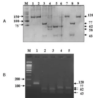
Differentiation of NDV by REA
In order to differentiate between vaccine strains and the recent genotype VI and VII field isolates, Hinf I and Pst I were selected and REA was conducted. 41 isolates and 2 reference vaccine strains, Ulster and La Sota, were employed for the REA. NDVs were grouped into 4 REA types, H
−/P
−(both enzyme recognition sites negative, 120 bp), H+/P
−( Hinf I recognition site positive, 62 bp/58 bp), H
−/P+ ( Pst I recognition site positive, 77 bp/43 bp) and H+/P+ (both enzyme recogni- tion sites positive, 62 bp/43 bp/15 bp) (Table 2, Fig. 2 &
4). Ulster, La Sota, SNU8050, SNU80143, SNU8419, SNU8426, SNU8465, SNU8490, SNU8550, SNU8557,
SNU8559, SNU8566, SNU85135, SNU9515, SNU96130 and SNU9983 were grouped into H
−/P
−(Table 2).
SNU90111 was digested only by Hinf I (H+/P
−) and SNU8876, SNU8887, SNU88139, SNU88147, SNU9033, SNU9550, SNU9575, SNU9586, SNU95107 and SNU 95124 were digested only by Pst I (H
−/P+) (Table 2).
SNU8861, SNU8871, SNU9222, SNU9227, SNU9229, SNU9257, SNU9275, SNU9280, SNU9282, SNU9283, SNU9284B, SNU92105, SNU9358GG, SNU9598, SNU 9969, SNU9970 and SNU9971 were grouped into H+/
Fig. 3. Phylogenetic tree based on the nucleotide sequences of the common HN (nucleotides 658-736) via neighbor- joining. The nucleotide sequences of the foreign NDV strains were obtained from the GenBank database, and their accession numbers are as follows. Ulster/67 (M24694), La Sota/46 (AF077761), ASTR/74 (Y19016), AUS/32 (M24712), Italien/45 (M24715), Miyadera/51 (M24713), sf02 (AF473851), zj/1/00/Go (AF456432), Taiwan95 (U62620), AF2240 (X79092), CA211472 (AY246050), V4/66 (J03911), Ck/Kenya/139/90 (AY288997), Ck/USA (CA)/1083 (Fontana)/72 (AY288992), Ck/Italy/3286/00 (AY288994).
Fig. 4. Differentiation of NDV strains via REA analysis.
Panel A (polyacrylamide gel electrophoresis and silver staining), M: size marker (25/100; Bioneer, Korea), lane 1:
Ulster, 2: La Sota, 3: SNU88139, 4: SNU90111, 5: SNU 9222, 6: SNU9358GG, 7: SNU9515, 8: SNU95124, 9: SNU 96130. Panel B (agarose gel electrophoresis and ethidium bromide staining), M: size marker (100 bp; iNtRON Biotech- nology, Korea), 1: Ulster, 2: SNU9969, 3: SUN9970, 4: SNU 95124, 5: SNU0021.
Fig. 5. Multiplex RT-PCR for differentiation of vaccine strains and pathogenic field isolates (velogens). Lanes 1:
SNU92105, 2: SNU9550, 3: SNU9575, 4: SNU9586, 5:
SNU9598, 6: SNU95124, 7: La Sota, 8: Ulster, 9: SNU
9515, 10: SNU96130, M: size marker (100 bp; iNtRON
Biotechnology, Korea).
P+ (Table 2). Based on the nucleotide sequences, SNU 80108, SNU9991, SNU99111 and SNU99123 (H
−/P
−), SNU9139 (H+/P
−), SNU9512, SNU9553, SNU95119, SNU95132, SNU2083, and SNU0021
−, SNU0202
−and SNU2108-like viruses (H
−/P+), and SNU9444 and SNU99105 (H+/P
−) were grouped into each of the REA groups (Table 2).
Differentiation of NDV via pathotype-specific RT- PCR The vaccine strains and field lentogens evidenced no bands on pathotype-specific RT-PCR, but the virulent field isolates did exhibit a specific band. All of the NDV isolates previously grouped into virulent genotypes VI and VII as velogens, as well as the new viruses in the present study, were identified as velogens and lentogens via pathotype-specific RT-PCR (Table 1). In addition, 2 primer sets were determined to be suitable for multiplex-RT-PCR (Fig. 5).
Discussion
NDV evolves only via nucleotide substitutions intro- duced by RNA-dependent RNA polymerase without recombination [23, 27]. The principal determinant of virulence is the proteolytic cleavability of the fusion
protein, but the results of a reverse genetic study have revealed that the HN gene is also relevant to virulence [11].
The presence of genotype VII dates back to 1984 and 1985, where it was identified in Taiwan and Japan, respectively [29]. The first genotype VII virus was isolated from imported peafowl in a zoo in 1984 [15], but the genotype VII viruses from chickens were initially reported in Korea, in 1995 [13]. Genotype VII, in addition to genotypes VI and IX, was identified in China [16]. Therefore, genotype VII viruses had already spread throughout the Far East in the 1990s [29].
According to the results of phylogenetic analyses with the HN gene, Korean NDV isolates obtained between 1980 and 2005 were grouped into the VI and VII genotypes. The genotype VI viruses were present from 1980 to 1999, but were rarely isolated after the beginning of 2000. Genotype VII first appeared in 1995 but since then, had never been isolated in poultry prior to 2000 [15]. They recurred in 2000, and have since become the only detectable genotype in Korea.
The Korean government has been exerting a great deal of effort to eradicate Newcastle disease, via the vacci- nation of the majority of one-day old chicks. The fact that genotype VI is no longer isolated can be attributed to this massive vaccination effort, but it appears that Table 2. Summary of restriction enzyme analysis (REA)
REA type
(Fragment size) No. of
viruses Vaccine strains and field isolates
H-/P-
(120) 20 Ulster, La Sota, SNU8050, SNU80108
a, SNU80143, SNU8419, SNU8426, SNU8465, SNU8490, SNU8550, SNU8557, SNU8559, SNU8566, SNU85135, SNU9515, SNU96130, SNU9983, SNU9991
a, SNU99111
a, SNU99123
aH+/P-
(62/58) 2 SNU90111, SNU9139
aH-/P+
(77/43) 66
SNU8876, SNU8887, SNU88139, SNU88147, SNU9033, SNU9512
a, SNU9550, SNU9553
a, SNU9575, SNU9586, SNU95107, SNU95119
a, SNU95124, SNU95132
a, SNU0021
a, SNU0036
a, SNU0079
a, SNU0086
a, SNU0105
a, SNU0117
a, SNU0118
a, SNU0119
a, SNU0123
a, SNU0125
a, SNU0129
a, SNU0164
a, SNU0165
a, SNU0169
a, SNU0202
a, SNU1024
a, SNU2001
a, SNU2009
a, SNU2017
a, SNU2026
a, SNU2028
a, SNU2048
a, SNU2049
a, SNU2063
a, SNU2064
a, SNU2078
a, SNU2080
a, SNU2083
a, SNU2084
a, SNU2091
a, SNU2100
a, SNU2102
a, SNU2108
a, SNU2117
a, SNU2124
a, SNU3058
a, SNU4152
a, SNU5005
a, SNU5009
a, SNU5047
a, SNU5062
a, SNU5063
a, SNU5064
a, SNU5065
a, SNU5070
a, SNU5074
a, SNU5076
a, SNU5079
a, SNU5081
a, SNU5084
a, SNU5085
a, SNU5100
aH+/P+
(62/43/15) 19 SNU8861, SNU8871, SNU9222, SNU9227, SNU9229, SNU9257, SNU9275, SNU9280, SNU9282, SNU9283, SNU9284B, SNU92105, SNU9358GG, SNU9598, SNU9969, SNU9970, SNU9971, SNU9444
a, SNU99105
aa