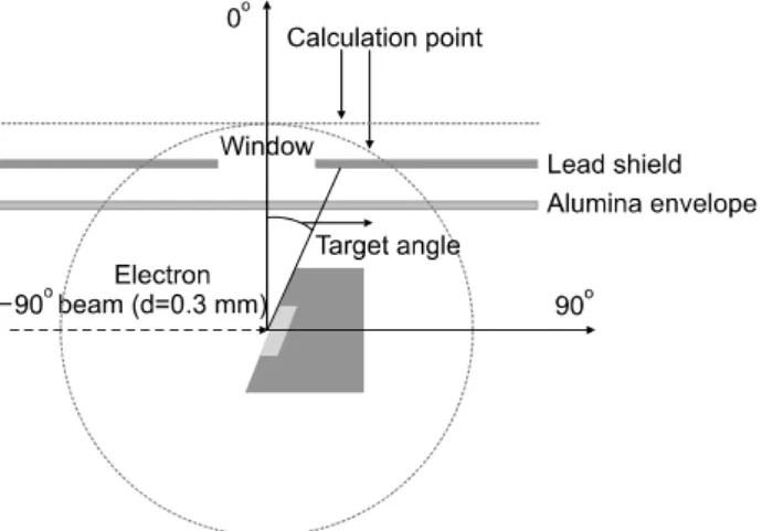
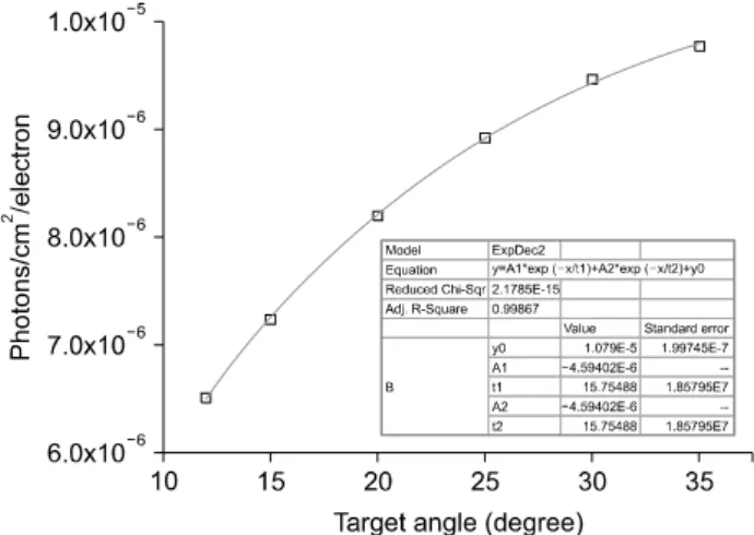
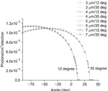
Effect of Target Angle and Thickness on the Heel Effect and X-ray Intensity Characteristics for 70 kV X-ray Tube Target
5
0
0
전체 글
(2)
(3)
(4)
(5)
수치
관련 문서
Excitation Detection X-ray photoelectron spectroscopy (XPS) Photons(X-ray) Electrons UV photoelectron spectroscopy (UPS) Photons (UV) Electrons
- The effect of the motif (one wide slit) is to alter the intensity of each main peak, but the position of the main peaks are unchangedD. - intensity envolope →
Most line searches used in practice are inexact: the step length is chosen to approximately minimize f along the ray {x + t∆x |t ≥ 0}, or to reduce f enough...
A is the number of photons per nuclear transformation – L x-rays for transuranium elements, and gamma rays for fission and
This research is to conduct comparative analysis on the rain intensity of expected rainfall depth of the research-use X-band dual-polarimetric radar with that of actually
The structure and film optical properties were investigated by X-ray diffraction(XRD), the particle size and thickness were investigated by scanning
Single crystal X-ray structure determination clearly revealed structural authenticity of each compound and alluded electronic alteration via investigation on the
Postoperative X-ray (A : ankle lateral view, B : calcaneal axial view), Böhler angle and Gissane angle have recovered (Böhler angle is 32.2°, Gissne angle is 105.5°,
