WKtKtK빼KtKtKKK빼KKtKH빼빼빼빼KK빼M
조골세포내 cAM P 농도 변호ᅡ가 파골세포 형성에 미치는 영향
전 윤 나 • 임 미 정 # 숙명여자대학교 약학대학
(Received January 19, 2005; Revised February 11,2005)
Regulatory Effects of Cyclic AMP on Osteoclast Formation
Yunna Chun and Mijung Yim#
College of Pharmacy, Sookmyung Women's University, Seoul 140-742, Korea
A bstract — In the present study, treatment of IBM X, a phosphodiesterase (PDE) inhibitor, alone induced osteoclast for
mation in co-cultures of mouse bone marrow cells and calvarial osteoblasts. However, treatment of IB M X in combination with prostaglandin E 2 (PGE2) inhibited osteoclast formation in a dose-dependent manner. Among various isozyme-specific PDE inhibitors, a PDE4 specific inhibitor, rolipram, showed similar effects as IBM X on osteoclast formation. To address the involvement of cyclic adenosine monophosphate (cAMP) in osteoclast formation, cAMP concentration in calvarial osteo
blasts was investigated. When calvarial osteoblasts were co-cultured with IBMX alone or in combination with PGE 2,the pat
terns of cAMP concentration in calvarial osteoblasts were differ each other, suggesting that cAMP in calvarial osteoblasts subtly regulates osteoclast formation.
Keywords □ osteoblast, osteoclast, cyclic adenosine monophosphate, phosphodiesterase, prostaglandin E 2
생물의 뼈조직은 파골세포(osteoclast)에 의한 뼈롭수와 조골 세포(osteoblast)에 의한 뼈형성이 동적인 평형상태를 유지하고 있다.1> 오래된 뼈조직을 새로운 뼈로 바꾸는 이러한 재형성(bone remodeling)은 평생을 거쳐 반복되며, 골다공증, 만성 관절 류마 티즘 등의 대시성 뼈질환은 파골세포에 의한 뼈 흡수가 이상 활 성화되어 평형이 깨짐으로써 나타나는 결과라고 할 수 있다.
파골세포는 조혈모세포(hematopoietic cell)에서 분화한 것으 로, 파골세포의 분화는 조골세포에 의해 엄격하게 조절되고 있 다.1외 조골세포는 1,25-dihydroxyvitamin D3[l,25(OH)2D3],부갑 상선 호르몬(PTH), prostaglandin E^PGEy 등의 자극에 의해 파골세포 분화인자인 TRANCE(TNF-related activation-induced cytokine, OPGL, ODR or RANKL)를 생성하는 한편, decoy receptor인 OPG(osteoprotogenin)를 분비하여 TRANCE를 억제 함으로써 파골세포의 분화를 조절한다.2체
이러한 조골세포■파골세포 상호작용에 있어서,cAMP는 중요 한 내부 신호전달자로 작용한다. PTH나 PGE2 등의 자극에 의
#본 논문에 관한 문의는 저자에게로 (전화) 02-710-9572 (팩스) 02-710-9871 (E-mail) [email protected]
해 조골 세포내 cAMP의 농도가 상승하면, protein kinase A (PKA)가 활성화되고 이를 통해 TRANCE의 발현이 증가되어 파 골세포 형성이 촉진된다고 알려져 있다.7ᅵ9) 세포내 cAMP의 농 도는 2가지 효소에 의해 항상 일정하게 유지된다. Adenylate cyclase는 ATP를 기질로 cAMP를' 합성하고,phosphodiesterase (PDE)는 cAMP를 5’-AMP로 분해시 킨다,10’u) 지금까지 PDE family는 PDE1 에서 11까지의 isozyme이 존재하는 것으로 밝혀 졌으며, 그 중 PDE1, 2, 3,4,7,8, 10, 11이 cAMP의 분해에 관여한다. 각각의 isozyme은 PDE1A,IB, 1C와 같은 subtype을 가진다.W
최근 본 저자는 PDE 저해제 pentoxyfylline이 초기 배양한 마 우스 조골세포에서 TRANCE의 발현을 유도함으로써 파골세포 분화를 촉진함을 보고한 바 있다.12) 이는 PDE 저해제 처리에 의 해 조골세포내 cAMP 농도가 상승함으로써 나타나는 효과라 할 수 있다. 본 연구에서는 파골세포 분화와 조골세포내 cAMP와의 연관성을 명확히 하기 위해 초기 배양한 마우스 조골세포와 골 수세포의 공배양계(co-culture)에 PDE 저해제를 단독으로, 또는 PGE2와 함께 병용 처리하였다. 본 연구 결과 공배양계에 PDE 저해제를 단독 처리하면 파골세포의 분화가 유도되는 것에 반해, PDE 저해제와 PGE2를 병용 처리하면 오히려 분화가 억제되는
109
것으로 나타났다. PDE 저해제 단독 및 PGE2와의 병용 처리에 의한 조골세포내 cAMP 농도 변화를 측정함으로써 , 조골세포내 cAMP 농도 변화는 파골세포 분화를 미묘하게 조절할 /[능성이 제시되었다.
실험 방법
마우스 조골세포의 초기 배양
생우 0~1일의 신생아 ddy mouse로부터 두개골 피부를 벗긴 후 두개골을 적출하였다. 이때 후두골 근육 부착부분은 제거하 였다. 부착된 근육, 혈구 등을 제거한 후 ot-MEM으로 가볍게 세 척하였다. 0.1% collagenase와 0.2% dispase 효소 용액에 넣어 37°C에서 5분간 진탕시킨 후 상등액을 버리고 새로운 효소 용액 을 가하였다. 37°(:에서 약 10분간 진탕하여 상동액을 모으는 조 작을 4회 반복하였다. 원심 분리한 후 10% FBS 함유 cc-MEM 으로 약 3~5X10⁴ 세포/ 100 mm plastic dish가 되도록 접종하였 다. 5% C 0 2, 37°C에서 3~4일간 배양한 세포를 이후 조골세포 로 실험에 人h용하였다.
Df우스 골수세포의 배양
ddy mouse(6~9주, 수컷)를 경추 탈골한 후 70% 에탄올로 소 독하였다. 경골 부분의 피부를 절개하여 부착 근육을 떼어냈다.
경골 원심부를 절단하고 슬개골을 탈골시켜 경골을 적출하였다.
뼈 잉끝을 조금 잘라 한 쪽 끝에 25G의 주사바늘을 꽂고 a-MEM 을 흘려보내 골수세포를 시험관에 모았다. 원심 분리한 후 (X -
M EM에 현탁하고 2배의 Gey’s solution을 가해 적혈구를 제거 했다. 원심 분리한 후 10% FBS가 함유된 a-MEM으로 재현탁 했다.
공배양(coculture)법을 이용한 파골세포의 분화유도
96 well plate에 초기 배양한 조골세포 5X 10³ 세포/well, 골수 세포 lx 105 세포/well을 10% FBS가 함유된 ot-MEM으로 공배 양했다. 배지는 3일에 한번씩 교환했다. 배양이 끝난 세포는 10%
formalin0.?. 10분■간 고정한 후 ethanol-aceton(l: 1)로 1분간 재 고정하여 TRAP(tartxate-resistant acid phosphatase) 염색을 했 다. 3개 이상의 핵을 가진 TRAP+ 세포를 다핵 파골세포로 판 정했다.
RT-PCR 분석
Total RNA 1 |igi- Superscript II(Invitrogen, CA, USA)로 역 전사하였다. 얻어진 cDNA의 일부를 Go Taq DNA polymerase (Promega, USA)로 PCR 증폭하였다. PCR 증폭에 사용한 primer 의 서열은 다음과 같다. PDE1A, 5'-tcacagtcttcaaactacgg-3' (forward), 5'-tgttcacacctatggacgtg-3' (reverse), PDE IB 5'-gacg-
tgacccagactgtcc-3' (forward), 5'-tgatgtcagcagcatgaag-3' (reverse), PDE1C, 5'-cagcatcaactcatcagatg-3' (forward), 5'-gagtgatccttct- tgtctgc-3' (reverse), PDE2, 5'-aaggtgtcccacaagttcac-3' (forward), 5'-tggtcactactgtgctcttg-3' (reverse), PDE3A, 5'-gcctgtgaaagcag- tatagc-3' (forward), 5'-gaaaatgtctgagacgagtc-3' (reverse), PDE3B, 5'-tgttcaggagaccgtcgttg-3' (forward), 5'-gatcccaccttgaacagtgc-3' (reverse), PDE4A, 5'-ggaactcacacacctgtcg-3' (forward), 5'-gttc- ttgtgctaagaggtcc-3'(reverse); PDE4B, 5'-tggaaatcctggctgccat-3' (forward), 5'-tccacagaagctgtgtgct-3' (reverse); PDE4C, 5'-tggt- atcagagtaggattcc-3' (forward), 5'-ctctgtgtaaaccttggctg-3' (reverse);
PDE4D, 5'-cggaactcgctctgatgt-3' (forward), 5-acagaggcgttgtgcttg- 3' (reverse), PDE5, 5'-tttgctgctctaaaagcaggc-3' (forward), 5'- gctatccgttgttgaatagg-3' (reverse), PDE7, 5'-actcaggccatgcactgttac- 3' (forward), 5'-ggactcacacccaaatggtac-3' (reverse), PDE8, 5'- acggcgtatttcctttccag-3’ (forward), 5'-gacttgggaatgctgcaggtg-3' (reverse), PDE9, 5'-taccagatcaatgcccgc-3' (forward), 5'-ggaaga- gcttggtcactg-3' (reverse), 및 GAPDH, 5'-gaaggtcggtgtgaacg- gatttggc-3' (forward), 5'-catgtaggccatgaggtccaccac-3' (reverse).
반응조건은 940C에서 3분 초기 반응 후, 940C에서 30초, 52°C 에서 45초, 그리고 720C에서 1분 간격으로 총 34주기 (CTR과 cathepsin K) 또는 28주기 (GAPDH)로 이루어졌다. 증폭된 PCR 산물들은 1% agarose gel에서 전기영동하여 분리하였다.
세포내 cAMP 측정
초기 배양 마우스 조골세포를 시간대별로 시약 처리한 후 세 포내 cAMP 의 양을 cAMP Biotrak Enzyme immunoassay (Amersham Biosciences, USA)로 측정하였다.
실험 결과 및 고찰
PDE 저해제의 단독 및 PGEa익의 병용 처리가 파골세포 분화에 미치는 영향
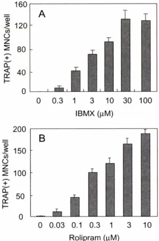
TRAP+ 다핵 파골세포는 초기 배양한 마우스 조골세포와 골 수세포의 공배양계에서 l ,25-dihydroxyvitamin D3[l,25(OH)2D3], 부갑상선 호르몬(PTH), prostaglandin E2(PGE2), interleukin-1 (IL-1), lipopolysaccharide(LPS) 둥의 자극에 의해 분화 유도된 다.1> 이러한 공배양계를 이용하여 PDE 저해제인 3-isobutyl-l- methylxanthine(IBMX)가 파골세포 분화에 미치는 효과를 조사 하였다. 기존에 발표된 것과 동일하게,12) 공배양계에 IBMX를 단 독 처리하였을 때 파골세포 분화가 농도 의존적으로 유도되었다 (Fig. 1A). 현재 11종류의 PDE isozyme이 알려져 있으며,u) IBM X는 모든 PDE isozyme을 비특이적으로 저해한다. 특정 PDE isozyme이 파골세포 분화에 관여하는지의 여부를 확인하 기 위해 다양한 PDE isozyme 특이적 저해제를 사용하여 그 효
R olipram (jiM) R o lip ra m (|iM ) Fig. 1 - Effect of PDE inhibitors alone on osteoclast formation.
Dose-dependent effect of IBMX (A) or rolipram (B) on osteoclast formation in co-cultures. Mouse bone marrow cells and calvarial osteoblasts were co-cultured in the presence of the indicated concentrations of IBMX or rolipram for 6 days. Cells were then fixed and stained for TRAP TRAP-positive (+) multinucleated cells (MNCs) were counted. Data are expressed as the mean+SD of triplicate cultures.
과를 살펴보았다. 그 결과, PDE4 저해제인 rolipram을 공배양계 에 단독 처리하였을 때 IBMX와 동일한 파골세포 분화 유도 효과 를 보였다(Fig. IB). 이에 반해 vinpocetine, EHNA, milrinone, dipyridamole, zaprinast(PDEl, 2,3,5 와 8,5와 9의 특이적 저 해제)는 파골세포 분화 유도 효과를 보이지 않았다(미발표 결과).
이는 PDE isozyme 중 주로 PDE4가 조골세포내 cAMP 농도를 조절함으로써 파골세포 분화에 관여함을 시사하는 결과라 할 수 있다.
조골세포는 IL-1, PTH, LPS 등의 자극에 반응하여 PGE2를 세포 밖으로 분비한다.1345) PGE2는 조골세포에 autocrine으로 작용하여, 세포내 cAMP 농도를 상승시킴으로써 파골세포 형성 을 촉진함이 알려져 있다.8} 공배양계에 IBMX와 PGE2를 병용 처리하였을 때 파골세포 분화에 미치는 영향을 살펴보았다. 단 독 처리했을 때 파골세포 분화를 유도한 것과 대조적으로, IBMX 는 PGE? 존재하에서 파골세포 분화를 농도 의존적으로 억제함
Fig. 2 - Effect of PDE inhibitors in combination with PGE2 on osteoclast formation. Dose-dependent effect of IBMX (A) or rolipram (B) in the presence of 10 |iM PGE2 on osteoclast formation in co-cultures.
이 밝혀졌다(Fig. 2A). 이러한 분화억제 효과는 PDE4 특이적 저해제인 rolipram을 처리했을 때에도 동일하게 관찰되었다 (Fig. 2B).
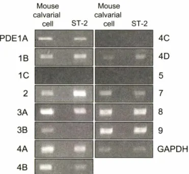
조골세포의 PDE isozyme 및 subtype 발현
각 PDE mRNA의 발현을 초기배양한 조골세포를 이용하여 RT-PCR로 확인하였다. 대부분의 PDE isozyme은 하나 이상의 subtype으로 구성되어 있다.11) 각 subtype를 인식할 수 있는 primer를 용하여 PCR를 시행하였으며 , Fig. 3에 그 결과를 표 시하고 있다. 조골세포는 PDE1A, PDE1B, PDE2, PDE3A, PDE3B, PDE4A, PDE4B, PDE4D, PDE7, PDE8, 그리고 PDE9 mRNA를 발현하고 있음을 알 수 있다. 이러한 PDE mRNA의 발현 패턴이 초기 배양한 조골세포에 국한되는 것이 아니라는 것을 확민하기 위해 마f 스 조골세포주 ST-2를 사용하 여 동일한 실험을 수행하였다. ST-2는 초기배양한 조골세포와 동 일한 PDE mRNA 발현 패턴을 보였으나,초기 배양한 조골세포 와 달리 PDE3B를 발현하지 않는 것으로 나타났다. 이는 초기 배양한 세포와 확립된 세포주 사이의 차이에 기인하는 것으로 추 0 0.3 1 3 10 30 100
IBMX (uM)
0 0.3 1 3 10 30 100 IB M X (나M)
120
160
A
11 I i i
쇼200
0
128
4
hi/so
n ia i
+)dvai)oo
o
o
o 0
8
6
4
2
T-
I
N IA
oj/VVSUI
(-ocdvylVol. 49’ No. I, 2005
Fig. 4 - Effect of PDE inhibitor alone or in combination with PGE2 on intracellular cAMP concentration. Calvarial osteoblasts were treated with 50 (xM of IBMX alone (A) or in combination with 10 |iM PGE2 (B) for indicated times.
Intracellular cAMP concentration was measured by EIA.
0 5 30min 1 3 9 24 48 hr 2000
3 9 24 48 72 hr Mouse Mouse 1000
calvarial calvarial cell ST-2 cell ST-2
4 B
4 C 4 D
G A P D H
Fig. 3 - RT-PCR analysis of PDE mRNAs. Total RNAs were prepared from mouse calvarial cells and ST-2 cell line.
Templates for PCR were synthesized in the presence of reverse transcriptase. mRNA expression was determinrd by RT-PCR using specific primers designed for each gene.
론되었다.
조골세포내 cAM P의 농도 변화
Fig. 1과 Fig. 2에서 공배양계에 PDE 저해제를 단독 처리하면 파골세포 분화가 유도되나,PGE2와 병용 처리하면 오히려 파골 세포 분화가 억제됨을 알 수 있다. PDE 저해제와 PGE2는 모두 세포내 cAMP의 농도를 상승시키는 cAMP-elevating agent이다.
띠라서 파골세포 분화에 대한 이러한 상반된 효과가 세포내 cAMP 농도와 관련되어 있음을 확인하기 위해 시약처리 후 시간대별로 cAMP의 농도를 측정하였다. Fig. 4A에서 보듯이, 조골세포에 PDE 저해제 IBMX를 처리하였을 때 cAMP 농도는 3시간 후 최 고에 달했으며 그 후 점차 감소하여 72시간 후에는 초기 농도로 떨어지는 양상을 보였다. 이에 반해 IBMX와 P G E # 병용 처리 하면 초반에 cAMP 농도가 증가하는 것은 동일하나,단독 처리 와 달리 48시간 후에도 비교적 높은 농도를 유지하였다(Fig. 4B).
이는 조골세포내 cAMP 농도 변화가 파골 세포 분화를 미묘하 게 조절하고 있을 가능성을 시사하는 결과이다.
결 론
본 연구에서는 초기 배양한 미우스 조골세포와 골수세포의 공 배양계를 이용하여, PDE 저해제 IBM X의 단독 처리 및 PGE2 와의 병용 처리에 의한 cAMP의 농도 변화가 파골세포 분화에 미치는 영향을 조사하였다. PDE 저해제 IBM X를 단독 처리하
였을 때 파골세포 분화가 촉진되었으나, PGE2와 병용 처리하였 을 때는 오히려 파골세포 분화가 억제되었다. 초기 배양한 조골 세포 및 ST-2 조골세포주는 다양한 PDE isozyme 및 subtype을 발현하고 있 헤 도 불구하고 PDE4 저해제 rolipram 만이 IBMX 와 동일한 효과를 보여, PDE4가 조골세포내 cAMP 조절에 주 로 관여하고 있음을 시사하였다. 조골세포내 cAMP 농도를 추적 한 결과 PDE 저해제를 단독 처리하였을 때와 PGE21~ 병용 처 리하였을 때 서로 다른 양상을 보여, 조골세포내 cAMP의 농도 변화는 파골세포 분화를 미묘하게 조절하고 있음을 시사하였다.
향후 추가적인 연구를 통한 cAMP와 파골세포 분화의 명확한 연 관성 규명은 골다공중 등의 대사성 뼈질환 치료제를 개발하기 위 한 기초 자료로 활용될 것으로 사료된다.
감사의 말씀
본 연구는 숙명여자대학교 2003년도 교내 연구비 지원에 의해 수행되었으므로 이에 감사드립니다.
문 헌
1) Takahashi, N., Akatsu, T, Udagawa, N., Sasaki, T., Yamaguchi, A., Moseley, J. M., Martin, T. J. and Suda, T. : Osteoblastic cells
i i l i n
LilUili
B00 00 00 00 16 128
4
ol M®
o/ Ef c CLS
V O
^ 800 Q)
호
o 600 g
^ 400 o 200
are involved in osteoclast formation. Endocrinology 123, 2600 (1988).
2) Suda, T,Takahashi, N., Udagawa, N., Jimi, E., Gillespie, M. T.
and Martin, T. J. : Modulation of osteoclast differentiation and function by the new members of the tumor necrosis factor receptor and ligand families. Endocr. Rev. 20, 345 (1999).
3) Wong, B. R., Rho, J., Arron, J., Robinson, E., Orlinick, J., Chao, M., Kalachikov, S., Cayani, E., Bartlett, E S. 3rd, Frankel, W.
N., Lee, S. Y. and Choi, Y. : TRANCE is a novel ligand of the tumor necrosis factor receptor family that activates c-Jun N- terminal kinase in T cells. J. Biol. Chem. 272,25190 (1997).
4) Yasuda, H., Shima, N., Nakagawa, N., Yamaguchi, K.,Kinosaki, M.,Mochizuki, S., Tomoyasu, A., Yano, K.,Goto, M.,
Murakami, A., Tsuda, E., Morinaga, T., Higashio, K., Udagawa, N.,Takahashi, N. and Suda, T. : Osteoclast differentiation factor is a ligand for osteoprotegerin/osteoclastogenesis- inhibitory factor and is identical to TRANCE/RANKL. Proc.
Natl. Acad. Sci. USA 95,3597 (1998).
5) Lacey, D. L.,Timms, E.,Tan, H. L., Kelley, M. J., Dunstan, C.
R., Burgess, T., Elliott, R.,Colombero, A., Elliott, G.,Scully, S., Hsu, H., Sullivan, J., Hawkins, N., Davy, E., Capparelli, C., Eli, A., Qian, Y. X., Kaufman, S., Sarosi, I., Shalhoub, V, Senaldi, G., Guo, J., Delaney, J. and Boyle, W. J. : Osteoprotegerin ligand is a cytokine that regulates osteoclast differentiation and activation. Cell 93,165 (1998).
6) Simonet, W. S., Lacey, D. L., Dunstan, C. R., Kelley, M .,Chang, M. S., Luthy, R.,Nguyen, H. Q.,Wooden, S., Bennett, L., Boone, T, Shimamoto, G.’ DeRose, M.,Elliott, R.’ Colombero, A., Tan, H. L.,Trail, G., Sullivan, J., Davy, E.,Bucay, N., Renshaw-Gegg, L., Hughes, T. M.,Hill, D., Pattison, W., Campbell, R and Boyle, W J. : Osteoprotegerin: a novel secreted protein involved in the regulation of bone density. Cell 89, 309 (1997).
7) Kondo, H., Guo, J. and Bringhurst, E R. : Cyclic adenosine monophosphate/protein kinase A mediates parathyroid
hormone/parathyroid hormone-related protein receptor regula
tion of osteoclastogenesis and expression of RANKL and osteoprotegerin mRNAs by marrow stromal cells. J. Bone Miner. Res. 17,1667 (2002).
8) Kaji, H., Sugimoto, T., Kanatani, M., Fukase, M., Kumegawa, M. and Chihara, K. : Prostaglandin E2 stimulates osteoclast
like cell formation and bone-resorbing activity via osteoblasts:
role of cAMP-dependent protein kinase. /. BoneMiner. Res. 11,
62 (1996).
9) Fu, Q., Jilka, R. L.,Manolagas, S. C. and O'Brien, C. A. : Parathyroid hormone stimulates receptor activator of NF-kB ligand and inhibits osteoprotegerin expression via protein kinase A activation of cAMP-response element-binding protein.
/• Biol. Chem. 277,48868 (2002).
10) Antoni, F. A. : Molecular diversity of cyclic AMP signaling.
Front Neuroendocrinol. 21,103 (2000).
11) Essayan, D. M. : Cyclic nucleotide phosphodiesterases. J.
Allergy Clin. Im munol. 108,671 (2001).
12) Kim, M., Chun, Y. and Yim, M. : Effect of pentoxifylline, a phosphodiesterase inhibitor, on osteoclast formation. Yakhak Hoeji 48,197 (2004).
13) Min, Y. K.,Rao, Y., Okada, Y, Raisz, L. G. and Pilbeam, C. C : Regulation of prostaglandin G/H synthase-2 expression by interleuki-1 in human osteoblast-like cells. J. Bone Miner. Res.
13,1066 (1998).
14) Tetradis, S., Pilbeam, C. C.,Liu, Y., Herschman, H. R. and Kream B. E .: Parathyroid hormone increases prostaglandin G/
H synthase-2 transcription by a cyclic adenosine 3',5'- monophosphate-mediated pathway in murine osteoblastic MC3T3-E1 cells. Endocrinology 138,3594 (1997).
15) Miyaura, C., Inada, M.,Matsumoto, C., Ohshiba, T, Uozumi, N.,Shimizu, T. and Ito, A. : An essential role of cytosolic phospholipase A2alpha in prostaglandin E2-mediated bone resorption associated with inflammation. /. Exp. Med. 197, 1303 (2003).
Vol. 49’ No. 1, 2005