Craniopharyngioma: : : :
Comparison of Tumor Characteristics Relevant with Initial Symptomatology between Children and Adults
Dong Hyuk Park, M.D., Jung Yul Park, M.D., Joo Han Kim, M.D., Yong Gu Jung, M.D., Hoon Kap Lee, M.D.,
Ki Chan Lee, M.D., Jung Keun Suh, M.D.
Department of Neurosurgery, Korea University, Seoul, Korea
= Abstract =
두개인두종:소아와 성인에서 초기 증상과 연관된 종양의 특징 비교
고려대학교 의과대학 신경외과학교실
박동혁ᆞ박정율ᆞ김주한ᆞ정용구ᆞ이훈갑ᆞ이기찬ᆞ서중근
bjectives:The craniopharyngioma is a benign tumor located at least in part in the suprasellar cistern. However, the symptoms and signs from this tumor may be determined not only by the location of the tumor but also by its size and the age of the patient. The objective of our study is to analyze retrospectively the clinical manifestations of craniopharyngiomas with regards to tumor characteristics in children and adults.
Material and Methods:Twenty-three patients(16 adults, 7 children) treated for craniopharyngioma between 1990 and 1999 were studied to demonstrate the relationship of tumor size, growth pattern, and its invasiveness with clinical symptoms. As part of the assessment, 16 adults(M:F=8:8, mean age:43.7 years) and 7 children(M:F=5:2, mean age:10.1 years) underwent magnetic resonance(MR) imaging and computerized tomography(CT) scanning with a three-dimensional volume acquisition sequence.
Results:The three major cardinal signs were defined to increased intracranial pressure, endocrine dysfunction, and visual problems. The tumor size in child group was larger than that in adult group. Also, visual problems, symptoms of increased intracranial pressure and hydrocephalus were more frequently observed in child group. However, endocrine dysfunction and neuropsychological symptoms related with hypothalamic connections to the thalamus, pituitary, frontal lobe, and other cortical areas were more frequent in adult group.
Conclusions:In our series, the tumor size and invasiveness of craniopharyngioma revealed to be relevent with initial symptoms of increased intracranial pressure and visual symptoms which were more frequent in child group. As for the growth pattern, we did not find major difference between adults and children.
KEY WORDS:Craniopharyngioma・Symptom・Invasiveness・Growth pattern.
Introduction
Erdheim in 1904 contributed the first correct histologic interpretation and adequate description of craniopharyn- giomas
16). He postulated that these tumors originated from
embryonic squamous cell rests of an incompletely involuted hypophyseal-pharyngeal duct. Since then several authors have noted differences in the histological patterns of adult and childhood craniopharyngiomas
10)14). It is speculated that an embryonic orign for tumors in adults is not required and craniopharyngiomas appearing later in life may arise
OOOO
from metaplasia of pituitary cells. Since squamous cell rests are rarely found in children, but show increasing frequency in each succeeding decade of life, it was suggested that the craniopharyngioma originates from metaplasia of mature cells of the anterior pituitary rather than from embryonic remnants
3). As it has been generally believed that the origin of craniopharyngioma in child is different from that in adult, the symptoms and signs caused by this tumor can be thought to be also different between age groups. The aim of this study is to analyze retrospectively the relationship of the clinical manifestations of craniopharyngiomas, with regards to initial symptoms and signs in children and adults, with tumor characteristics(e.g., location, size and growth pattern of the tumor).
Material and Method
1. Patients characteristics
Twenty-three patients who were treated for cranioph- aryngioma in our institution were included in this study. Of these, 7 patients were children of age less than 16 years at the time of diagnosis. There were 5 males and 2 females.
The mean age in child group was 10.1 years with range between 3 to 14 years. There were 8 males and 8 females in adult group. The mean age in adult group was 43.7 years (range 17-79 years).
2. Methods
We defined the the tumor characteristics as location, size, direction of growth, and invasiveness by brain MR images.
Initial symptoms and signs were defined as those related to increased intracranial pressure, endocrine dysfunction, and
visual problems. Locations of tumor were described as suprasellar, sellar, and combined
8). Tumor size were exp- ressed as tumor volume, determined from MR images, and is measured by following formula in which A, B, C re- present the three orthogonal planes on MR images
2);
A is the largest diameter on the axial slice with the largest area of tumor. B is the diameter that is 90 to diameter A and is measured at the midpoint of diameter A. Diameter C(vertical diameter) is the largest diameter on the saggital image with the largest area of tumor(Fig. 1):
V = A×B×C 2 (cm
3)
Direction of tumor expansion included prechiasmatic, subchiasmatic, retrochiasmatic and lateral. Also, we defined the invasive craniopharyngioma as large craniopharyn- giomas extending either superiorly into the third ventricle, or inferiorly into the sphenoid sinus and nasopharynx, or anteriorly into frontal fossa, or laterally into the temporal fossa, or posteriorly into the posterior fossa.
Symptoms and signs related to increased intracranial pressure included headache, vomiting, papilledema. Sym- ptoms and signs related to the endocrine dysfunction were described into hormonal dysfunction(e.g., growth failure, diabetes insipidus, delayed puberty, sexual problems). Ne- uropsychological problems(e.g., personality change, ment- ation deficit) were also described. Visual problems included decreased visual acuity, visual field defect and eye ball mo- tion defect.
These signs and symptoms were analyzed retrospectively with regards to tumor characteristics in both child and adult group.
Fig.1. A is the largest diameter on the slice with the largest area of tumor. B is the diameter that is 90°
to diameter A and is measured at
the midpoint of diameter A. Dia-
meter C(vertical diameter) is the
largest diameter on the sagittal im-
age with the largest area of tumor.
Results
The tumor characteristics, initial symptoms and signs in adult and child group are listed in(Table 1, 2) Five(71%) of 7 patients in child group had headache and vomiting.
Whereas, eleven(69%) of 16 patients in adult group had these symptoms. The hydrocephalus was seen in two(29%) of 7 patients in child group, and as disease progressed, 3
patients also developed in child group. However, only one (6%) of 16 patients in adult group developed the hydroce- phalus in whom ventriculo-peritoneal(VP) shunt was ne- cessary. Visual problems as mentioned above were noted in six(86%) patients in child group, and ten(62%) of 16 pa- tients in adult group. Four(57%) of 7 patients showed pa- pilledema in child group, and two(13%) of 16 patients in adult group. One patient developed total blindness as dis- ease progressed. Interestingly, two(29%) of 7 patients in
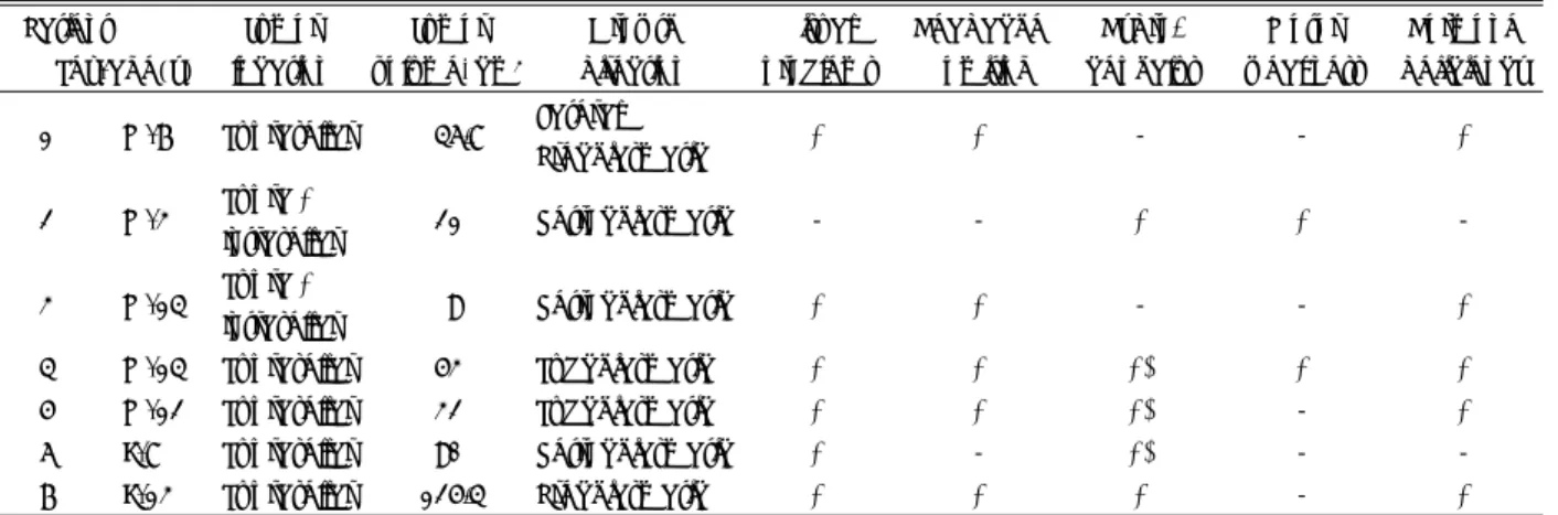
Table 1. Tumor characteristics, initial symptoms and signs in adult group
Patient Tumor Tumor Growth Visual Headache Hydro- General Psychologic/hormonal Sex/age(y) location volume, cm
3direction problems Vomiting cephalus weakness problems
1 M/34 Suprasellar 12.2 Subchiasmatic + + - - -/+
Supra + 2 M/44
Intrasellar 60 Prechiasmatic + + - - -/- Prechiasmatic
3 M/79 Suprasellar 55.2
+ Lateral - + - + -/?
4 M/67 Suprasellar 29.4 Retrochiasmatic + - - - +/+
5 M/56 Suprasellar 21.6 Retrochiasmatic - - - + +/+
6 M/64 Suprasellar 23.1 Retrochiasmatic + + - - -/+
7 M/32 Suprasellar 56 Retrochiasmatic + + - - -/+
8 M/30 Suprasellar 24.5 Retrochiasmatic - + - - +/+
Supra + 9 F/41
Intrasellar 3.7 Subchiasmatic + + - - +/+
10 F/22 Suprasellar 22.2 Retrochiasmatic - + + + -/+
11 F/65 Suprasellar 45 Retrochiasmatic + - - + -/+
12 F/35 Suprasellar 24.3 Subchiasmatic + - - - -/+
13 F/30 Suprasellar 14 Retrochiasmatic + + - - -/+
14 F/46 *Intrasellar 6.75 Subchiasmatic - + - - -/+
Supra + 15 F/55
Intrasellar 28.6 Prechiasmatic + - - - -/+
16 F/49 Suprasellar 1.7 Retrochaismatic - + - - -/?
*:Tumor was initially intrasellar, however, when recurred, it occupied both intrasellar and suprasellar region
?:Hormone studies were incomplete
Table 2. Tumor characteristics, initial symptoms and signs in children group
Patient Tumor Tumor Growth Visual Headache Hydro- Motor Hormone Sex/age(y) location volume, cm
3direction problems Vomiting cephalus weakness deficiency
Lateral 1 M/7 Suprasellar 46.8
Prechiasmatic + + - - +
Supra + 2 M/3
Intrasellar 21 Retrochiasmatic - - + + -
Supra + 3 M/14
Intrasellar 9 Retrochiasmatic + + - - +
4 M/14 Suprasellar 53 Subchiasmatic + + +* + +
5 M/12 Suprasellar 32 Subchiasmatic + + +* - +
6 F/8 Suprasellar 90 Retrochiasmatic + - +* - -
7 F/13 Suprasellar 125.4 Prechiasmatic + + + - +
*:Hydrocephalus developed with disease progression
child group showed motor weakness, including the he- miparesis, and four(25%) of 16 patients in adult group com- plained generalized weakness.
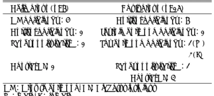
Hormonal dysfunction is listed in(Table 3) Seventy-one percent of child group presented with growth hormone deficiency, but only one(14%) patient showed deficiency of cortisol. Unlike children seven(44%) of 16 patients showed deficiency of cortisol and one(6%) patient presented hypothyroidism. Two(33%) of 8 male patients showed de- ficiency of sex hormone and one of them presented hypo- gonadism. All three premenopausal women of 8 female had low level of lutenizing hormone(LH) and follicular stimulating hormone(FSH) except 2 perimenopausal women and 3 postmenopausal women. Two premenopausal women had no mensturation and one postmenopausal woman had early menopause at 32 years old. Two(12.5%) of 16 patients showed panhypopituitarism in adult group. One(14%) of 7 patients presented it in child group, and the boy had no secondary sexual development. Four(25%) of 16 patients developed preoperative or postoperative central diabetes insipidus(DI) in adult group, and one(14%) of 7 patient developed it in child group. Four(25%) of 16 patients in adult group presented neuropsychological symptoms and signs that were disorientation, personality change and me- mory dysfunction, but one(14%) of 7 children developed irritability like neuropsychological symptoms and signs in adult. Two female patients developed unilateral hearing defect and walking disturbance. A large cystic tumor ex- tended into cerebellopontine angle and foramen magnum was seen in one and a huge cystic suprasellar mass that ex- tended into subfrontal area in the other.
Radiologic evaluation
In child group, the largest tumor size was 7.6×6.6×5cm (volume:125.4cm
3) and the smallest tumor size was 3×
2.5×2.4cm(volume:9cm
3). The mean volume of tumor in child group was 54cm
3. In adult group, the largest tumor
size was 6×4×5cm with volume of 60cm
3. The smallest tumor size was 1.5×1.5×1.5cm with volume of 1.7cm
3. The mean volume was 26.8cm
3.
In child group, tumor location in 5(71%) cases were on suprasellar and 2(29%) on both supresellar and sellar re- gions. None of cases in child group showed tumor location limited to sellar area. In adult group, twelve(75%) of 16 tumors were located in suprasellar area and three(19%) cases involved both suprasellar and sellar area. One(6%) case had tumor located within sellar at first operation. Ho- wever, tumor was located both suprasellar and intrasellar sites after 4 years.
As for the tumor extension, three(43%) of 7 tumors in child group extended into retrochiasmatic area, and two (28.5%) into subchiasmatic area. One case showed prech- iasmatic and expanded laterally into temporal fossa, and another case located only prechiasmatic area. In adult group, nine(56%) of 16 tumors into retrochiasmatic area, and four(25%) cases extended into subchiasmatic area. Three (18.7%) cases involved prechiasmatic area. In one case, tumor spreaded into temporal fossa.
As for invasiveness, four(57%) of 7 tumors were shown to be invasive craniopharyngioma in child group. In the contrary, two(12.5%) of 16 tumors in adult group were sh- own to be invasive craniopharyngioma.
Discussion
Tumor size and volume in child group were greater than that in adult group. The mean volume of tumor in child group was twice as in adult group. Since craniopharyngiomas are slow growing extra-axial tumors, it can be expected that these become quite large before they cause symptoms, esp- ecially in children. In the majority of cases the time interval between onset of symptoms and diagnosis of tumor ranged from 1-2 years. Invasive craniopharyngiomas in child group were observed more frequently than in adult group.
All except one patient with invasive craniopharyngioma had symptoms and signs of increased intracranial pressure and visual problems. The suprasellar area was the most commonly involved location and retrochiasmatic expansion was the most common type of growth. However, there was no definite difference in location and pattern of growth in paragangliomas between childeren and adults in our series.
Several authors reported that headache was common complaint in patients with retrochiasmatic tumors whereas
Table 3. Hormonal dysfunction
Child group(n=7) Adult group(n=16) GH deficiency:5 Cortisol deficiency:7 Cortisol deficiency:1 Thyroid hormone deficiency:1 Panhypopituitarism:1 Sex hormone deficiency:2(M) 3(F) Central DI:1 Panhypopituitarism:2
Central DI:4
GH:Growth hormone, DI:Diabetus insipidus
M:male, F:female
hydrocephalus, and visual problems were more common complaint in patients with prechiasmatic tumors
3)9)16). In our series, headache and vomiting in child group were more common than in adult group and it was correlated with size of tumor. Visual problems were more frequently ob- served in children than adults although they were not co- rrelated to mass location on both groups. It is generally believed that visual deficits may result both from direct compression of the optic pathways by the tumor and se- condarily from intracranial hypertension
5)16). According to several authors and from our experience, children often tolerate a high degree of visual loss without complaint, and continue their school works or watch television without arousing the suspicion from parents and teachers
3)5)9)16). In our series, only three of 6 patients who diagnosed high visual loss by opthalmologic examination complained their visual discomforts at initial visit.
As for the hormonal dysfunction, eighteen patient(78%) showed various types of hormonal dysfunction. Growth hormone deficiency was more common in child group, whereas cortisol deficiency was more frequently observed in adult group. Deficiency of sex hormone was more often observed in adult group with decreased sexual drive in men(50%) and amenorrhea in women(100%). The chil- dren with deficiency of sex hormone showed no secondary sexual development. The central diabetes insipius(DI) was more commonly observed in adult group, and caused lon- ger hospital stay and higher morbidity due to postoperative sepsis and decreased immunity in case. These endocrine changes are believed to be caused by compression of the hypothalamic-hypo- physeal axis
16). However, difference in the pattern of hormonal deficiency between children and adults in our series did not correlate with tumor growth patterns.
Neuropsychological symptoms and signs such as per- sonality and cognition changes were more evident in adult group. These were commonly seen in adults with tumor mass extended into frontal lobe. Therefore, it is believed that neuropsychological symptoms related with hypothal- amic connections to the thalamus, pituitary, frontal lobe, and other cortical areas are more frequent clinical findings in adult group
16). Motor weakness in the form of monopar- esis or hemiparesis were observed in two children in our cases. However, no definite motor weakness was seen in adult group.
Although craniopharyngiomas account for a great per-
centage of the intracranial tumors of childhood, they acc- ount for only 1 to 3% of intracranial tumors in all ages
14)17). Pathogenesis of craniopharyngioma is explained by two opposing hypotheses. These tumors may arise from ectopic embryonic cell remains of enamel organs. Also, they may represent the residual metaplastic squamous epithelium found in the adenohypophysis and anterior infundibulum
1). Petito et al.
14)reported that the great majority of tumors were located in the suprasellar area(94%) at the time of diagnosis. Only rarely, do these tumors extend into the an- terior fossa(5%), middle fossa(2%), or posterior fossa(4%).
However, a study from postmortem examination revealed higher incidence of extension into these fossa has been found(12%)
14). According to Yasargil, the tumor, with its expansive and infiltrative behavior, affects not only the pituitary-hypothalamic axis and visual pathways, but also the frontal lobes, striocapsulothalamic areas, mammillary bodies, and limbic system
17). The three major clinical syn- dromes associated with craniopharyngioma are related to increased intracranial pressure, endocrine dysfunction, and visual problems
16). Children frequently present with sym- ptoms of increased intracranial pressure(65-75%) such as headache and vomiting due to enlarging intracranial mass
16). Visual deficits are commonly well tolerated by children.
Only 20-30% of them complain of visual problems, usually
when almost complete visual loss has already taken place
13).
Endocrine dysfunctions are present in one half of the chil-
dren, and frequently manifest as short stature and diabetes
insipidus. One third of children with craniopharyngioma
have growth failure at the time of diagnosis. Delayed puberty
is present in one half of the adolescents, and polydipsia and
polyuria due to diabetes insipidus in 20% of cases. The hy-
pothalamic-pituitary complex is essential for endocrine, au-
tonomic, and behavioral performance
4)15). Disturbance of
hypothalamic connections to the thalamus, frontal lobes,
and other cortical areas have been related to some of the
psychological and social problems seen among affected
patients
6)7)12). Visual deficits and endocrine dysfunction are
the most frequent clinical findings in adults
3). Signs of
increased intracranial pressure are less common than in
children
16). Large subfrontal masses may cause neuropsyc-
hological symptoms, mentation deficits and memory loss,
as well as incontinence, apathy, and Korsakoff's syndrome
11).
Large subtemporal masses extending laterally to the sylvian
fissure may produce complex psychomotor seizures
16).
Conclusion
The characteristics of craniopharyngiomas in reported and our series, such as the location of tumor, its size, invasi- veness and direction of tumor expansion were compar- atively similar. However, the symptoms and signs from these tumors, may be determined not only by the location of the tumor but also revealed to be affected by its size and the age of the patient.
In this study, as tumors were more larger and more in- vasive in child group, the symptoms and signs related to increased intracranial pressure and visual problems were more frequently observed compared with adult group. Ho- wever, with regards the growth pattern of tumor, there was no distinctive difference between child and adult group.
Tumors in children were different from that in adults with respect to endocrine dysfunction resulting from compre- ssion of the hypothalamic-hypophyseal axis, but the cause for the difference of endocrine dysfunction between children and adults remains undetermined.
•
논문접수일:2001년 3월 20일•
심사완료일:2001년 8월 22일•
책임저자:박 정 율136-701 서울 성북구 안암동 5가 126-1 고려대학교 의과대학 신경외과학교실
전화:02) 920-5729, 전송:02) 928-9703 E-mail:[email protected]
References
1) Adamson TE, Wiestler OD, Kleihues P, Yasargil MG:Co-
rrelation of clinical and pathological features in surgically treated craniopharyngiomas. J Neurosurg 73 : 12-17, 1990
2) Broderick JP, Brott T, Zuccarello M:Management of Intr-acerebral Hemorrhage, in Batjer HH, Caplan LR, Friberg L ( eds ): Cerebrovascular disease, ed 1. Philadelphia : Lipp- incott - Raven, 1997, pp615
3) Carmel PW:Brain tumors of disordered embryogenesis, in
Youmans JR ( ed ): Neurological surgery, ed 4, Philadelphia : Saunders, 1996, Vol 4, pp2761-2771
4) Carmel PW:Surgical syndromes of the hypothalamus. Clin
Neurosurg 27 : 133-159, 1980
5) Dejardin V, Jaumain P, Stragier A, Boschi A:Craniophary-
ngioma : ophthalmological manifestations and prognosis after treatment. Bull Soc Belge Ophtalmol 271 : 85-90, 1999
6) Fischer EG, Welch K, Shillito J, Winston KR, Tarbell NJ:Craniopharyngiomas in children. Long-term effects of conser- vative surgical procedures combined with radiation therapy. J Neurosurg 73 : 534-540, 1990
7) Galatzer A, Nofar E, Beit-Halachmi N, Aran O, Shalit M, Roitman A, Lason Z:Intellectual and psychosocial functions
of children, adolescents and young adults before and after operation for craniopharyngioma. Child Care Health Dev 7 : 307-316, 1981
8) Hershey BL:Suprasellar masses
: Diagnosis and Differential Diagnosis. Semin Ultrasound CT MR 14 ( 3 ): 215-231, 1993
9) Hoffman HJ, Drake JM, Stapleton SR:Craniopharyngiomasand pituitary tumors, in Choux M, Di Rocco C, Hockley A ( eds ): Pediatric Neurosurgery, ed 1. London : Churchill Li- vingstone, 1999, pp531-544
10) Kahn EA, Gosch HH, Seeger JF, Hicks SP:Forty-five years
experience with the craniopharyngiomas. Surg Neurol 1 : 5- 12, 1973
11) Kahn EA, Crosby EC:Korsakoff's syndrome associated with
surgical lesions involving the mammilary bodies. Neurology 22 : 117-125, 1972
12) Palm L, Nordin V, Elmqvist D, Blennow G, Persson E, Wes- tgren U:Sleep and wakefulness after treatment for cranioph-
aryngioma in childhood ; influence on the quality and matur- ation of sleep. Neuropediatrics 23 : 39-45, 1992
13) Pang D:Surgical management of craniopharyngioma, in Se-
khar LN, Janecka IP ( eds ) Surgery of cranial base tumors, New York : Raven Press, 1993, pp787-807
14) Petito CK, De Girolami U, Earle KM:Craniopharyngiomas
: A clinical and pathological review. Cancer 37 : 1944-1952, 1976
15) Plum F, van Uitert R:Nonendocrine diseases and disordersof the hypothalamus, in Reichlin S ( ed ) The hypothalamus, New York : Raven Press, 1978, pp415
16) Samii M, Tatagiba M:Craniopharyngioma, in Kaye AH, Laws
ER Jr ( eds ): Brain tumor, ed 1. London : Churchill Livingstone, 1995, pp873-892
17) Yasargil MG, Curcic M, Kis M, Siegenthaler G, Teddy PJ, Roth P:Total removal of craniopharyngiomas. J Neurosurg
73 : 3-11, 1990
두개인두종:소아와 성인에서 초기 증상과 연관된 종양의 특징 비교
고려대학교 의과대학 신경외과학교실
박동혁ᆞ박정율ᆞ김주한ᆞ정용구ᆞ이훈갑ᆞ이기찬ᆞ서중근