내측두엽간질 환자에서 전측두엽절제술이 지능과 기억기능에 미치는 효과: 1년 추적
대구대학교 재활과학대학 재활심리학과, 계명대학교 의과대학 신경과학교실a, 신경외과학교실b,
대구가톨릭대학교 의과대학 신경과학교실c
김홍근 이상도a 손은익b 김지언c
Effects of Anterior Temporal Lobectomy for Mesial Temporal Lobe Epilepsy on Intellectual and Memory Functions: 1-Year Follow-up
Hongkeun Kim, Ph.D., Sangdoe Yi, M.D.a, Eun Ik Son, M.D.b, Jieun Kim, M.D.c
Department of Rehabilitation Psychology, Daegu University, College of Rehabilitation Science, Daegu;
Departments of Neurology
aand Neurosurgery
b, Keimyung University, College of Medicine, Daegu;
Department of Neurology, Catholic University of Daegu, College of Medicine
c, Daegu, Korea
Background: The aim of this study was to identify factors predicting intellectual and memory changes following
anterior temporal lobectomy (ATL) for mesial temporal lobe epilepsy (TLE).Methods: The sample consisted of 31 patients who underwent ATL for treatment of medically intractable TLE. All
patients were administered intellectual and memory tests preoperatively and postoperatively.Results: All statistically significant intellectual and memory changes at 1-year follow-up were in the direction of
improvement. Left vs. right ATL had significantly differential effects on verbal intelligence and verbal memory, reflecting greater decline (or less improvement) following a left ATL. A later onset age of seizures, an older age at surgery, and a higher presurgical cognitive performance predicted a greater cognitive decline following an ATL.Conclusions: At 1-year after ATL, most cognitive functions showed either no significant changes or significant
changes in a favorable direction. Decreased verbal functions following a left ATL was the area of greatest potential neuropsychological morbidity associated with ATL. Greater cognitive decline following ATL was predicted by later onset age of recurrent seizures, older age at time of surgery, and higher presurgical cognitive performance.J Korean Neurol Assoc 24(4):347-355, 2006
Key Words: Temporal lobe epilepsy, Anterior temporal lobectomy, Epilepsy surgery, Intelligence, Memory
Received September 22, 2005 Accepted January 13, 2006
*Hongkeun Kim, Ph.D.
Department of Rehabilitation Psychology, Daegu University, College of Rehabilitation Science
2288 Daemyung3-dong, Nam-gu, Daegu, 705-714, Korea Tel: +82-53-650-8295 Fax: +82-53-650-8259
E-mail: [email protected]
*This research was supported, in part, by the Daegu University Research Grant 2003.
INTRODUCTION
Anterior temporal lobectomy (ATL) is a well-established
method of treatment for medically intractable epilepsy of temporal origin. Research during the past half-century has found potentially important demographic, clinical, and surgical variables that predict variability in cognitive changes following ATL. However, there are still a number of unresolved issues in this area of research. Thus, the aim of the present study was to further determine the factors influencing the cognitive effects of ATL. The following issues were of particular interest.
Firstly, the majority of previous studies that assessed
the effects of ATL on cognitive functions used a relatively
short-term (i.e., weeks, months) follow-up interval. For example, a recent meta-analytic review
1of the literature on the effects of ATL on memory functions has included 33 individual studies. Twenty-six of the 33 studies employed a follow-up interval less than 1 year. However, most cognitive deficits that are detected at a short-term follow-up interval may resolve to or even improve over baseline at a longer-term follow-up.
2,3Long-term, stable cognitive changes following ATL could be of more clinical importance compared with short-term, transient cognitive changes. Thus, we investigated the cognitive effects of ATL using a relatively long-term (i.e., 1 year) follow-up interval.
Secondly, it is well established that left vs. right ATL has differential effects on material-specific cognitive functions at least at a short-term follow-up. For example, left ATL, but not right ATL reliably incurs reduction in verbal cognitive functions such as verbal intelligence, verbal memory, and confrontation naming.
4We investigated whether these differential effects of left vs. right ATL on material-specific cognitive functions are also present at 1-year postoperative follow-up. To the extent that acute decline in material-specific cognitive functions following ATL recovers progressively over time, the differential effects of left vs. right ATL may be weaker or even absent at 1-year postoperative follow-up.
Lastly, in virtually all studies that examined the effects of ATL, there were wide individual differences in direction and degree of cognitive changes.
5Identification of the factors predicting these individual differences is of keen clinical interest, because the factors could be used to forecast postsurgical cognitive changes. Thus, we examined the ability of three variables - onset age of recurrent seizures, age at time of surgery, and pre- operative cognitive abilities - to predict cognitive changes following ATL. We focused on the three predictors, because their values are readily available before surgery and their abilities to predict individual cognitive outcome were relatively well supported in prior studies. Results of prior studies indicate that patients who are older,
6more able preoperatively,
7,8and had a later onset of recurrent seizures
4,9tend to show more
deterioration (or less improvement) following ATL. Thus, we predicted an inverse relationship between each pre- to postoperative change score (computed as postoperative minus preoperative) and each predictor variable.
MATERIALS AND METHODS
1. Research Participants
The study sample consisted of 31 patients who under- went left (n=16) or right (n=15) ATL for treatment of medically intractable epilepsy of unilateral temporal origin. They were drawn from a consecutive series of patients undergoing ATL at ** Hospital in Korea. All subjects met the following inclusion criteria: (a) completion of relevant neuropsychological testing (described below) preoperatively and postoperatively, (b) age greater than 15 years, (c) no evidence of space-occupying structural lesions (e.g., tumor, arteriovenous malfor- mation, infarct) on magnetic resonance imaging (MRI) other than hippocampal atrophy, and (d) seizure-free status (Class I
10) at 1-year postoperative follow-up. The presurgical evaluation included seizure semiology, prolonged interictal and ictal video-electroencephalo- graphy (EEG) from scalp/sphenoidal electrodes, MRI scanning, interictal single-photon emission computed tomography, intracarotid amobarbital procedure (IAP), neuropsychological testing, and if necessary (n=4), EEG from chronically implanted bilateral subdural strip electrodes.
The mean age at time of surgery was 27.5 years (SD=
7.0). The mean onset age of recurrent seizures was 12.2
years (SD=6.7). Twenty-nine of 31 subjects were right-
handed. The demographic and clinical characteristics of
the patients who underwent left ATL vs. right ATL are
presented in Table 1. Group differences were examined by
t tests for continuous variables and by χ
2test for
categorical variables. Statistical analyses revealed no
significant differences between the left vs. right ATL
groups in terms of age, education, gender ratio, onset
age of recurrent seizures, duration of seizures, incidence
of febrile convulsions, or incidence of mesial temporal
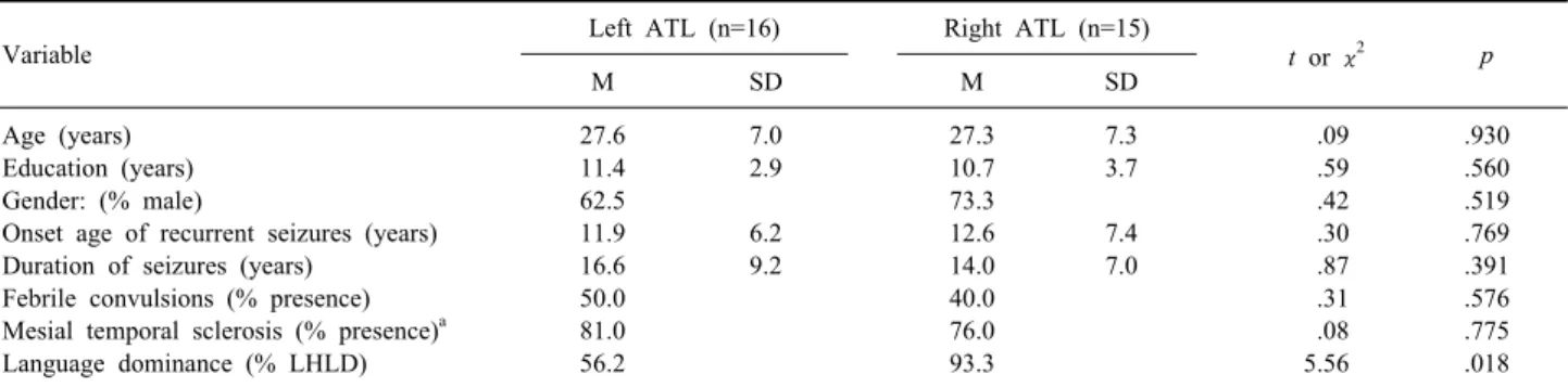
Variable Left ATL (n=16) Right ATL (n=15)
t or χ2 p
M SD M SD
Age (years) Education (years) Gender: (% male)
Onset age of recurrent seizures (years) Duration of seizures (years)
Febrile convulsions (% presence) Mesial temporal sclerosis (% presence)
aLanguage dominance (% LHLD)
27.6 11.4 62.5 11.9 16.6 50.0 81.0 56.2
7.0 2.9 6.2 9.2
27.3 10.7 73.3 12.6 14.0 40.0 76.0 93.3
7.3 3.7 7.4 7.0
.09 .59 .42 .30 .87 .31 .08 5.56
.930 .560 .519 .769 .391 .576 .775 .018 LHLD; left hemisphere language dominance,
afrom pathological examination of surgical specimen and available in 29 of 31 patients, P values are two-tailed.
Table 1. Comparison of the left vs. right anterior temporal lobectomy (ATL) groups in demographic and clinical variables
sclerosis as determined by pathological examination of surgical specimen. As expected, the incidence of non- left-hemisphere language dominance, as determined by the intracarotid amobarital procedure (IAP; see below), was significantly greater in the left ATL group than in the right ATL group ( p =.018)
2. Neuropsychological Evaluation
Preoperative neuropsychological testing was administered as a part of comprehensive presurgical work-up. Post- operative neuropsychological testing was administered approximately 1 year (M=13.0 months, range=10.5-16.4 months) after surgery.
1) Intelligence Test
The K-Wechsler Adult Intelligence Scale (K-WAIS),
11a Korean version of the WAIS-R,
12was administered. Only 9 of 11 subscales of the K-WAIS, excluding Vocabulary and Picture Arrangement subscales, were administered to shorten the testing time. For the purposes of this study, the following four measures were examined: full scale IQ (FSIQ), verbal comprehension index (VCI), perceptual organization index (POI), and freedom-from-distracti- bility index (FDI). The VCI was derived from Information, Comprehension, and Similarities subscales, the POI was derived from Picture Completion, Block design, and Object Assembly subscales, and the FDI was derived from Digit Span, Arithmetic, and Digit Symbol subscales. All
three indices had a normative mean of 100 and a SD of 15,
2) Memory Test
The Rey-Kim Memory Test (RKMT)
13was administered.
In RKMT, verbal memory performance is assessed by K-Auditory Verbal Learning Test (KAVLT), a Korean version of Rey Auditory Verbal Learning Test, and nonverbal memory performance is assessed by K-Complex Figure Test (KCFT), a Korean version of Rey Complex Figure Test. KAVLT required serial learning of a list of 15 unrelated words over 5 consecutive trials, each trial followed by immediate recall. After a delay period of 20 min, the patient was again required to recall the 15 words and then, to choose the 15 words from a list of 50 words.
KCFT was essentially identical to a standard version of Rey Complex Figure Test. For the purposes of this investigation, the following five measures were examined:
memory quotient (MQ), KAVLT immediate recall (KAVLT- IR; sum of trial 1 to 5), KAVLT delayed recall (KAVLT-DR), KCFT immediate recall (KCFT-IR), and KCFT delayed recall (KCFT-DR). The MQ, which had a normative mean of 100 and a SD of 15, was a summary measure derived from both KAVLT and KCFT. Raw scores were used for the other four measures.
3. IAP
The IAP was conducted, with the patient supine,
immediately following angiography. Amobarbital 125 mg
in a 10% solution was injected into the internal carotid artery using a transfemoral catheter over a 4~5 s interval.
Left and right hemisphere injections were done on the same day with a minimum of 40 min between the two injections. Following demonstration of hemiplegia, the patient was presented with a series of language tasks that lasted approximately 60 to 90s. The ‘core’ language tasks were: following a series of simple commands, reading a short sentence, naming a picture, and repeti- tion of a phrase spoken by the examiner. Left- hemisphere language dominance (LHLD) was ‘conser- vatively’ defined by classifying only patients who showed competence in all ‘core’ language tasks following right hemisphere injection but failed all core tasks following left hemisphere injection. All other patients were classified as having non-left-hemisphere language dominance (NLHLD).
4. Epilepsy Surgery
A tailored ATL, including both anterolateral temporal resection and amygdalohippocampectomy, was performed on all patients. We routinely varied the extent of hippocampal resections, depending on the intraoperative interictal epileptiform abnormalities on preresection electrocorticography (ECoG) recorded from the hippocampus and parahippocampal gyrus. After initial removal, postresection recording of ECoG from the remaining hippocampus and parahippocampal gyrus was repeated to decide whether further hippocampal resection was needed or not. The mean extent of lateral resection (measured from temporal pole along the middle temporal gyrus) was 4.0 cm (SD=0.6) for left ATL and 4.6 cm (SD
=0.8) for right ATL. The mean extent of hippocampal resection (measured from anterior tip) was 2.6 cm (SD=
0.6) for left ATL and 3.0 cm (SD=1.0) for right ATL. More detailed description of our surgical techniques is provided elsewhere.
145. Data Analyses
Three different types of analyses were conducted.
First, for comparisons of pre- to postoperative cognitive performances, paired t test was used ( p <.05, two-tailed).
Second, to investigate whether left vs. right ATL has differential effects on cognitive functions, for each cog- nitive measure, we first calculated a change score following ATL. The change score was computed as post- operative score minus preoperative score. Thus, a positive change score reflected cognitive improvement, whereas a negative change score reflected cognitive decline. Then, we compared the left vs. right ATL groups in each change score using an independent t test ( p <.05, two-tailed). Preliminary analyses indicated no significant differences in any preoperative cognitive scores between the left vs. right ATL groups. Thus, preoperative cog- nitive score was not entered as a covariate in comparisons of the left vs. right ATL groups in cognitive change scores.
Third, Pearson correlational analyses ( p <.05, one-tailed) were performed between each cognitive change score and three predictor variables of interest - onset age of recurrent seizures, age at time of surgery, preoperative score. One-tailed test was used because, based on results of prior relevant studies,
4,6-9we predicted an inverse relationship between each pre- to postoperative change score and each predictor variable.
RESULTS
1. Pre- to Postoperative Changes
For each cognitive measure, a comparison between the
preoperative vs. postoperative score was performed using
a paired t test. The results are shown in Table 2. When
both left and right ATL groups (n=31) were considered
together, the comparison showed a significant improve-
ment in FSIQ, POI, FDI, MQ, KAVLT-IR, and KAVLT-DR
following ATL (all p s<.05), and no other significant
changes. Within the left ATL group (n=16), the com-
parison revealed no significant change in any intellectual
and memory measures. Within the right ATL group
(n=15), the comparison revealed a significant improve-
Variable Preoperative scores Postoperative scores
t p
M SD M SD
Both left and right ATL (n=31) FSIQ
VCI POI FDI MQ
KAVLT immediate recall KAVLT delayed recall KCFT immediate recall KCFT delayed recall
84.8 86.4 84.7 88.1 84.2 39.5 7.0 15.7 15.5
15.1 17.7 18.4 13.2 15.8 9.1 3.8 8.0 7.4
87.8 87.2 89.1 91.1 87.9 45.4 8.5 15.3 15.7
16.0 19.3 18.8 14.1 15.2 10.6 3.7 7.0 6.9
2.75 0.59 2.85 2.07 2.59 3.63 3.03 -0.46 0.32
.010 .559 .008 .047 .015 .001 .005 .649 .753 Left ATL (n=16)
FSIQ VCI POI FDI MQ
KAVLT immediate recall KAVLT delayed recall KCFT immediate recall KCFT delayed recall
87.6 88.8 88.3 90.4 85.9 39.9 6.6 17.9 17.0
14.8 16.4 18.9 14.6 16.1 9.1 4.4 7.6 7.8
89.8 88.3 91.6 93.4 87.8 42.6 7.4 17.5 18.1
17.2 20.1 19.5 14.5 13.3 10.0 3.8 6.8 7.2
1.30 -0.19 1.62 1.44 0.93 1.14 1.00 -0.40 0.83
.213 .849 .127 .171 .370 .271 .333 .694 .417 Right ATL (n=15)
FSIQ VCI POI FDI MQ
KAVLT immediate recall KAVLT delayed recall KCFT immediate recall KCFT delayed recall
81.9 83.9 80.9 85.6 82.4 39.0 7.5 13.3 13.8
15.4 19.3 17.7 11.5 15.8 9.4 3.1 8.0 6.9
85.8 86.1 86.4 88.6 88.1 48.4 9.7 13.0 13.2
15.0 19.1 18.3 13.6 17.5 10.8 3.4 6.7 5.8
2.76 1.31 2.36 1.45 2.93 4.70 4.04 -0.24 -0.49
.015 .213 .033 .169 .011 .000 .001 .811 .632 FSIQ; full-scale IQ, VCI; verbal comprehension index, POI; perceptual organization index, FDI; freedom-from-distractability index, MQ;
memory quotient, K-AVLT; K-auditory verbal learning test, K-CFT; K-complex figure test, P values are two-tailed.
Table 2. Comparison of preoperative vs. postoperative intelligence and memory scores
ment in FSIQ, POI, MQ, KAVLT-IR, and KAVLT-DR (all p s<.05), and no other significant changes. Thus, regard- less of whether all patients were considered together or separately for the left vs. right ATL groups, at 1-year follow-up, all statistically significant (and most non- significant) changes following ATL were in the direction of improvement.
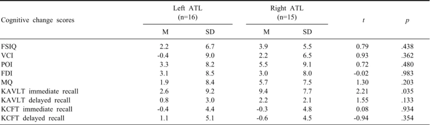
2. Effects of Surgical Side
We compared the left vs. right ATL groups in each change score using an independent t test. The results are shown in Table 3. These analyses revealed a significant difference in KAVLT-IR change score [ t (29)=-2.21, p =
.035] and no other significant differences. As depicted in Fig. 1, the significant group difference in KAVLT-IR change score reflected less improvement of verbal memory functions in the left ATL group relative to the right ATL group. When a similar set of analyses was repeated using only patients with LHLD, the left ATL (n
=9) vs. right ATL groups (n=14) showed a significant
difference in VCI change score [t(21)=2.15, p =.044], and
no other significant differences. In addition, the differ-
ence in KAVLT-IR change score approached statistical
significance [ t (21)=1.98, p =.061]. This difference would
remain significant with a one-tailed test ( p =.030), which
would be a more proper statistical test given a priori
expected direction of difference, namely, more decline in
Cognitive change scores
Left ATL (n=16)
Right ATL
(n=15)
t pM SD M SD
FSIQ VCI POI FDI MQ
KAVLT immediate recall KAVLT delayed recall KCFT immediate recall KCFT delayed recall
2.2 -0.4 3.3 3.1 1.9 2.6 0.8 -0.4 1.1
6.7 9.0 8.2 8.5 8.4 9.2 3.0 4.4 5.1
3.9 2.2 5.5 3.0 5.7 9.4 2.2 -0.3 -0.6
5.5 6.5 9.1 8.0 7.5 7.7 2.1 4.8 4.5
0.79 0.93 0.72 -0.02 1.30 2.21 1.55 0.08 -0.94
.438 .362 .480 .983 .203 .035 .133 .934 .354 Abbreviations are as in Table 2. P values are two-tailed.
Table 3. Comparison of the left vs. right anterior temporal lobectomy (ATL) groups in cognitive change scores (computed as
postoperative score minus preoperative score)Figure 1. Mean preoperative and postoperative K-auditory
verbal learning test-immediate recall (KAVLT-IR) score for the left ATL (n=16) and right ATL groups (n=15). Error bars represent±1 SE.verbal functions following left than right ATL. The significant differences again reflected less improvement of verbal functions following ATL in the left ATL group.
3. Predictors of Individual Patient Changes
Pearson correlational analyses were performed between each cognitive change score and three predictor variables of interest - onset age of recurrent seizures, age at time of surgery, preoperative score. These analyses were performed using all subjects (n=31). The correlations
Cognitive change score
Predictor variables Onset age
of recurrent seizures
Age at time of surgery
Preoperative score
FSIQ VCI POI FDI MQ
KAVLT immediate recall KAVLT delayed recall KCFT immediate recall KCFT delayed recall
-.410
a-.327
a-.232 -.314
a-.191 -.185 .028 -.070 -.179
-.305
a-.125 -.277 -.348
a-.297 -.353
a-.116 -.124 -.140
-.052 -.015 -.179 -.196 -.327
a-.315
a-.374
a-.488
b-.428
bAbbreviations are as in Table 2.
ap<.05, bp<.01 (all ps are one-tailed).
Table 4. Pearson correlations between cognitive change scores
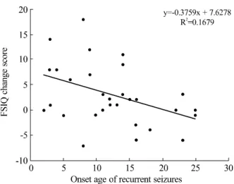
(computed as postoperative score minus preoperative score) and predictor variables (n=31)obtained from these analyses are listed in Table 4.
Statistical significance of each correlation was evaluated
with α=.05, one-tailed. Eleven of the 27 correlations
were statistically significant. All significant (and most
non-significant) correlations were in the expected
direction (i.e., inverse). Onset age of recurrent seizures
showed a significant inverse relationship with FSIQ, VCI,
and FDI change scores; age at time of surgery with FSIQ,
FDI, and KAVLT-IR change scores; and preoperative
score with MQ, KAVLT-IR, KAVLT-DR, KCFT-IR, and
KCFT-DR change scores. The significant correlations
indicated greater postoperative cognitive decline for
Figure 2. Scatterplot of the relationship between onset age of
recurrent seizures (x-axis) and Full-scale intelligence quotient (FSIQ) change score (y-axis). The change score was computed as postoperative FSIQ minus preoperative FSIQ.patients who are older, more able preoperatively, and had a later onset of recurrent seizures. As an example, the correlation between FSIQ change score and onset age of recurrent seizures is illustrated in Fig. 2.
DISCUSSION
There were three main sets of findings. First, at 1-year followup, all statistically significant (and most non-significant) changes following ATL were in the direction of improvement. The statistically significant improvements were found in both intellectual (FSIQ, POI, FDI) and memory measures (MQ, KAVLT-IR, KAVLT- DR). These cognitive improvements may reflect several factors such as practice effects, tailored surgery, seizure-free status, a decrease in the number or dosage of medications, and possibly for certain measures, release of non-epileptogenic hemisphere functions from inter- fering effects of epileptogenic hemisphere. More impor- tantly for the present purpose, the cognitive improve- ments may also reflect gradual recovery from acute or short-term cognitive decline following ATL. Consistent with this view, prior studies using a relatively long-term follow-up interval also reported mostly favorable cognitive outcome.
3,15The present findings and similar
prior findings underscore the fact that short-term cognitive deficits following ATL could overestimate deleterious effects of ATL on cognitive functions.
Second, left vs. right ATL had significantly differential effects both on verbal intelligence (VCI) and verbal memory (KAVLT-IR). Both verbal functions tended to show less improvement (or more deterioration) following left than right ATL. While these findings are replication of prior results, they are of importance in showing that greater risk to verbal functions following left ATL is apparent even at a relatively long-term follow-up. Thus, acute or short-term decline in verbal functions following left ATL, though recovering progressively over time, may not fully resolve to baseline level even at a long-term follow-up. In fact, within the left ATL group, no intellectual and memory measures showed significant improvement following ATL. By contrast, the right ATL group showed a significant improvement in five cognitive measures (i.e., FSIQ, POI, MQ, KAVL-IR, and KAVLT- DR). There were no significant differential effects of left vs. right ATL on nonverbal cognitive functions. These results may reflect use of “nonverbal” materials that can be easily verbalized.
1Alternatively, lateralization of nonverbal cognitive functions to the right hemisphere may be genuinely weaker in degree than lateralization of verbal functions to the left hemisphere.
Third, the present study investigated abilities of three
variables - onset age of recurrent seizures, age at time of
surgery, and preoperative cognitive abilities - to predict
individual cognitive changes after ATL. As expected on
the basis of prior literature,
4,6-9all significant (and most
non-significant) correlations between the change scores
and the predictor variables were inverse in direction. The
inverse correlations indicated greater postoperative
cognitive decline for patients who are older, more able
preoperatively, and had a later onset of recurrent
seizures. An increasingly influencing view of cognitive
changes following ATL is the functional adequacy model.
16According to this model, the more functional the to-
be-resected temporal lobe, the more likely it is that
cognitive decline will occur. Both later onset age of
recurrent seizures and higher preoperative performance
are associated with relatively intact state of the to-be-resected temporal lobe.
17-19Thus, the association of later onset age of recurrent seizures and higher preoperative performance with greater cognitive decline is consistent with the functional adequacy model. The association of older age at time of surgery with greater postoperative cognitive decline may reflect worse cerebral plasticity or worse capacity to compensate for the deleterious effects of ATL.
There are several limitations of the present study.
First, the sample size was relatively small. Thus, the present results need to be replicated using a larger sample size. Second, the present sample was restricted to those patients who are seizure-free following ATL. Thus, generalization of the present findings should be limited to the TLE population who are seizure-free following surgery. Third, a pre- to postoperative increase in cognitive score could reflect practice effects as well as
“genuine” cognitive improvement. In a research design lacking a control group as in the present study, it is impossible to rule out the possibility that an improvement in cognitive score is due to practice effects. Finally, most subjects received two or three antiepileptic drugs (AEDs;
e.g., carbamazepine, valproic acid, phenytoin) before surgery. The number and/or dosage of medications were typically reduced following surgery. This factor may account for, at least in part, the cognitive improvements found in the present study.
In summary, the present results indicate that at 1-year after ATL, most cognitive functions show either no significant change or a significant change in the direction of improvements. This finding indicates that short-term cognitive deficits following ATL could overestimate deleterious effects of ATL on cognitive functions. Left ATL had more deleterious effects on both verbal intellectual and verbal memory functions than right ATL.
Thus, decreased verbal functions following left ATL may represent the area of greatest potential neuropsycho- logical morbidity associated with ATL. Greater cognitive decline (or less cognitive improvement) following ATL was predicted by later onset age of recurrent seizures, older age at time of surgery, and higher preoperative perfor-
mance.
REFERENCES
1. Lee TM, Yip JT, Jones-Gotman M. Memory deficits after resection from left or right anterior temporal lobe in humans: a meta-analytic review. Epilepsia 2002;43:283-291.
2. Selwa LM, Berent S, Giordani B, Henry TR, Buchtel HA, Ross DA. Serial cognitive testing in temporal lobe epilepsy: longitudinal changes with medical and surgical therapies. Epilepsia 1994;35:
743-749.
3. Gleissner U, Sassen R, Lendt M, Clusmann H, Elger CE, Helmstaedter C. Pre- and postoperative verbal memory in pediatric patients with temporal lobe epilepsy. Epilepsy Res 2002;
51:287-296.
4. Hermann BP, Seidenberg M, Haltiner A, Wyler AR. Relationship of age at onset, chronologic age, and adequacy of preoperative performance to verbal memory change after anterior temporal lobectomy. Epilepsia 1995;36:137-145.
5. Sawrie SM, Chelune GJ, Naugle RI, L?ders HO. Empirical methods for assessing meaningful neuropsychological change following epilepsy surgery. J Int Neuropsychol Soc 1996;2:556-564.
6. Davies KG, Bell BD, Bush AJ, Wyler AR. Prediction of verbal memory loss in individuals after anterior temporal lobectomy.
Epilepsia 1998;39:820-828.
7. Chelune GJ, Naugle RI, Lüders H, Awad IA. Prediction of cognitive change as a function of preoperative ability status among temporal lobectomy patients seen at 6-month follow-up. Neurology 1991;41:399-404.
8. Stroup E, Langfitt J, Berg M, McDermott M, Pilcher W, Como P. Predicting verbal memory decline following anterior temporal lobectomy (ATL). Neurology 2003;60:1266-1273.
9. Bell B, Hermann B, Seidenberg M, Davies K, Cariski D, Rosenbek J, et al. Ipsilateral reorganization of language in early- onset left temporal lobe epilepsy. Epilepsy Behav 2002;3:158-164.
10. Engel J, Van Ness PC, Rasmussen TB, Ojemann LM. Outcome with respect to epileptic seizures. In: Engel J, Surgical treatment of
the epilepsies. 2nd ed. New York: Raven Press. 1993;609-621.
11. Yeom Y, Park Y, Oh K, Kim J, Lee Y. Korea-Wechsler Adult
Intelligence Scale. Seoul, South Korea: Korea Guidance, 1992.
12. Wechsler D. Wechsler Adult Intelligence Scale-Revised. San Antonio, TX: The Psychological Corporation, 1981.
13. Kim H. Rey-Kim Memory Test. Daegu, South Korea: Neuro- psychology Press, 1999.
14. Son EI, Howard MA, Ojemann GA, Lettich E. Comparing the extent of hippocampal removal to the outcome in terms of seizure control. Stereotact Funct Neurosurg 1994;62:232-237.
15. Engman E, Andersson-Roswall L, Malmgren K. Pre- and post- operative general neurocognitive status and memory in 70 epilepsy surgery patients. Acta Neurol Scand 2001;103:351-359.
16. Chelune GJ. Hippocampal adequacy versus functional reserve:
predicting memory functions following temporal lobectomy. Arch
Clin Neuropsychol 1995;10:413-432.
17. Sass KJ, Spencer DD, Kim JH, Westerveld M, Novelly RA, Lencz T. Verbal memory impairment correlates with hippocampal
pyramidal cell density. Neurology 1990;40:1694-1697.
18. Rausch R, Babb TL. Hippocampal neuron loss and memory scores before and after temporal lobe surgery for epilepsy. Arch Neurol 1993;50:812-817.
19. Davies KG, Hermann BP, Dohan FC Jr, Foley KT, Bush AJ, Wyler AR. Relationship of hippocampal sclerosis to duration and age of onset of epilepsy, and childhood febrile seizures in temporal lobectomy patients. Epilepsy Res 1996;24:119-126.