J. Exp. Biomed. Sci. 10 (2004) 429–434
Effects of High Taurocholic Acid Load on Liver Lysosomal Cathepsin B and D, and Acid Phosphatase Activities in Rats with
Choledocho-Caval Shunt
Hye-Jung Choi, You-Hee Kim and Chun-Sik Kwak
†Department of Biochemistry, Keimyung University, School of Medicine, Daegu, 700-712, Korea
The effects of intravenous administration of high concentration of taurocholic acid (TCA) on cathepsin B and D, and acid phosphatase activities in rat liver lysosome were studied. These liver lysosomal enzymes were determined from the experimental rats with choledocho-caval shunt (CCS). The activities of liver lysosomal cathepsin B and D, and acid phosphatase were found to be significantly increased in the CCS plus TCA injection group than in control group, such as group of CCS alone group. However, these hepatic enzyme activities did not change in the CCS plus tauroursodeoxycholic acid injection group. The above results suggest that TCA stimulates the biosynthesis of the lysosomal cathepsin B and D, and acid phosphatase in the liver.
Key Words: Acid phosphatase, Cathepsin B, Cathepsin D, Choledocho-caval shunt, Taurocholic acid
서 론
담즙울체간에서 활성도가 변동되는 효소가 많다는 사실 은 잘 알려져 있으며 그 활성도 변동은 담즙울체에 의한 taurocholic acid (TCA)의 간 내 농도 증가로 효소들의 유전 자 발현률이 변동되어 나타난 현상 (Han et al., 1997; Kim et al., 1997; Rhee et al., 2000; Kim et al., 2002; Rhee et al., 2002;
Rhee et al., 2004)이라 한다. 이런 현상의 파악을 위해서는 실 험적 담즙울체간을 만들어야하며 이를 만드는 동물 모델은 두 가지가 있다 (Ogawa et al., 1990; Park et al., 1999). 즉, 첫째 모델은 쥐에게 총담관 결찰 (common bile duct ligation)로 담 관을 폐쇠 시킨 후 TCA를 혈중에 주입하는 모델이며 둘째 모델은 쥐에게 총담관 대정맥문합 (choledocho-caval shunt)을 시킨 후 TCA를 혈중에 주입하는 모델이다. 이 두 모델은 시간이 경과 될수록 간 내 담즙산의 농도가 증가되는 점은 동일하나 다른점은 그 증가 속도가 첫째 모델이 둘째 모델 보다 훨씬 빠르다 (Toyota et al., 1984; Ogawa et al., 1990)는 것 이다. 이와 같이 첫째 모델이 둘째 모델보다 시간이 경과 될 수록 간 내 담즙산 농도가 빠르게 증가 되는 것은 담관 폐 쇄로 담관 내 수압이 증가되어 담관 내 담즙산이 간 세포 내
로 강제 역류되고 또한 간 내 담즙산의 간 외 배출이 억제 되기 때문에 나타난 현상이며 (Tayota et al., 1984), 둘째 모델 은 담관 내 수압이 증가 되지 않기 때문에 담즙산의 간세 포 내로의 역류가 심하지 않다 (Toyota et al., 1984). 따라서 첫 째 모델인 총담관 결찰 직후 TCA를 상대정맥 내 주입한 모 델로는 간 내 TCA의 고농도 증가에 따른 효소 활성도 변동 에 대한 TCA의 효과를 알아낼 수가 있는 것 (Ogawa et al., 1990; Park et al., 1999)이고 둘째 모델인 총담관 대정맥문합 직후 TCA를 상대 정맥내 주입한 모델로는 비교적 낮은 간 내 TCA 농도 증가에 따른 TCA효과를 알아낼 수가 있는 것 (Ogawa et al., 1990; Park et al., 1999)이며, 특히 이 모델의 특 징은 첫째 모델 보다 TCA 효과를 더욱 민감하게 알아 낼 수가 있는 것이다.
쥐를 사용한 실험적 담즙울체간을 만드는 모델 중 한가 지인 총담관 대정맥문합을 시킨 후 TCA를 혈중에 주입 하여 각종 효소들의 활성도 변동을 조사한 연구결과, 담즙울체간 에서 그 활성도가 변동되는 효소들은 다음과 같다. 즉, alkaline phosphatase (Ogawa et al., 1990), γ-glutamyl transpeptidase (Kim et al., 1997), catalase, alcohol dehydrogenase, 마이크로솜 ethanol oxidizing system, aldehyde dehydrogenase (Kim et al., 2002), aryle- sterase (Han et al., 1997), arylamine N-methyltransferase (Rhee et al., 2000), thiol methyltransferase (Rhee et al., 2002) 및 thiosulfate sulfurtransferase (Rhee et al., 2004) 등이며 TCA가 이들 효소의 유전자 발현율을 변동시켜 이들 효소의 합성 속도를 변동 시킨다는 것이다. 따라서 총담관 대정맥문합과 함께 TCA를 혈중에 주입 시킨 동물모델의 간에서 그 활성도가 변동되는
*논 문 접 수: 2004년 10월 4일 수정재접수: 2004년 12월 7일
†교신저자: 곽춘식, (우) 700-712 대구광역시 중구 동산동 194, 계명대학교 의과대학 생화학교실
Tel: 053-250-7461, Fax: 053-250-7461 e-mail: [email protected]
효소들에 대해서는 TCA의 효과를 더욱 분명하게 알 수가 있을 것이다.
Cathepsin B (EC 3. 4. 22. 1)는 thiol proteinase에 속하는 효소 며 (Barrett, 1977b) cathepsin D (EC 3. 4. 23. 5)는 carboxyl (acid) proteinase에 속하는 효소이다 (Barrett, 1977a). 이 두가지 효 소는 프레단백질들을 가공하고 세포 내외의 단백질 분해와 더불어 각종 단백질의 교체 속도를 조절하고 있다 (Ansorge et al., 1977; Quinn et al., 1978; Barat et al., 1979; Graf et al., 1979;
Satav et al., 1981). 또한 acid phosphatase [orthophosphoric -monoester phosphohydrolase (acid optimum), EC 3. 1. 3. 2]는 pH 5 부근에서 orthophosphoric monoester들을 가수분해하여 알콜 과 인산을 유리시키는 효소이다 (Noel et al., 1987). 이와 같 은 3종의 효소는 간의 라이소솜에 풍부히 국재되어 있는 효소이며 (Turk et al., 1984a & 1984b; Himeno et al., 1988), 담즙 울체간에서는 이들 라이소솜 효소들의 활성도가 증가됨이 (Ihm et al., 1994) 알려져 있을 뿐만 아니라 TCA에 의해서 이들 효소의 유전자 발현률이 증가된다고 추정하고 있다 (Park et al., 2001).
이 연구는 간 세포의 라이소솜에 풍부히 국재되어 있는 cathepsin B 및 D와 acid phosphatase의 활성도가 담즙울체간 에서 왜 증가되었는지 그 기전을 분명하게 알아내기 위하 여 시행하였으며 쥐에게 총담관 대정맥문합을 시킨 직후 TCA 와 간의 효소 합성에 영향을 미치지 않는다 (Ogawa et al., 1990; Kim et al., 1997; Rhee et al., 2000)고 알려진 tauroursodeo- xycholic acid (TUDCA)를 각각 상대정맥 내에 주입한 후 경 시적으로 간에서 cathepsin B 및 D와 acid phosphatase의 활성 도를 측정하여 이들 효소의 활성도 변동 기전을 밝혀 보고 자 하였다.
재료 및 방법
1. 시 약
Nα-benzoyl-DL-arginine-β-naphtylamide, mersalyl acid, 4-nitro- phenol, polyoxyethylene-2, 3-lauayl ether, β-naphthylamine, 4-am- ino-2, 3-dimethylazobenzene, disodium-ρ-nitrophenyl phosphate, L-tyrosine, hemoglobin (from bovine erythrocyte, substrate powder), Triton X-100, TCA (from ox bile, sodium salt), TUDCA (sodium salt), cathepsin B (from bovine spleen, C 6286), cathepsin D (from bovine spleen, C 3138), acid phosphatase (AcP Lin-Trol®) 및 단백 질 표준액 (10 g/100 ml, bovine serum albumin) 등은 Sigma사 (St. Louis, 미국)의 제품을 사용하였으며 그 외 일반 시약들 은 특급 또는 일급품을 사용하였다.
2. 동물 및 처치
동물은 4주 이상 같은 조건으로 사육한 체중 280~320 g
이 되는 Sprague-Dawley 종의 숫쥐를 사용하였으며 1군을 5 마리로 하여 다음과 같이 9개군으로 나누었다. 즉 정상군 1 군, 가수술군은 가수술 후 1일 및 2일에 각각 희생시킨 군 총 2군으로 하였고 총담관 대정맥문합 군은 총담관 대정맥 문합 후 1일 및 2일에 각각 희생시킨 군 총 2군, 총담관 대 정맥문합과 함께 TCA를 주입한 군은 총담관 대정맥문합 직 후 Ogawa et al. (1990)과 Park et al. (1999)의 방법에 따라 TCA (체중 100 g당 45 µmoles)를 상대정맥 내에 주입한 후 1일 및 2일에 각각 희생시킨 군 총 2군, 총담관 대정맥문합과 함께 TUDCA를 주입한 군은 총담관 대정맥문합 직후 Ogawa et al.
(1990)과 Park et al. (1999)의 방법에 따라 TUDCA (체중 100 g당 45 µmoles)를 상대정맥 내에 주입한 후 1일 및 2일에 각각 희생시킨 군 총 2군으로 나누었다. 각 실험군은 개별 분 리 수용하였으며 실험 전후에 일정한 조건으로 사육하였다.
총담관 대정맥문합 수술 및 가수술은 효소 활성의 일중 변 동을 고려하여 쥐를 오후 2시에서 5시 사이에 희생시킬 수 있도록 수술 시간을 조절하였으며 12시간 금식시킨 후 이터 마취하에서 실시하였다. 총담관 대정맥문합은 상대정맥과 총 담관을 medical grade silicon tube를 사용하여 연결하였으며 가 수술은 단순 개복술만 시행하였다. TCA 및 TUDCA 액의 상 대정맥 내 주입은 syringe pump (model 341A, Sage instruments, 미국)를 사용하여 15분 동안 주입하였다.
3. 간적출 및 시료 조제
모든 실험군에서 간의 적출은 12시간 금식시킨 후 이터 마 취 하에서 시행하였으며 복부 대동맥으로부터 채혈하여 쥐 를 실혈사시켰다. 그리고는 간문맥에 삽관한 후 4℃의 0.25 M sucrose 액으로 관류하여 간에 남아 있던 혈액을 제거한 다음 간을 적출하였다. 적출한 간은 면포로 균등히 압박하여 간에 남아 있던 sucrose 액을 가능한 한 모두 제거하였다. 간 라이 소솜 효소 시료의 조제를 위하여서는 적출하여 0.25 M sucr- ose 액으로 관류한 간을 즉시 2~4℃로 냉각시킨 후 잘게 썰 어서 절편으로 만들고 잘 혼합하여 사용하였다. 간 절편의 마 쇄를 위해서는 Teflon pestle galss homogenizer (chamber clear- ance 0.005~0.007 inches, Thomas사, 미국)를 사용하였으며, 간 절편의 마쇄는 2~4℃를 유지하면서 400 rpm 속도로 마쇄를 하였다. 간 라이소솜의 cathepsin B와 D 활성도 측정용 시료 는 0.25 M sucrose 액으로 제조한 10% (w/v) 간 마쇄액을 ultr- asonic dismembrator로 2~4℃를 유지하면서 20±4 Kcycle/sec 의 조건으로 2분씩 5회 초음파 마쇄를 하여 (Barrett, 1972;
Barrett, 1977a) 사용하였다. 간 라이소솜의 acid phosphatase 활성도 측정용 시료의 조제는 먼저 Graham (1984)법으로 라 이소솜을 분리한 후 Triton X-100을 처리한 다음 사용하였 다. 즉 0.25 M sucrose 액으로 만든 10% (w/v) 간 마쇄균질액 약 10 ml을 취하여 1,000×g에서 20분간 원심분리하여 침사
를 얻었으며 이 침사를 10% (w/v) sucrose 액에 현탁시켜 그 일정량을 37~72% (w/v) sucrose linear density gradient 용액을 넣은 원심관 관저에 부하시켰다. 그리고는 이 원심분리관을 95,000×g에서 2시간 원심분리하여 51~57% (1.23~1.26 g/cm) sucrose 액층 부위에 형성된 pellet을 취한 후 이 pellet을 0.25 M sucrose 액으로 1회 세척한 것을 0.1% Triton X-100으로 2 배 희석하여 진탕한 액을 사용하였다. 위의 효소 시료 조제 과정에서 모든 조작은 2~4℃에서 시행하였으며 사용한 원 심분리기는 Du Pont Sorvall사 (미국)의 RC-5B refrigerated su- perspeed centrifuge와 OTD-65B ultracentrifuge였다. 그리고 su- crose density gradient 용액의 제조는 gradient former (model 570, ISCO, 미국)를 사용하여 제조하였다.
4. 효소 활성도 측정
간의 라이소솜 cathepsin B의 활성도 측정은 Nα-benzoyl- DL-arginine-β-naphthylamide를 기질로 사용하여 40℃에서 10 분간 반응시켜 생성된 β-naphthylamine을 측정하는 방법인 Barrett (1972)의 방법에 따랐으며 단위는 1분간에 1 mg의 단 백질이 반응하여 생성한 β-naphthylamine의 양을 nmol로 나 타내었다. 그리고 간의 라이소솜 cathepsin D의 활성도 측정 은 hemoglobin을 기질로 사용하여 37℃에서 10분간 반응시 키는 동안에 생성된 tyrosine을 측정하는 방법인 Barrett (1977a)
법에 의하였으며 단위는 1분간에 1 mg의 단백질이 반응하 여 생성한 tyrosine을 nmol로 나타내었다. 간의 라이소솜 acid phosphatase 활성도 측정은 disodium-4-nitrophenylphosphate를 기질로 사용하여 효소액과 함께 37℃에서 30분간 반응시키 는 동안에 생성된 4-nitrophenol을 측정하는 Moss (1984)의 법으로 측정하였으며 단위는 1분간에 1 mg의 단백질이 반 응하여 생성한 4-nitrophenol을 nmol로 나타내었다. 이 실험 에서 채택한 효소 활성도 측정법들의 정확도를 높이기 위 하여 Sigma사의 정제 효소들을 사용하여 검정하였으며 같 은 시료에 대하여 2회 측정하여 그 평균치를 취하였다. 이 실험에서 효소 활성도 측정에 사용한 분광광도계는 computer controlled enzyme spectrophotometer (Cary 210, Varian, 미국) 였다.
5. 단백질 정량
효소 시료 중의 단백질 정량은 0.5 M perchloric acid와 me- thanol-ether 혼합액 (3:1)으로 단백질을 정제하는 Greenberg et al. (1957)법으로 효소 시료 중의 단백질을 정제한 다음 biuret 법 (Gornall et al., 1949)으로 정량하였다.
6. 성적 검정
유의성 검정은 Student's t-test로 하였으며 유의수준은 0.05
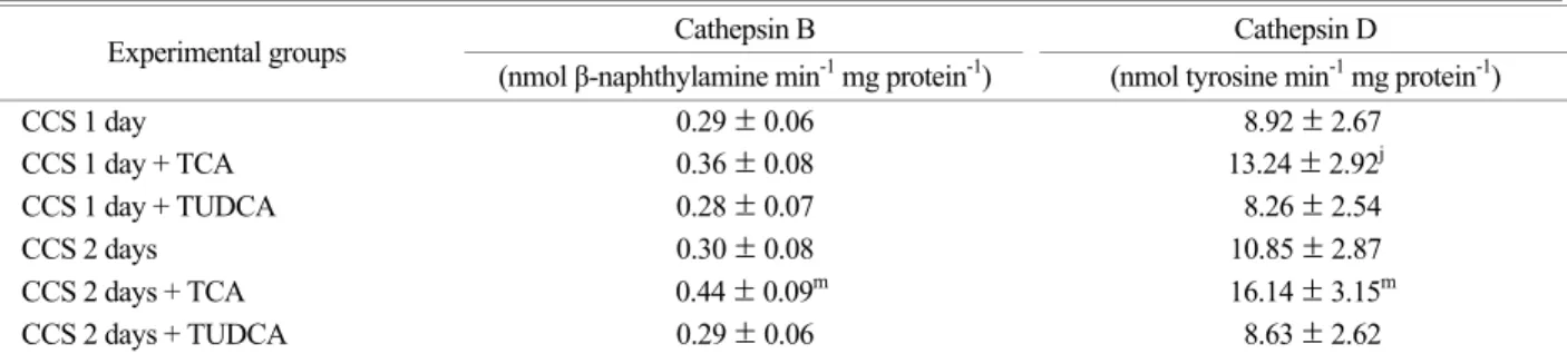
Table 2. Effects of taurocholic acid (TCA), and tauroursodeoxycholic acid (TUDCA) infusions after choledocho-caval shunt (CCS) on liver lysosomal cathepsin B and D activities in rats
Cathepsin B Cathepsin D
Experimental groups
(nmol β-naphthylamine min-1 mg protein-1) (nmol tyrosine min-1 mg protein-1)
CCS 1 day 0.29 ± 0.06 8.92 ± 2.67
CCS 1 day + TCA 0.36 ± 0.08 13.24 ± 2.92j
CCS 1 day + TUDCA 0.28 ± 0.07 8.26 ± 2.54
CCS 2 days 0.30 ± 0.08 10.85 ± 2.87
CCS 2 days + TCA 0.44 ± 0.09m 16.14 ± 3.15m
CCS 2 days + TUDCA 0.29 ± 0.06 8.63 ± 2.62
The data are expressed as mean ± SD with 5 rats in each group; CCS 1 day or CCS 2 days, sacrificed 1st or 2nd day after choledocho -caval shunt; One of the following bile acids, TCA and TUDCA (45 µmol/100 g body weight) was intravenously administered through the superior vena cava. j, P<0.05 vs. CCS 1 day; m, P<0.05 vs. CCS 2 days
Table 1. Effects of choledocho-caval shunt (CCS) on liver lysosomal cathepsin B and D activities in rats
Cathepsin B Cathepsin D
Experimental groups
(nmol β-naphthylamine min-1 mg protein-1) (nmol tyrosine min-1 mg protein-1)
Normal 0.28 ± 0.05 8.02 ± 2.48
Sham 1 day 0.28 ± 0.07 8.05 ± 2.56
Sham 2 day 0.29 ± 0.06 8.08 ± 2.42
CCS 1 day 0.29 ± 0.06 8.92 ± 2.67
CCS 2 days 0.30 ± 0.08 10.85 ± 2.87
The data are expressed as mean ± SD with 5 rats in each group; Sham 1 day or Sham 2 days, sacrificed on the 1st or 2nd day after sham operation; CCS 1 day or CCS 2 days, sarificed on the 1st or 2nd day after choledocho-calval shunt.
이하로 하였다.
결 과
1. 쥐에서 총담관 대정맥문합 시 TCA 또는 TUDCA 주 입이 간 라이소솜의 cathepsin B와 cathepsin D 활성도에 미치는 영향
쥐에게 총담관 대정맥문합을 시킨 후 1일 및 2일째 (결과 Table에서 CCS 1 day 및 CCS 2 days) 간 라이소솜의 cathepsin B 및 D의 활성도는 변동을 나타내지 않았다 (Table 1). 쥐에 게 총담관 대정맥문합을 시킨 직후 TCA를 주입시켜 2일 경 과 (결과 Table에서 CCS 2 days + TCA)시켰을 때 간 라이소솜 의 cathepsin B 활성도는 대조군인 총담관 대정맥문합만 시킨 군에 비해 통계학적으로 유의한 증가를 나타내었다. 즉 총담 관 대정맥문합 직후 TCA를 주입시키고 2일 후 간 라이소솜 의 cathepsin B 활성도는 대조군인 총담관 대정맥문합만 시킨 군보다 약 47% (P<0.05)의 증가를 나타내었다 (Table 2).
쥐에게 총담관 대정맥문합을 시킨 직후 TCA를 주입시켜 1일 및 2일 경과 (결과 Table에서 CCS 1 day + TCA 및 CCS 2 days + TCA) 후 간 라이소솜의 cathepsin D 활성도는 대조군 인 총담관 대정맥문합만 시킨 군 (결과 Table에서 CCS 1 day 및 CCS 2 days)에 비해 통계학적으로 유의한 증가를 나 타내었다. 즉 총담관 결찰 직후 TCA를 주입시키고 1일 및 2일째 간 라이소솜의 cathepsin D 활성도는 대조군인 총담관 대정맥문합만 시킨 군보다 각각 약 48% (P<0.05) 및 약 49%
(P<0.05)의 증가를 나타내었다. 그러나 총담관 대정맥문합을 시킨 직후 TUDCA를 주입시킨 간 (결과 Table에서 CCS 1 day + TUDCA 및 CCS 2 days + TUDCA)에서 이들 효소의 활 성도는 대조군과 차이가 없었다 (Table 2).
2. 쥐에서 총담관 대정맥문합 시 TCA 또는 TUDCA 주 입이 간 라이소솜의 acid phosphatase 활성도에 미치는 영향
쥐에게 총담관 대정맥문합을 시킨 후 1일 및 2일째 (결
과 Table에서 CCS 1 day 및 CCS 2 days) 간 라이소솜의 acid phosphatase 활성도는 변동을 나타내지 않았다 (Table 3).
쥐에게 총담관 대정맥문합을 시킨 직후 TCA를 주입시켜 1일 및 2일 경과 (결과 Table에서 CCS 1 day + TCA 및 CCS 2 days + TCA)시켰을 때 간 라이소솜의 acid phosphatase 활성 도는 대조군에 비해 통계학적으로 유의한 증가를 나타내었 다. 즉 총담관 대정맥문합 직후 TCA를 주입시키고 1일 및 2일 경과시켰을 때 간 라이소솜의 acid phosphatase 활성도 는 대조군인 총담관 대정맥문합만 시킨 군 (결과 Table에서 CCS 1 day 및 CCS 2 days)보다 각각 약 20% (P<0.05) 및 약 29% (P<0.05)의 증가를 나타내었다. 그러나 총담관 대정맥문 합을 시킨 직후 TUDCA를 주입시켜 1일 및 2일 경과 (결과 Table에서 CCS 1 day + TUDCA 및 CCS 2 days + TUDCA)시켰 을 때 간의 이 효소 활성도는 대조군과 별 차이가 없었다 (Table 4).
고 찰
원발성 담즙성 간경변증, 담즙울체형 간염, 담관염 등의 질환에 이환되거나 선천성 담도폐쇄, 종양 및 담석에 의해 담 도가 폐쇄되었을 때 간은 담즙울체가 야기된다 (Halsted, 1976;
Sherlock et al., 2002). 이러한 담즙울체성 간담도 질환 시 담 즙울체의 시간이 경과하면 간조직은 괴사, 지방변성, 담도 증 식, 섬유화, 경화성 변화 등 형태학적인 변화가 연속적으로 나타남 (Desmet, 1994)과 동시에 간세포는 기능 장애가 초래 되며 (Halsted, 1976; Sherlock et al., 2002) 이때 담즙울체간에 서는 각종 효소들의 활성도가 변동되는 것은 이미 잘 알려 진 사실이다. 특히 쥐를 사용하여 담즙울체간을 만드는 모 델 실험에서 쥐 담즙울체간의 조직학적소견을 관찰한 Kou- ntourase et al. (1984)의 보고에서 쥐에게 심한 담즙울체를 야 기 시킨 후 12시간이 경과되었을 때는 많은 간세포들이 괴 Table 3. Effects of choledocho-caval shunt (CCS) on liver lysoso-
mal acid phosphatase activity in rats
Acid phosphatase Experimental
groups (nmol 4-nitrophenol min-1 mg protein-1)
Normal 183.6 ± 15.2
Sham 1 day 182.8 ± 16.4 Sham 2 day 183.5 ± 16.7 CCS 1 day 196.3 ± 17.8 CCS 2 days 206.7 ± 19.2
The data are expressed as mean ± SD with 5 rats in each group.
Experimental groups are described in Table 1 and text
Table 4. Effects of taurocholic acid (TCA), and tauroursodeo- xycholic acid (TUDCA) infusions after choledocho-caval shunt (CCS) on liver lysosomal acid phosphatase activity in rats
Acid phosphatase Experimental groups
(nmol 4-nitrophenol min-1 mg protein-1)
CCS 1 day 196.3 ± 17.8
CCS 1 day + TCA 234.7 ± 26.4j CCS 1 day + TUDCA 192.5 ± 16.6 CCS 2 days 206.7 ± 19.2 CCS 2 days + TCA 267.3 ± 41.2m CCS 2 days + TUDCA 198.6 ± 17.2
The data are expressed as mean ± SD with 5 rats in each group;
Experimental groups are described in Table 2 and text. j, P<0.01 vs. CCS 1 day; m, P<0.05 vs. CCS 2 days
사 현상을 나타내었으며 동시에 담도세포도 증식되기 시작 하였다고 하였다. 그리고 1일 후에는 간의 전부위에 괴사 현 상이 확산되고 괴사 부위에는 염증 세포의 침윤이 보였으 며 2주경에는 괴사 현상이 약간 감소되는 반면에 담도세포 의 증식이 증가되고 섬유화가 시작되었으며 6주부터는 초기 의 경화성 변화가 나타났다고 보고하였다. 이와 같이 담즙 울체로 간손상을 받는 모델 실험에서는 여러 효소들의 활 성도가 변동되는 것이 잘 알려져 있다. 그리고 쥐의 담즙울 체간에서 그 활성도가 증감되는 효소들의 활성도 변동 기 전에 대해서 현재까지 알려져 있는 것은 다음과 같다. 즉, arylesterase (Han et al., 1997), carboxylesterase (Han et al., 1998), cholinesterase (Park et al., 1999), alcohol dehydrogenase, catalase (Kim et al., 2002) 및 thiosulfate sulfurtransferase (Rhee et al., 2004)는 담즙울체간에서 그 활성도가 감소되며 그 기전은 담즙울체로 간세포내에 증가된 TCA가 유전자 발현을 억제 시켜 이들 효소의 합성을 억제시킨다는 것이고 alkaline pho- sphatase (Ogawa et al., 1990), 5'-nucleotidase (Kim et al., 2001b), γ-glutamyl transpeptidase (Kim et al., 1997), 마이크로솜 ethanol oxidizing system, aldehyde dehydrogenase (Kim et al., 2002), ben- zoyltransferase (Kim et al., 2001a), arylamine N-methyltransferase (Rhee et al., 2000) 및 thiol methyltransferase (Rhee et al., 2002)는 담즙울체간에서 그 활성도가 증가되며 그 기전은 담즙울체 로 간세포 내에 증가된 TCA가 유전자 발현을 자극시켜 이 들 효소의 합성을 촉진시킨다는 것이다.
Ihm et al. (1994)은 쥐를 모델로 한 동물실험에서 cathepsin B 및 D와 acid phosphatase 활성도가 총담관 결찰 후의 담즙 울체간에서 증가되었다고 보고하였다. 그러나 이번 실험에서 총담관 대정맥문합만 시행한 간에서는 이들 효소의 활성도는 변동이 없었다. 이 현상은 간에 담즙울체의 정도가 미약해서 나타난 결과가 아닌가 생각된다. Park et al. (2001)은 쥐를 사 용한 실험에서 총담관 결찰 후 혈중에 TCA를 주입한 결과 간에서 cathepsin B 및 D와 acid phosphatase의 활성도가 모두 증가되었으며 이 결과는 TCA가 간에서 이들 효소의 합성을 유도하여 나타난 결과라고 추론하였다. 이번 실험은 이 사실 을 더욱 분명하게 알아내기 위하여 시행한 것이며 또한 이 실험에서는 주입하는 담즙산의 종류가 다르면 이들 효소 활 성도에 미치는 영향도 달라지는가를 알기 위하여 담즙울체간 에서 효소들의 합성에 영향을 주지 않으며 (Ogawa et al., 1990;
Kim et al., 1997; Rhee et al., 2000) 담즙산의 간 독성에 대해 보 호 효과를 가진다는 TUDCA (Poupon et al., 1987; Ogawa et al., 1990; Heuman et al., 1991)를 쥐에게 총담관 대정맥문합을 시 킨 직후 상대정맥 내에 다량 주입시켜 효과를 관찰하였다.
이 실험에서 쥐에게 총담관 대정맥문합을 시킨 직후 TCA 를 주입시켜 1일 및 2일 경과시켰을 때 간 라이소솜의 cathe- psin D와 acid phosphatase 활성도 그리고 2일 경과 시켰을 때
간 라이소솜의 cathepsin B의 활성도는 대조군인 총담관 대 정맥문합만 시킨 군보다 통계학적으로 유의한 증가를 나타내 었다. 이 결과로 보아 담즙울체 시 TCA는 간 라이소솜의 ca- thepsin B 및 D와 acid phosphatase의 합성을 자극한다고 확신 할 수 있었으며, 이런 결과는 담즙울체로 손상을 받은 간세 포를 자가 분해시키기 위해 일어난 하나의 현상이라는 Park et al. (2001)의 생각을 더욱 뒷받침 해주는 결과라 생각된다.
이 실험에서 쥐에게 총담관 대정맥문합을 시킨 직후 TU- DCA를 주입시켜 1일 및 2일 경과시켰을 때 간의 라이소솜 에서 이 실험에서 측정한 효소들의 활성도는 모두 대조군과 별 차이가 없었다. 이 결과를 볼 때 TUDCA는 이 실험에서 관찰한 라이소솜 효소들의 유전자 발현에는 관여하지 않는 다고 추정 할 수가 있었다.
이상 실험의 결과들과 문헌상의 지견을 볼 때 담즙울체 간에서 활성도가 증가되는 라이소솜의 cathepsin B 및 D와 acid phosphotase의 활성도 증가는 담즙산 중 TCA에 의해 이 들 효소의 합성이 자극되어 나타난 결과로 생각된다.
REFERENCES
Ansorge S, Kirschke H, Friedrich K. Conversion of proinsulin into insulin by cathepsin B and L from rat liver lysosomes.
Acta Biol Med Germ. 1977. 36: 1723-1727.
Barat E, Patthy A, Graf L. Action of cathepsin D on human β -lipotropin: A possible source of human β-melanotropin. Proc Natl Acad Sci USA. 1979. 76: 6120-6123.
Barrett AJ. A new assay for cathepsin B1 and other thiol protei- nases, Anal Biochem. 1972. 47: 280-293.
Barrett AJ. Cathepsin D and other carboxyl proteinases in Protei- nases mammalian cells and tissues (Barrett AJ. Ed). 1977a.
pp 209-248. Elsevier, North-Holland Biomedical Press. Am- sterdam, The Netherlands.
Barrett AJ. Cathepsin B and other thiol proteinases in Proteinases mammalian cells and tissues (Barrett AJ. Ed). 1977b. pp 181 -208. Elsevier, North-Holland Biomedical Press. Amsterdam, The Netherlands.
Desmet VJ. Cholestasis: Extrahepatic obstruction and secondary biliary cirrhosis in Pathology of the liver, 3rd ed. (MacSween RNM, Anthony PP, Scheuer PJ, Burt AD, Portman BC. Eds).
1994. pp 425-474. Churchill Livingstone. NY, USA.
Gornall AG, Bardawill CJ, David MM. Determination of serum protein by means of biuret reaction. J Biol Chem. 1949. 177:
751-766.
Graf L, Kennessey A, Patthy A, Grybaum A, Marks N, Lajtha A.
Cathepsin D generates γ-endorphin from β-endorphin. Arch
Biochem Biophys. 1979. 193: 101-109.
Graham J. Isolation of subcellular organelles and membranes in Centrifugation, a practical approch, 2nd ed. (Rickwood D. Ed).
1984. pp 161-182. IRL Press. Oxford, UK.
Greenberg DM, Rothstein M. Method for isolation and degra- dation of labelled compounds in Method in enzymology (Co- lowick SP, Kaplan NO. Eds). 1957. Vol 4, pp 708-731. Aca- demic Press. NY, USA.
Halsted JA. The laboratory in clinical medicine. interpretation and application. 1976. pp 426-429. Saunders. London, UK.
Han BH, Kim YH. Effect of high taurochoclate load on activity of rat liver arylesterase. Korean J Hepatol. 1997. 3: 154-169.
Han BH, Kim YH. Effect of high taurocholate load on activity of rat liver carboxylesterase. Keimyung Med J. 1998. 17: 487-503.
Heuman DM, Mills AS, McCall J, Hylemon PB, Pandak WM, Vla- hcevic ZR. Conjugates of ursodeoxycholate protect againt cho- lestasis and hepatocellular necrosis caused by more hydro- phobic bile salts: In vivo studies in the rat. Gastroenterology.
1991. 100: 203-211.
Himeno M, Koutoku H, Tsuji H, Kato K. Purification and chara- cterization of acid phosphatase in rat liver lysosomal contents.
J Biochem. 1988. 104: 773-776.
Ihm JS, Mun KC, Kwak CS. Cathepsin B, D, H and acid pho- sphatase activities in cholestatic rat liver. Korean J Biochem.
1994. 26: 203-207.
Kim IK, Kim YH, Kwak CS. Induction of hepatic benzoyltransfe- rase by bile acid in rats. Keimyung Med J. 2001a. 20: 20-30.
Kim SK, Kim YH, Kwak CS. Induction of cholestatic rat liver 5'-nucleotidase by taurocholic acid load. Keimyang Med J.
2001b. 20: 129-139.
Kim SK, Kim YH. Induction of rat liver γ-glutamyl transpeptidase by bile acid load. Korean J Hepatol. 1997. 3: 210-226.
Kim YH, Shin MJ. Effects of high taurocholate load on activities of hepatic alcohol metabolizing enzymes. Exp Mol Med.
2002. 34: 123-130.
Kountouras J, Billing BH, Scheuer PJ. Prolonged bile duct obstru- ction: A new experimental model for cirrhosis in the rats. Br J Exp Pathol. 1984. 65: 305-311.
Moss DW. Acid phosphatase in Method of enzymatic analysis, 3rd ed. (Bergmeyer HU, Bergmeyer J, Graβ1 M. Eds). 1984. Vol IV, pp 92-105. Verlag Chemie GmbH. Weinheim, Germany.
Noel SA, Lott JA. Acid phosphatase, total in Methods in clinical
chemistry (Pesce AJ, Kaplan LA. Eds). 1987. pp 683-690.
The C. V. Mosby Company. ST. Louis, USA.
Ogawa H, Mink J, Hardison WGM, Miyai K. Alkaline phospha- tase activity in hepatic tissue and serum correlates with am- ount and type of bile acid load. Lab Invest. 1990. 62: 87-95.
Park SK, Kim YH, Kwak CS. Effect of intravenous administra- tion of taurocholate on liver lysosomal cathepsin B and D, and acid phosphotase activities in rats with extrahepatic cho- lestasis. Keimyung Med J. 2001. 20: 145-153.
Park SK, Kwak CS. Repression of rat hepatic cholinesterase by bile acid load. Keimyung Med J. 1999. 18: 204-217.
Poupon R, Poupon RE, Calmus Y, Chritien Y, Ballet F, Darnis F.
Is ursodeoxycholic acid an effective treatment for primary bi- lliary cirrhosis? Lancet. 1987. 1: 834-836.
Quinn PS, Judah JD. Calcium dependent Golgi-vesicle fusion and cathepsin B in the conversion of proalbumin into albumin in rat liver. Biochem J. 1978. 172: 301-309.
Rhee BW, Kwak CS. Induction of hepatic arylamine N-methyltra- nsferase by a taurocholate load in rats. J Korean Surg Soc.
2000. 59: 141-153.
Rhee BW, Kwak CS. Effects of intravenous administration of tau- rocholate on hepatic thiol methyltransferase activity in chole- static rat. J Korean Surg Soc. 2002. 63: 1-10.
Rhee BW, Kwak CS. Effects of intravenous adminstration of tau- rocholate on liver and serum thiosulfate sulfurtransferase acti- vities in cholestatic rat. J Korean Surg Soc. 2004. 66: 359-366.
Satav JG, Katyare SS. Thyroid hormones and cathepsin D activity in the rat liver, kidney and brain. Experientia. 1981. 37: 100-102.
Sherlock S, Dooley J. Diseases of the liver biliary system. 11th ed.
2002. pp 219-398. Blackwell Scientific Publications. Oxford, UK.
Toyota N, Miyai K, Hardison WGM. Effect of biliary pressure versus high bile acid flux on the permeability of hepatoce- llular tight junction. Lab Invest. 1984. 50: 536-542.
Turk V, Kregar I. Cathepsin B, Cathepsin H, Cathepsin L in Me- thod of enzymatic analysis, 3rd ed. (Bergmeyer HU, Bergm- eyer J, Graβ1 M. Eds). 1984a. Vol V, pp 195-209. Verlag Che- mie GmbH. Weinheim, Germany.
Turk V, Lah T, Kregar I. Cathepsin D, Cathepsin E in Method of enzymatic analysis, 3rd ed. (Bergmeyer HU, Bergmeyer J, Graβ1 M. Eds). 1984b. Vol V, pp 211-221. Verlag Chemie GmbH. Weinheim, Germany.