Received: September 16, 2019 / Revised: November 15, 2019 / Accepted: November 19, 2019
Corresponding author: Hyun Kim, Department of Psychiatry, Ilsan Paik Hospital, Inje University College of Medicine, 170 Juhwa-ro, Ilsanseo-gu, Goyang 10380, Korea
Tel:031) 910-7260·Fax:031) 910-7268·E-mail:[email protected]
알츠하이머형 치매 및 경도인지장애 환자에서 인지기능 및 행동심리증상과 내측두엽 위축의 연관성
인제대학교 의과대학 일산백병원 정신건강의학교실
정재윤
·이강준
·김 현
Association between Cognitive function, Behavioral and Psychological Symptoms of Dementia and Temporal Lobe Atrophy in Patients
with Alzheimer’s Disease and Mild Cognitive Impairment
Jae Yoon Jeong, M.D., Kang Joon Lee, M.D., Ph.D., Hyun Kim, M.D., Ph.D.
Department of Psychiatry, Ilsan Paik Hospital, Inje University College of Medicine, Goyang, Korea
ABSTRACT
Objectives:The aim of this study was to compare severity, neurocognitive functions, and behavioral and psy- chological symptoms of dementia (BPSD) according to the degree of temporal lobe atrophy (MTA) in Ko- rean patients with dementia due to Alzheimer’s disease and mild cognitive impairment due to Alzheimer’s disease.
Methods:Participants were 114 elderly subjects diagnosed with Alzheimer’s disease or mild cognitive impair- ment in this cross-sectional study. MTA in brain MRI was rated with standardized visual rating scales (Scheltens scale) and the subjects were divided into two groups according to Scheltens scale. Severity was evaluated with Clinical Dementia Rating (CDR) and Global Deterioration Scale (GDS). Neurocognitive functions was evaluated with the Korean version of Short Blessed Test (SBT-K) and the Korean version of the Consortium to Establish a Registry for Alzheimer’s Disease assessment packet (CERAD-K). BPSD was evaluated with the Korean version of the Neuropsychiatric Inventory (K-NPI). Independent t-test was performed to compare severity, neurocogni- tive functions, and BPSD between two groups.
Results:The group with high severity of MTA showed significantly lower scores in CDR, SBT-K, MMSE-KC, modified Boston naming test, word list recognition, and word list memory (p<0.05). There were no differences in K-NPI scores between two groups.
Conclusions:Severity and neurocognitive functions of dementia had significant positive association with MTA, but BPSD had no association with MTA. Evaluating MTA seems to have potential benefit in diagnosing and treating neurocognitive impairments in the elderly. Further evaluation is needed to confirm the association between certain brain structures and BPSD.
KEY WORDS: Alzheimer’s disease ㆍMild cognitive impairment ㆍMedial temporal lobe atrophy ㆍ Neurocognitive function ㆍBehavioral psychological symptoms of dementiar.
https://doi.org/10.22722/KJPM.2019.27.2.155 ISSN 1225-6471
서 론
최근 고령화시대에 들어섬에 따라 치매로 진단받는 환자 의 비율이 늘고 있다. 2017년 중앙치매센터에서 발간된 연 차보고서에 따르면, 현재 우리나라에는 70만명 정도의 치매 환자가 있을 것으로 추정되고 있다. 이에 따라 치매를 조기 에 발견하고 치료하는 것의 필요성이 어느 때보다도 크게 대 두되고 있는 실정이다.
치매는 대뇌 피질의 신경퇴행성 변화가 나타나 기억력, 판 단력, 실행기능 등의 인지기능 장애 및 망상, 초조, 불안, 배 회 등의 행동심리증상(Behavioral and Psychological Symp- toms of Dementia, BPSD)을 보이는 장애이다. 많은 환자들 은 인지기능장애 뿐만 아니라 BPSD를 보이는데 알츠하이 머병 환자에서 BPSD의 유병률은 75~90% 정도로 높게 보 고되고 있으며,1) 무감각, 우울, 불안 및 이상 운동 등이 흔하 게 나타나는 증상들이다.2)
치매는 다양한 원인에 의해 발생할 수 있는데 가장 대표 적이고 흔한 것은 알츠하이머형 치매로 전체 치매의 약 50~
60%를 차지할 정도로 그 비율이 높다.3) 흔히 치매의 전단계 로 여겨지는 경도인지장애 또한 관심의 초점이 되고 있다.
경도인지장애란 객관적으로 인지기능 저하가 확인되지만 일상생활에 큰 지장이 없는 상태로 정의된다. 경도인지장애 를 가진 사람이 알츠하이머형 치매로 진행하는 비율은 매년 10~15% 정도로 일반인에 비해 높은 편이다.3)
최근의 연구들은 알츠하이머형 치매와 내측두엽 위축 사 이의 연관성에 대한 결과를 제시하고 있다. 내측두엽 위축은 알츠하이머형 치매의 중요한 해부학적 특징 중 하나이자4) 민감한 진단적 표지자이고,5) 경도인지장애가 알츠하이머형 치매로 진행될 가능성을 예측할 때 이용되기도 한다.6) 내측 두엽 위축이 인지기능 변화에 미치는 영향에 대해서는 이전 부터 다양한 연구들이 진행되어 왔다. 치매 및 경도인지장애 에 이환된 환자는 이름을 잊어버리는 등의 삽화 기억의 결 함을 보이는 동시에,7) 해마(hippocampus) 및 내후각피질 (entorhinal cortex)의 위축을 보이는데,8) 이는 두 부위가 삽 화 기억의 중추라는 기존 연구결과에 상응하는 것이다.9)
치매 환자의 간병에 큰 영향을 미치는 BPSD와 뇌구조 사 이의 연관성에 대해서도 여러 연구가 진행되었는데 몇몇 연
구들10,11)은 내측두엽 위축과 BPSD 관련 증상 사이의 연관성
을 보고하였으나, 다른 연구들12,13)은 둘 사이에서 연관성을 나타내지 못하였다. 이렇듯 연구 결과가 상반되거나 일관적 이지 않아 추가적 연구가 필요한 실정이다.
내측두엽 위축을 평가하는 방법은 다양하지만, 그 중에서
도 임상의가 직접 뇌 영상 검사 결과를 보면서 수행하는 시 각 기반 정성 평가는 적절한 훈련 뒤에 간편하게 적용할 수 있다는 장점을 가진다. 그러나 이를 이용한 평가의 유용성 및 진단적 의의에 대해서는 아직 연구가 많이 부족한 상황 이다. 따라서 본 논문은 알츠하이머형 치매 및 경도인지장애 환자군을 대상으로 시각 기반 정성 평가의 하나인 Scheltens 척도를 시행하여 내측두엽 위축이 경도 이하인 군과 중등도 이상인 군으로 나누고, 두 군의 신경인지기능과 행동심리증 상에 차이가 있는지 알아보고자 하였다.
방 법
1. 대 상
본 연구는 2011년 1월부터 2017년 12월까지 기억력 저하 를 주소로 인제대학교 일산백병원 정신건강의학과 치매클 리닉을 방문하여 뇌 자기공명영상검사, 정신상태검사, 신체 검사, 신경인지기능검사를 시행한 환자들 중 알츠하이머형 치매(NINCDS-ADRDA 진단기준14) 상 유력 알츠하이머형 치매(probable Alzheimer’s disease)로 진단된 환자)과 경도 인지장애(Petersen 진단기준15))로 진단된 120명의 환자를 대 상으로 한 단면 연구이다.
알츠하이머형 치매 이외에 다른 원인에 의한 치매로 진단 받았거나, 두부 외상 또는 뇌 손상의 과거력, 파킨슨병이나 헌팅턴병 등의 신경퇴행성 질환, 약물 남용의 과거력이 있거 나 기타 인지기능에 장애를 줄 수 있는 내과적 문제가 있는 환자 및 동반된 정신병적 혹은 기분 장애가 있는 환자는 연 구에서 제외하였다. 본 연구는 인제의대 일산백병원 임상연 구 윤리위원회(Institutional Review Board, IRB)의 승인 (2019-04-026)을 받았다.
2. 뇌 자기공명영상 및 내측두엽위축 평가
모든 피험자는 인제대학교 일산백병원에서 뇌 자기공명 영상검사(MAGNETOM Avanto 1.5T, SIEMENS, Erlan- gen, Germany)를 받았다. 뇌의 퇴행성 변화를 평가하기 위 해 대상자의 임상 정보를 모르는 2명의 정신건강의학과 의 사가 다음 표준화된 시각 기반 척도를 이용하여 내측두엽 위축을 평가하였으며 각 척도의 점수에 따라 두 군으로 나 누어 통계적 분석을 실시하였다. 내측두엽 위축의 정도는 Scheltens 척도를 사용하여 T1-강조 관상면 영상(Coronal view)에서 측뇌실(Lateral ventricle)의 맥락틈새(choroid fissure), 측두각(temporal horn) 그리고 해마의 높이를 0~4 점 척도로 평가하였다(MTA-0, 위축 없음 ; MTA-1, 극소 ;
MTA-2, 경도 ; MTA-3, 중등도 ; MTA-4, 심한 위축). Schel- tens 척도의 절단값에 대해서는 기존에 다양한 연구결과들 이 있었다. Claus 등16)의 연구 및 Ferreira 등17)의 연구는 나 이에 따라 1.0부터 2.0까지 다양한 범위의 절단값이 분포할 수 있음을 보였다. 한편 Velickaite 등18)은 75세 인구를 대상 으로 한 연구에서 나이, 교육, 성별에 따라 절단값이 다르다 는 결과를 보이기도 했다. 국내에서는 최린 등19)이 60세 이 상, CDR 0.5점 이상의 주요신경인지장애 환자들을 절단값 2점 기준으로 나누어 연구를 수행한 바 있다. 본 논문에서는 경도 이하의 환자군(Scheltens 척도 0~1)과 중등도 이상의 환자군(Scheltens 척도 2~4)의 두 군으로 분류하여 연구를 진행하였다.
3. 증증도와 신경인지기능 및 행동심리증상의 평가 연구에 참여한 114명의 피험자에 대해 중증도의 평가를 위해 CDR 및 GDS가 시행되었으며 신경인지기능의 평가를 위해 SBT-K, CEARD-K가 시행되었다. 한편 행동심리증상 의 경우 63명의 피험자에 대해 K-NPI가 시행되었다. 중증 도와 신경인지기능 및 행동심리증상의 평가는 모두 동일한 날에 시행되었으며 MRI 촬영 날짜와 각각의 검사 시행 날짜 사이의 평균 간격은 15±3일이었다.
치매 환자에서 중증도의 정도를 평가하는 CDR은 환자와 보호자의 면담을 통해 인지기능 및 사회기능을 측정하는 도 구이다. 이 척도는 기억력, 지남력, 사회활동, 판단력과 문제 해결능력 외에도 집안생활과 취미, 위생 및 몸치장의 항목을 포함한다. 이렇게 구성된 총 6개의 세부 항목을 각각 0, 0.5, 1, 2, 3점으로 수치화하고 이를 바탕으로 전체 점수를 결정 한다.20) GDS의 경우 총 7단계에 걸쳐 치매의 임상단계를 평 가하는 도구로 인지기능 뿐 아니라 행동 이상 및 일상생활의 정도를 측정할 수 있으며, 단계가 높아질수록 인지기능 저하 정도는 증가하는 것으로 본다.21)
신경인지기능을 평가하는 항목의 하나인 SBT-K는 크게 지남력, 기억력, 집중력의 3가지 인지영역에 대한 문항들로 구성되어 있으며, Fillenbaum 등22)은 Short Blessed Test (SBT) 의 각 항목이 간이정신상태검사(Mini-Mental State Exam- ination, MMSE)의 항목들과 개념적으로 일치하며 두 검사 사이에 높은 상관관계가 있음을 밝힌 바 있다. 세부 항목은 연도, 월 및 시간에 대한 지남력, 숫자 거꾸로 세기, 월 이름 을 거꾸로 말하기, 주소 및 사람 이름에 대한 지연 회상의 6 개 문항으로 이루어져 있으며, 신뢰도와 타당도에 대한 검증 이 이루어진 바 있다.23)
상기한 검사들에 비해 비교적 시간이 많이 걸리는 검사인
CERAD-K는 한국판 간이정신상태검사(Mini-Mental State Examination in the Korean version of the Consortium to Establish a Registry for Alzheimer’s Disease assessment packet, MMSE-KC), 언어 유창성 검사(verbal fluency test), 단축형 보스턴 이름대기 검사(modified Boston naming test), 단어목록기억 검사(word list memory), 구성행동 검사(con- struction praxis), 단어목록회상 검사(word list recall), 단어 목록재인 검사(word list recognition), 구성행동 및 회상 검 사(constructional praxis recall)의 8가지 검사로 이루어져 있 으며 신뢰도와 타당도 검증을 거친 심리 평가 방법이다.24) 흔 히 치매의 선별 검사로 이용되는 MMSE-KC의 경우 MMSE 의 한국어판을 참고하되 CERAD에 포함된 MMSE의 내용, 방법 및 채점 기준 등을 반영하여 구성되었다. 이는 총 30문 항으로 구성되어 있으며, 시간지남력, 공간지남력, 기억등 록, 기억회상, 주의집중 및 계산, 언어 기능, 이해 및 판단의 7가지 범주에 따라 전반적 인지기능을 측정한다. 언어 유창 성 검사의 경우 의미 기억(semantic memory)을 주로 반영하 며 단축형 보스턴 이름대기 검사는 시각적 이름붙이기(vi- sual naming), 직면 후 단어 인출과 단어 찾기(confrontational word retrieval and word finding) 기능을 반영한다. 3가지 단 어목록 검사의 경우 모두 언어와 관련된 삽화 기억(episodic memory)의 기능을 반영하며, 구성행동 검사는 시공간 분석 능력 및 구성 능력을, 구성회상 검사는 삽화 기억 및 시각적 지연회상을 평가한다.
미국의 Cummings 등25)이 개발한 신경정신행동검사(Neu- ropsychiatric Inventory, NPI)의 경우 가장 흔하게 사용되는 BPSD의 평가 도구 중 하나로 망상, 환각, 초조/공격성, 우 울, 불안, 고양된 기분, 무감동, 탈억제, 이자극성, 이상운동, 수면, 식욕의 12개의 영역에 걸쳐 평가를 진행하며 심각도 와 빈도를 측정한 뒤 두 값을 곱해 총점(최고 114점)을 계산 한다.
4. 통계학적 분석
환자들의 MRI axial FLAIR image 상에서 내측두엽 위축 의 정도를 확인하기 위하여 Scheltens 척도를 측정하였다.
위축의 정도에 따라 전체 대상을 Scheltens 0~1군, Schel- tens 2~4군으로 분류하고 카이제곱검정(Chi-square test)을 통해 두 군 사이에 유의미한 성별 분포의 차이가 있는지 알 아보았다. 또한 독립표본 t-test를 통해 두 군 사이에 각각 나이, 교육 수준(총 교육년수), 그리고 노인 우울 척도의 평균 점수(the Korean version of the Geriatric Depression Scale, GDS-K)의 차이가 있는지 분석을 시행하였다. 두 군의 CDR,
GDS, SBT-K, 그리고 CERAD-K 세부검사결과의 차이와 K-NPI 점수 차이에 대한 분석에도 독립표본 t-test를 사용 하였다. 통계적 유의성은 p<0.05를 기준으로 하고, 모든 통 계 분석은 SPSS (25.0 version, SPSS Inc, Chicago, IL, USA)를 사용하였다.
결 과
1. 인구통계학적 정보
본 연구의 대상으로 선정된 120명 중, 6명이 제외되었다 [뇌종양 1명, 뇌수종 1명, 파르병(Fahr’s disease) 1명, 뇌수막 종 1명, 신경인지기능검사 시행 불가 1명, 관상면 영상의 부 재 1명]. 총 114명의 환자 중 알츠하이머병 치매 환자는 80명, 경도인지장애 환자는 34명이었으며 평균 연령은 76.56세였 다. 내측두엽 위축의 정도에 따라 76명이 Scheltens 척도 0~
1군으로[알츠하이머병 치매 환자 50명(65.8%), 경도인지장 애 환자 26명(34.2%)], 38명이 Scheltens 척도 2~4군[알츠하 이머병 치매 환자 30명(78.9%), 경도인지장애 환자 8명(21.1%)]
으로 분류되었다.
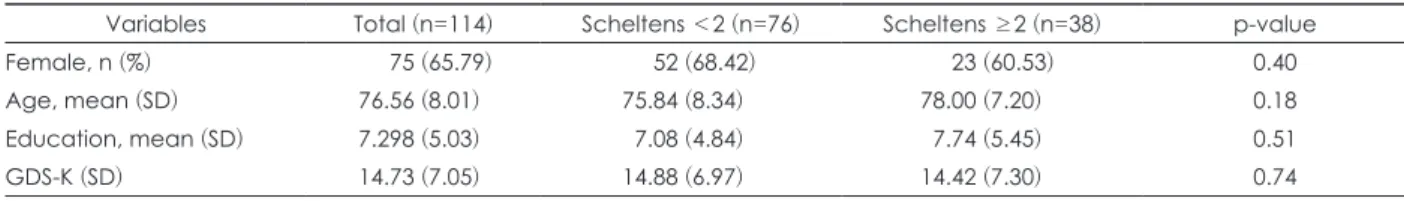
Scheltens 0~1군의 평균 연령은 75.84세, 평균 교육 연수 는 7.08년이었으며 Scheltens 2~4군의 평균 연령은 78.00세, 평균 교육 연수는 7.74년으로 통계적으로 유의한 차이를 나 타내지 않았다(각각 p=0.176, p=0.513). 두 군의 성별 및 GDS-K (p=0.749)의 평균 점수 또한 유의한 차이를 보이지 않았다. Table 1에 114명의 환자에 대한 기본적인 특성을 제 시하였다. K-NPI를 시행한 63명의 피험자들의 경우도 두 군 사이의 평균 연령, 평균 교육 연수, 성별 및 평균 GDS-K 점수가 통계적으로 유의한 차이를 보이지 않았다(Table 2).
2. 내측두엽 위축의 정도에 따른 중증도 및 영역별 인지기 능과 BPSD의 차이
중증도를 평가하는 CDR의 경우 Scheltens 척도 0~1군과 Scheltens 척도 2~4군 사이에서 유의한 차이가 있었으나(p=
0.000), GDS의 경우 두 군 사이에서 유의한 점수 차이가 없 었다(p=0.915).
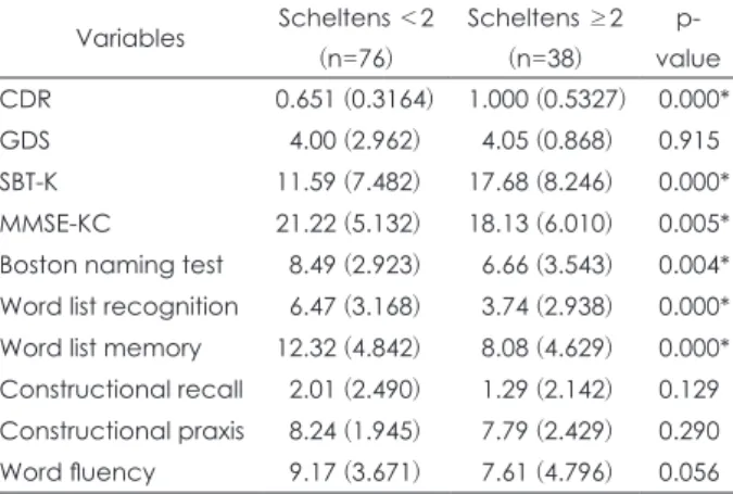
인지기능 항목 중에서 CERAD-K의 하위 항목인 단어 목 록 회상 검사(word list recall)의 경우 등분산의 검정이 기각 되어 평가 항목에서 제외되었다. 인지기능 항목 중에서 두 군 사이에 유의한 점수 차이를 보인 항목은 SBT-K (p=0.000) 이었으며 CERAD-K 항목 중에서는 MMSE-KC (p=0.005), 단축형 보스턴 이름대기 검사(modified Boston naming test, p=0.004), 단어 목록 재인 검사(word list recognition, p=0.000) 단어 목록 기억 검사(word list memory, p=0.000)이었다. 이 외에 CERAD-K 항목 중 구성 회상 검사(constructional re- call, p=0.129), 구성 행동 검사(constructional praxis, p=
0.290), 언어 유창성 검사(word fluency, p=0.056)에서는 두 군 사이에 유의한 차이가 나타나지 않았다(Table 3). BPSD 의 경우 K-NPI를 수행한 63명을 대상으로 두 군 사이의 평 균을 비교하였으나 K-NPI의 총점 및 하위항목 점수 모두 두 군 사이에서 유의한 차이를 보이지 않았다(Table 4).
고 찰
본 연구는 알츠하이머형 치매 및 경도인지장애 환자군에 서 나타나는 내측두엽 위축의 정도를 시각 기반 척도 중 하 나인 Scheltens 척도를 통해 측정하고, 내측두엽 위축의 정 도와 인지기능 및 BPSD 간의 관계를 확인해보고자 하였다.
Scheltens 척도는 내측두엽 위축을 평가하기 위한 시각 기
Table 1. Demographic characteristics of patients with neurocognitive function test scores (n=114)
Variables Total (n=114) Scheltens <2 (n=76) Scheltens ≥2 (n=38) p-value
Female, n (%) 75 (65.79) 52 (68.42) 23 (60.53) 0.40
Age, mean (SD) 76.56 (8.01) 75.84 (8.34) 78.00 (7.20) 0.18
Education, mean (SD) 7.298 (5.03) 7.08 (4.84) 7.74 (5.45) 0.51
GDS-K (SD) 14.73 (7.05) 14.88 (6.97) 14.42 (7.30) 0.74
* : Significant at p<0.05 by Chi-square test or independent t-test. GDS-K : the Korean version of the Geriatric Depression Scale
Table 2. Demographic characteristics of patients with K-NPI scores (n=63)
Variables Total (n=63) Scheltens <2 (n=40) Scheltens ≥2 (n=23) p-value
Female, n (%) 43 (68.25) 27 (67.50) 16 (69.57) 0.87
Age, mean (SD) 77.11 (8.30) 75.80 (8.97) 79.39 (6.54) 0.10
Education, mean (SD) 6.76 (4.40) 7.08 (4.30) 6.22 (4.60) 0.46
GDS-K (SD) 15.32 (7.65) 15.63 (7.87) 14.78 (7.40) 0.68
* : Significant at p<0.05 by Chi-square test or independent t-test. GDS-K : the Korean version of the Geriatric Depression Scale
반 척도 중 가장 대표적인 것이다. 이는 1992년 Scheltens 등26) 에 의해 고안된 것으로 측뇌실의 맥락막틈새, 측두각, 그리 고 해마의 높이를 0~4점 척도로 평가하는 방식이다. 높은 점 수일수록 위축의 정도가 더 심함을 의미한다. 내측두엽 위축 의 시각적 평가(visual assessment of Medial Temporal lobe Atrophy, vaMTA)의 유용성에 대해서는 다양한 연구 결과 가 제시되었는데, Ten Kate 등27) 및 Harper 등28)은 알츠하이 머형 치매를 예측할 수 있는 생체표지자로서 vaMTA의 진단 적 유용성을 보고하였다.
본 논문에서는 Scheltens 척도에 따라 나눈 두 군에서 GDS 점수는 유의한 차이를 보이지 않았는데, 내측두엽 위축의 정 도에 따라 GDS 점수의 차이 여부를 알아본 연구는 많지 않 아 본 논문의 결과와 비교하는 것이 어려웠다. 반면 CDR 점
수는 유의한 차이를 보였는데, 이는 CDR을 이용해 진단된 경도인지장애와 내측두엽 위축 점수 사이의 연관성을 보였던 Dhikav 등29)의 연구와 유사한 결과이다. 한편 Persson 등30) 은 알츠하이머형 치매 및 경도인지장애 환자에서 vaMTA와 CDR-SB 사이에 연관성을 발견하기 어렵다는 결과를 발표 했는데, 이와 같이 대비되는 결과가 나타나는 이유는 CDR 점수의 경우 기억력에 가중치가 적용되는 반면 CDR-SB 점 수의 경우 모든 영역에 걸쳐 동일한 가중치가 적용31)되므로 계산 방식에서 차이가 나타나기 때문인 것으로 보인다.
인지기능 평가 검사 중 두 군 사이에 유의한 점수 차이를 보인 항목은 SBT-K, 그리고 CERAD-K의 하위 항목 중 MMSE-KC, 보스턴 이름대기 검사, 단어 목록 재인 검사, 단어 목록 기억 검사였고 그 외의 다른 검사들에서는 유의 한 차이를 보이지 않았다.
기억은 크게 삽화 기억(episodic memory), 의미 기억(se- mantic memory), 작업 기억(working memory), 절차 기억 (procedural memory)으로 이루어져 있는데 내측두엽은 특 히 삽화 기억과 밀접한 관련성을 가진다.32) 내측두엽은 구조 적으로 CA 구역(CA fields), 치아이랑(dentate gyrus), 구상 회복합체(subicular complex)를 포함하는 해마 영역 그리고 인접 후각주위피질(perirhinal cortex), 내후각피질, 해마곁 피질(parahippocampal cortex)로 구성되어 있으며, 이 부위 들은 기억의 등록 및 공고화를 담당하는 것으로 알려져 있 다.9) 알츠하이머형 치매 환자의 인지기능과 뇌의 변화를 조 사한 과거 여러 연구에서 내측두엽 위축, 특히 해마의 위축 은 전반적 인지 상태(general cognitive status) 및 삽화 기억 의 저하와 연관이 있다는 일관된 결과를 보여 왔다. 최린 등19) 은 2점을 절단점으로 나눈 MTA 0~1군과 MTA 2~4군 사이
Table 4. Comparison of total score and subset score of K-NPI according to the degree of MTA (n=63)
Variables Scheltens <2 (n=40) Scheltens ≥2 (n=23) p-value
Item 1 (delusions) 1.25 (3.38) 1.04 (2.10) 0.80
Item 2 (hallucinations) 0.88 (2.691) 1.17 (2.37) 0.66
Item 3 (agitation/aggression) 1.70 (2.92) 1.83 (2.81) 0.87
Item 4 (depression) 1.88 (2.67) 1.48 (2.27) 0.55
Item 5 (anxiety) 2.00 (3.34) 1.00 (1.382) 0.18
Item 6 (elation/euphoria) 0.38 (1.51) 0.22 (0.60) 0.64
Item 7 (apathy/indifference) 2.65 (4.14) 2.09 (2.75) 0.56
Item 8 (disinhibition) 1.85 (3.49) 0.65 (1.37) 0.12
Item 9 (irritability) 2.45 (3.66) 1.26 (2.24) 0.16
Item 10 (aberrant motor behavior) 1.23 (3.13) 1.30 (2.36) 0.92
Item 11 (sleep and nightime behavior disorders) 2.05 (3.57) 2.04 (2.71) 0.99
Item 12 (appetite and eating disorders) 2.30 (3.50) 1.96 (2.84) 0.69
Total score 20.60 (21.99) 16.04 (14.61) 0.38
* : Significant at p<0.05 by independent t-test. K-NPI : The Korean version of the Neuropsychiatric Inventory Table 3. Comparison of CDR, GDS, SBT-K, MMSE-KC, and other
CERAD-K subtest score according to the degree of MTA (n=114) Variables Scheltens <2
(n=76)
Scheltens ≥2 (n=38)
p- value
CDR 0.651 (0.3164) 1.000 (0.5327) 0.000*
GDS 4.00 (2.962) 4.05 (0.868) 0.915
SBT-K 11.59 (7.482) 17.68 (8.246) 0.000*
MMSE-KC 21.22 (5.132) 18.13 (6.010) 0.005*
Boston naming test 8.49 (2.923) 6.66 (3.543) 0.004*
Word list recognition 6.47 (3.168) 3.74 (2.938) 0.000*
Word list memory 12.32 (4.842) 8.08 (4.629) 0.000*
Constructional recall 2.01 (2.490) 1.29 (2.142) 0.129 Constructional praxis 8.24 (1.945) 7.79 (2.429) 0.290 Word fluency 9.17 (3.671) 7.61 (4.796) 0.056
* : Significant at p<0.05 by independent t-test. CDR : Clinical Dementia Rating, GDS : Global Deterioration Scale, SBT-K : the Korean version of Short Blessed Test, MMSE-KC : Mini-Mental State Examination in the Korean version of the Consortium to Establish a Registry for Alzheimer’s Disease (CERAD) assessment packet
에서 MMSE 및 단어 목록 재인 검사 항목의 유의한 차이가 나타남을 보였다. Laakso 등33)은 알츠하이머형 치매 환자에 서 좌측 해마의 위축이 낮은 MMSE 점수와 유의한 상관관 계를 가지고 있음을 보였으며, Hsu 등13)은 내측두엽 위축과 낮은 MMSE 점수 사이의 연관성을 발표하였다. Stout 등34) 은 알츠하이머형 치매 환자에서 내측두엽의 위축이 심할수 록 삽화 기억능력이 떨어진다는 것을 보였다. Beck 등35)은 경 도인지장애 및 알츠하이머형 치매 환자에서 삽화 기억 기능 을 담당하는 해마 및 내후각피질이 유의미하게 위축된다고 발표하였다. Dos Santos 등36)은 CERAD를 이용해 인지기 능 저하와 내측두엽 위축 사이에 연관성이 있음을 보였다.
SBT의 경우 직접적으로 내측두엽 위축과의 연관성을 보인 연구는 드물었으나, 많은 연구들에서 치매 진단 시 높은 민 감성과 특이성을 보였고,37) MMSE와 높은 상관관계를 보였 다. 이러한 결과들은 본 연구에서의 결과와 상응하는 면이 많다고 볼 수 있겠다.
한편 K-NPI 점수를 통해 측정된 BPSD의 정도는 두 군 사 이에 유의한 차이를 보이지 않았다. K-NPI의 경우 총점뿐 아니라 각각의 세부항목 점수도 비교하였는데, 세부항목 점 수 역시 두 군 사이에 차이를 나타내지 않았다. 기존의 논문들 은 주로 백질고강도신호38) 또는 기저신경절(basal ganglia),39) 전두엽(frontal lobe)의 병변40) 등과 BPSD의 연관성을 다루 었는데 이들 병변의 심각도와 BPSD는 양의 상관관계를 나 타냈다. BPSD와 내측두엽 위축의 연관성을 다룬 연구는 많 지 않았는데, Burns 등10)은 내측두엽 위축과 공격성 사이에 연관성이 있음을 보였으며 Garcia-Alberca 등11)은 Schel- tens 척도에 의해 측정된 내측두엽 위축의 정도가 심할수록 무감동 및 탈억제와 같은 BPSD 관련 증상의 빈도가 높다는 것을 밝혔다. 반면, Staekenborg 등12)은 내측두엽 위축과 BPSD 사이에서 연관성을 발견하지 못했다. Hsu 등13)은 내 측두엽 위축과 후두피질 위축(posterior atrophy)의 차이를 언급하면서, 내측두엽 위축은 MMSE와, 후두피질 위축은 NPI 중 초조/공격성 소항목과 연관되어 있다는 것을 보이 기도 하였는데, 이는 MMSE로 대표되는 인지기능의 이상과 NPI로 대표되는 BPSD 중 전자만이 내측두엽 위축과의 관 련성을 보였던 본 연구의 결과와 상응하는 면이 많다.
본 연구의 제한점으로는 첫째, 시각 기반 척도의 특성상 정 량적 평가에 비해 정확도가 낮을 수밖에 없었다는 점이다. 둘 째, 연구 대상이 한 병원의 환자들에만 국한되었기 때문에 모집단 자체가 편향되어 있을 수 있다는 점이다. 셋째, 다른 뇌혈관 질환 및 전반적 뇌의 퇴행 정도가 고려되지 않은 상 태에서 Scheltens 척도만을 사용했던 본 연구의 특성상 뇌의
위축의 정도를 충분하게 반영하지 못했다는 것이다.
그럼에도 불구하고, 본 연구는 임상의들이 비교적 간편하 게 사용할 수 있는 시각 기반 척도를 이용하여 치매 환자에 서 나타나는 인지기능 이상 및 BPSD와 뇌의 구조적 변화 사이의 연관성을 확인할 수 있었다는 점에서 큰 의의를 가 진다. 내측두엽 위축을 평가할 때 전용 소프트웨어를 사용해 결과를 수치화하는 것은 진단적 정밀성을 높인다는 장점을 가지지만, 분석을 위해 전문 인력이 필요하며 장시간이 소요 된다는 제한이 있다. 반면 임상의가 직접 뇌 영상 검사 결과 를 보면서 수행하는 시각 기반 정성 평가는 적절한 훈련 뒤 에 임상에 간편하게 적용할 수 있다는 장점을 갖고 있다. 연 구에 따르면 시각 기반 척도는 복셀 기반 형태 계측(voxel- based morphometry)과 같은 정량적인 평가 결과와 비교적 유사한 정확도를 보이고 있으며,41) 평가를 수행하는 사람이 훈련을 통해 진단의 정확도를 개선시킬 수 있고28) 연구 재현 성이 높다는 면에서 매우 유용한 진단적 도구가 될 수 있다.
이와 같은 시각 기반 척도를 이용하면 알츠하이머형 치매 환 자 개개인별로 변화의 정도를 손쉽게 평가할 수 있으며, 임 상 의사간 뇌 영상 결과에 대한 정보의 상호 교류가 용이할 것이다. 본 연구의 결과 내측두엽 위축은 인지기능 저하와 유의한 상관관계를 보인 반면 BPSD와는 유의한 상관관계 를 보이지 않았다. 따라서 우리는 Scheltens 척도를 통해 시 각적으로 퇴행성 변화를 관찰함으로써 임상 실제에서 인지 기능 저하 환자들의 진단 및 예후 평가에 도움을 받을 수 있 을 것으로 기대할 수 있겠다.
Conflicts of Interest
The authors have no financial conflicts of interest.
REFERENCES
(1) Lyketsos CG, Lopez O, Jones B, Fitzpatrick AL, Breitner J, DeKosky S. Prevalence of neuropsychiatric symptoms in de- mentia and mild cognitive impairment: Results from the car- diovascular health study. Jama 2002;288:1475-1483.
(2) Piccininni M, Di Carlo A, Baldereschi M, Zaccara G, Inzi- tari D. Behavioral and psychological symptoms in Alzheim- er’s disease: Frequency and relationship with duration and se- verity of the disease. Dement Geriatr Cogn Disord 2005;19:
276-281.
(3) Sadock BJ, Sadock VA, Ruiz P. Kaplan and Sadock’s Synop- sis of Psychiatry: Behavioral Sciences/Clinical Psychiatry. 11th ed. Philadelphia: Wolters Kluwer;2016. p.704-740.
(4) Yilmaz R, Pilotto A, Roeben B, Preische O, Suenkel U, Heinzel S, Metzger FG, Laske C, Maetzler W, Berg D.
Structural Ultrasound of the Medial Temporal Lobe in Al- zheimer’s Disease. Ultraschall Med 2017;38:294-300.
(5) Braak H, Braak E. Neuropathological stageing of Alzheim- er-related changes. Acta Neuropathol 1991;82:239-259.
(6) Overdorp EJ, Kessels RP, Claassen JA, Oosterman JM. Cog- nitive impairments associated with medial temporal atrophy and white matter hyperintensities: an MRI study in memory clinic patients. Front Aging Neurosci 2014;6:98.
(7) Jonker C, Launer LJ, Hooijer C, Lindeboom J. Memory com- plaints and memory impairment in older individuals. J Am Geriatr Soc 1996;44:44-49.
(8) Krasuski JS, Alexander GE, Horwitz B, Daly EM, Murphy DG, Rapoport SI, Schapiro MB. Volumes of medial temporal lobe structures in patients with Alzheimer’s disease and mild cognitive impairment (and in healthy controls). Biol Psychia- try 1998;43:60-68.
(9) Squire LR, Stark CE, Clark RE. The medial temporal lobe.
Annu Rev Neurosci 2004;27:279-306.
(10) Burns A, Jacoby R, Levy R. Psychiatric phenomena in alzheim- er’s disease. Iv: Disorders of behaviour. Br J Psychiatry 1990;
157:86-94.
(11) García-Alberca JM, Florido M, Cáceres M, Sánchez-Toro A, Lara JP, García-Casares N. Medial temporal lobe atrophy is independently associated with behavioural and psychological symptoms in Alzheimer’s disease. Psychogeriatrics 2019;19:
46-54.
(12) Staekenborg SS, Gillissen F, Romkes R, Pijnenburg YA, Barkhof F, Scheltens P, van der Flier WM. Behavioural and psychological symptoms are not related to white matter hy- perintensities and medial temporal lobe atrophy in Alzheim- er’s disease. Int J Geriatr Psychiatry 2008;23:387-392.
(13) Hsu JL, Lee WJ, Liao YC, Lirng JF, Wang SJ, Fuh JL. Pos- terior Atrophy and Medial Temporal Atrophy Scores Are As- sociated with Different Symptoms in Patients with Alzheim- er’s Disease and Mild Cognitive Impairment. PLoS One 2015;
10:e0137121.
(14) McKhann G, Drachman D, Folstein M, Katzman R, Price D, Stadlan EM. Clinical diagnosis of Alzheimer’s disease:
report of the Nincds-Adrda Work Group under the auspices of Department of Health and Human Services Task Force on Al- zheimer’s Disease. Neurology 1984;34:939-944.
(15) Winblad B, Palmer K, Kivipelto M, Jelic V, Fratiglioni L, Wahlund LO, Nordberg A, Bäckman L, Albert M, Almkvist O, Arai H, Basun H, Blennow K, de Leon M, DeCarli C, Erkinjuntti T, Giacobini E, Graff C, Hardy J, Jack C, Jorm A, Ritchie K, van Duijn C, Visser P, Petersen RC. Mild cog- nitive impairment--beyond controversies, towards a consen- sus: report of the International Working Group on Mild Cogni- tive Impairment. J Intern Med 2004;256:240-246.
(16) Claus JJ, Staekenborg SS, Holl DC, Roorda JJ, Schuur J, Koster P, Tielkes CEM, Scheltens P. Practical use of visual medial temporal lobe atrophy cut-off scores in Alzheimer’s disease: Validation in a large memory clinic population. Eur Radiol 2017;27:3147-3155.
(17) Ferreira D, Cavallin L, Larsson EM, Muehlboeck JS, Me- cocci P, Vellas B, Tsolaki M, Kłoszewska I, Soininen H, Lovestone S, Simmons A, Wahlund LO, Westman E; Add- NeuroMed consortium and the Alzheimer’s Disease Neu- roimaging Initiative. Practical cut-offs for visual rating scales of medial temporal, frontal and posterior atrophy in alzheim- er’s disease and mild cognitive impairment. J Intern Med 2015;
278:277-290.
(18) Velickaite V, Ferreira D, Cavallin L, Lind L, Ahlström H, Kilander L, Westman E, Larsson EM. Medial temporal lobe atrophy ratings in a large 75-year-old population-based cohort:
gender-corrected and education-corrected normative data. Eur Radiol 2018;28:1739-1747.
(19) Choi L, Joo SH, Lee CU, Paik IH. Association between Global Cortical Atrophy, Medial Temporal Atrophy, White Matter Hyperintensities and Cognitive Functions in Korean Alzheimer’s Disease Patients. Korean J Biol Psychiatry 2015;
22:140-148.
(20) Morris JC. The Clinical Dementia Rating (Cdr): current ver- sion and scoring rules. Neurology 1993;43:2412-2414.
(21) Reisberg B, Ferris SH, de Leon MJ, Crook T. Global Dete- rioration Scale (Gds). Psychopharmacol Bull 1988;24:661-663.
(22) Fillenbaum GG, Heyman A, Wilkinson WE, Haynes CS.
Comparison of two screening tests in Alzheimer’s disease.
The correlation and reliability of the Mini-Mental State Ex- amination and the modified Blessed test. Arch Neurol 1987;
44:924-927.
(23) Lee DY, Yoon JC, Lee KU, Jhoo JH, Kim KW, Lee JH, Woo JI. Reliability and Validity of the Korean Version of Short Blessed Test(Sbt-K) as a Dementia Screening Instrument. J Korean Neuropsychiatr Assoc 1999;38:1365-1375.
(24) Lee JH, Lee KU, Lee DY, Kim KW, Jhoo JH, Kim JH, Lee KH, Kim SY, Han SH, Woo JI. Development of the Korean version of the Consortium to Establish a Registry for Alzheim- er’s Disease Assessment Packet (Cerad-K): clinical and neu- ropsychological assessment batteries. J Gerontol B Psychol Sci Soc Sci 2002;57:47-53.
(25) Cummings JL, Mega M, Gray K, Rosenberg-Thompson S, Carusi DA, Gornbein J. The Neuropsychiatric inventory:
comprehensive assessment of psychopathology in dementia.
Neurology 1994;44:2308-2314.
(26) Persson K, Barca ML, Cavallin L, Brækhus A, Knapskog AB, Selbæk G, Engedal K. Comparison of automated volu- metry of the hippocampus using Neuroquant® and visual as- sessment of the medial temporal lobe in Alzheimer’s disease.
Acta Radiol 2018;59:997-1001.
(27) Ten Kate M, Barkhof F, Boccardi M, Visser PJ, Jack CR Jr, Lovblad KO, Frisoni GB, Scheltens P; Geneva Task Force for the Roadmap of Alzheimer’s Biomarkers. Clinical valid- ity of medial temporal atrophy as a biomarker for Alzheimer’s disease in the context of a structured 5-phase development framework. Neurobiol Aging 2017;52:167-182.
(28) Harper L, Barkhof F, Fox NC, Schott JM. Using visual rat- ing to diagnose dementia: a critical evaluation of MRI atrophy scales. J Neurol Neurosurg Psychiatry 2015;86:1225-1233.
(29) Dhikav V, Sethi M, Anand KS. Medial temporal lobe atrophy in Alzheimer’s disease/mild cognitive impairment with de- pression. Br J Radiol 2014;87:20140150.
(30) Persson K, Barca ML, Eldholm RS, Cavallin L, Šaltytė Benth J, Selbæk G, Brækhus A, Saltvedt I, Engedal K. Vi- sual Evaluation of Medial Temporal Lobe Atrophy as a Clini- cal Marker of Conversion from Mild Cognitive Impairment to Dementia and for Predicting Progression in Patients with Mild Cognitive Impairment and Mild Alzheimer’s Disease. Dement Geriatr Cogn Disord 2017;44:12-24.
(31) Postuma RB, Lang AE, Gagnon JF, Pelletier A, Montplai- sir JY. How does parkinsonism start? Prodromal parkinson- ism motor changes in idiopathic REM sleep behaviour disor- der. Brain 2012;135:1860-1870.
(32) Matthews BR. Memory dysfunction. Continuum (Minneap Minn) 2015;21:613-626.
(33) Laakso MP, Soininen H, Partanen K, Helkala EL, Harti- kainen P, Vainio P, Hallikainen M, Hänninen T, Riekkin- en PJ Sr. Volumes of hippocampus, amygdala and frontal lobes in the MRI-based diagnosis of early Alzheimer’s dis- ease: correlation with memory functions. J Neural Transm Park Dis Dement Sect 1995;9:73-86.
(34) Stout JC, Bondi MW, Jernigan TL, Archibald SL, Delis DC, Salmon DP. Regional cerebral volume loss associated with verbal learning and memory in dementia of the Alzheim- er type. Neuropsychology 1999;13:188-197.
(35) Beck IR, Gagneux-Zurbriggen A, Berres M, Taylor KI, Monsch AU. Comparison of verbal episodic memory mea- sures: consortium to establish a registry for Alzheimer’s dis- ease--Neuropsychological Assessment Battery (Cerad-Nab) versus California Verbal Learning Test (CVLT). Arch Clin Neuropsychol 2012 27:510-519.
(36) Dos Santos V, Thomann PA, Wüstenberg T, Seidl U, Essig M, Schröder J. Morphological cerebral correlates of CERAD test performance in mild cognitive impairment and Alzheim- er’s disease. J Alzheimers Dis 2011;23:411-420.
(37) Lorentz WJ, Scanlan JM, Borson S. Brief screening tests for dementia. Can J Psychiatry 2002;47:723-733.
(38) Kandiah N, Chander R, Zhang A, Yee CC. Cerebral white matter disease is independently associated with BPSD in Al- zheimer’s disease. J Neurol Sci 2014;337:162-166.
(39) Palmqvist S, Sarwari A, Wattmo C, Bronge L, Zhang Y, Wah- lund LO, Nagga K. Association between subcortical lesions and behavioral and psychological symptoms in patients with Alzheimer’s disease. Dement Geriatr Cogn Disord 2011;32:
417-423.
(40) Ogama N, Sakurai T, Saji N, Nakai T, Niida S, Toba K, Umegaki H, Kuzuya M. Frontal White Matter Hyperinten- sity Is Associated with Verbal Aggressiveness in Elderly Wom- en with Alzheimer Disease and Amnestic Mild Cognitive Im- pairment. Dement Geriatr Cogn Dis Extra 2018;8:138-150.
(41) Kang DW, Lim HK. Current Knowledge and Clinical Appli- cation of Brain Imaging in Alzheimer’s Disease. J Korean Neu- ropsychiatr Assoc 2018;57:12-22.
국 문 초 록
연구목적
본 연구의 목적은 알츠하이머형 치매 및 경도인지장애 환자에서 내측두엽 위축 정도의 차이에 따른 중증도, 신경인지기능 및 행동심리증상을 비교하는 것이다.
방 법
본 연구는 단면 연구로서, 알츠하이머형 치매 및 경도인지장애로 진단받은 114명을 대상으로 하였다. 내측두 엽 위축은 표준화된 시각 기반 척도(Scheltens scales)에 의해 평가되었으며, 대상군은 두 그룹으로 분류되었다.
중증도는 임상치매평가척도(Clinical dementia rating, CDR)와 전반적 퇴화 척도(Global deterioration scale, GDS)로 평가되었으며 신경인지기능은 한국어판 간이 블레스드 검사(The Korean version of the short blessed test, SBT-K), 한국형 CERAD (The Korean version of the consortium to establish a registry for Alzheimer’s disease, CERAD-K), 행동심리증상은 한국형 신경정신행동검사(The Korean version of the Neuropsychiatric Inventory, K-NPI)로 평가되었다. 내측두엽 위축의 정도에 따른 중증도와 신경인지기능 및 행동심리증상의 차 이를 분석하기 위해 독립표본 t-test를 시행하였다.
결 과
내측두엽 위축의 중증도가 높은 군은 낮은 군에 비하여 CDR, SBT-K, 그리고 CERAD-K의 하부항목 중 네 가지 항목에서 유의하게 낮은 점수를 보였다(p<0.05).
결 론
내측두엽 위축과 중증도 및 신경인지기능 사이에는 유의한 양의 상관관계가 나타났으나 행동심리증상과는 유의한 상관관계가 나타나지 않았다. 내측두엽 위축의 평가는 인지장애의 진단과 치료에 잠재적 이점을 제공 할 것으로 생각된다. 행동심리증상과 뇌구조의 연관성에 대해서는 추가적인 연구가 필요할 것으로 보인다.
중심 단어:알츠하이머형 치매·경도인지장애·내측두엽 위축·신경인지기능·행동심리증상.