인제대학교 의과대학 내과학교실, 방사선학교실
구호석1, 김태균1, 박성길1, 최상분1, 김애란1, 최상봉1, 정 훈1, 박이내1, 허진원1, 이혁표1, 염호기1, 최수전1, 최석진2, 이현경1
A Case of Tuberculous Pleural Effusion Developed after Percu- taneous Needle Biopsy of a Solitary Pulmonary Nodule
Ho Seok Koo, M.D.1, Tae Kyun Kim, M.D.1, Sung Kil Park, M.D.1, Sang Bun Choi, M.D.1, Ae Ran Kim, M.D.1, Sang Bong Choi, M.D.1, Hoon Jung, M.D.1, I-Nae Park, M.D.1, Jin-Won Hur, M.D.1, Hyuk Pyo Lee, M.D.1, Ho-Kee Yum, M.D.1, Soo Jeon Choi, M.D.1, Suk-Jin Choi, M.D.2, Hyun-Kyung Lee, M.D.1
1Departments of Internal Medicine and 2Radiology, College of Medicine, Inje University, Busan, Korea
A tuberculous pleural effusion may be a sequel to a primary infection or represent the reactivation of pulmonary tuberculosis. It is believed to result from a rupture of a subpleural caseous focus in the lung into the pleural space.
It appears that delayed hypersensitivity plays a large role in the pathogenesis of a tuberculous pleural effusion. We encountered a 52 years old man with pleural effusion that developed several days after a CT guided percutaneous needle biopsy of a solitary pulmonary nodule. He was diagnosed with TB pleurisy. It is believed that his pleural effusion probably developed due to exposure of the parenchymal tuberculous focus into the pleural space during the percutaneous needle biopsy. This case might suggest one of the possible pathogeneses of tuberculous pleural effusion.
(Tuberc Respir Dis 2007; 63: 268-272)
Key Words: Tuberculous pleural effusion, Percutaneous needle biopsy, Solitary pulmonary nodule.
Address for correspondence: Hyun-Kyung Lee, M.D.
Department of Internal Medicine, Pusan Paik Hospital, College of Medicine, Inje University, 633-165,
Gae-gum-2-dong, Jin-gu, Busan, 614-735, Korea Phone: 82-51-890-6270, Fax: 82-51-892-0273 E-mail: [email protected]
Received: Aug. 7. 2007 Accepted: Aug. 30. 2007
서 론
결핵성 흉수는 폐의 건락성 병소가 흉막강내로 노 출되어 발생되는 것으로 알려져 있으며1, 지연성 과민 반응이 흉수발생에 관여함을 보여주는 여러 실험적 연구가 있었다2-6. 그러나 결핵성 흉수의 발생기전을 실제 임상상황에서 시사해 주는 증례는 거의 없었다.
저자들은 우연히 발견된 단일 폐결절의 진단을 위해 시행한 흉부 전산화단층촬영 유도하 경피 바늘생검 후 수일이 지나 발생한 결핵성 흉수 1예를 경험하여 보고한다.
증 례 환 자: 김 O 우, 남자 52세
주 소: 우측 흉통
현병력: 한 달 전 검진으로 시행한 흉부X-선에서 우연히 오른쪽 상부폐에 1.6 cm 크기의 단일 폐결절 이 발견되었다(Figure 1). 기침, 가래 호흡곤란 등의 호흡기 증상은 없었다. 흉부전산화단층촬영에서 결절 주변에 서너개의 작은 위성결절이 발견되었다. 흉부 전산화단층촬영 유도하에 경피적으로 종괴에 대한 바 늘생검을 시행하였다(Figure 2A, B). 조직검사에서는 단핵염증세포만 보였고, 결핵이나 악성의 증거가 없 어 외래추적 예정이었다. 환자는 조직검사 2주 후 우 측 흉통을 느껴 외래를 방문하였다.
과거력: 특이사항 없음
사회력: 음주력 없음, 흡연력: 30 갑년
이학적 소견: 혈압 110/70 ㎜Hg, 맥박수 분당 88회, 호흡수 분당 20회, 체온은 37℃였으며, 우측 폐하부에 호흡음이 감소되어 있었다.
검사실 소견: 혈액검사상 헤모글로빈 15.0 g/dL, 백 혈구 8,900×10^6/L, 혈소판 274×10^9/L, 단백질/알부 민 7.1/4.1 g/dL, 총빌리루빈 0.5 g/dL, AST/ALT 32/22 U/L, LDH 593 IU/L였다. 동맥혈가스검사는 pH 7.5, PaCO2 29.7 mmHg, PaO2 61.8 mmHg, HCO3
21.2 mEq/L, SaO2 95.7%였다. 흉수 세포 및 생화학검
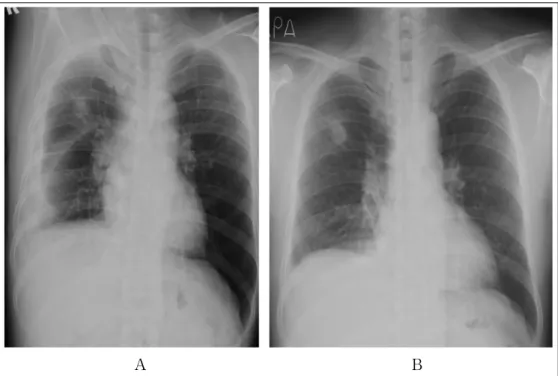
A B
Figure 2. CT-guided percutaneous biopsy. CT-guided biopsy was done in apical segment of right upper lobe with prone position (A, B).
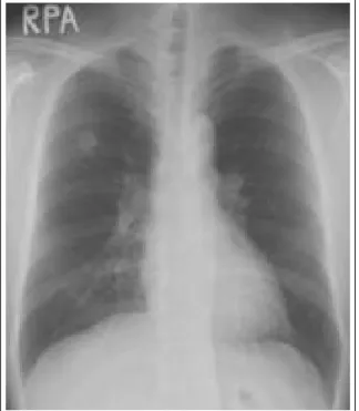
Figure 1. Chest X-ray (initial). Chest x-ray shows a well defined round nodule in right upper lobe.
사는 pH 8.0, 백혈구 6500/mm3(호중구 15%, 임파구 85%), 단백질 4.8 g/dL, LDH 736 IU/L, ADA 78 IU/L 이었다.
방사선 검사: 이전에 관찰되던 우상엽 단일폐결절 의 크기 및 모양에 변화는 없었으나 결절주변에 여러 개의 작은 경화성 병변이 관찰되었다(Figure 3A). 또 한 오른쪽 늑골횡격막각이 왼쪽에 비해 둔해진 소견
및 횡격막 상승소견이 관찰되어 오른쪽 흉수가 의심 되었다. 오른쪽 옆누운 자세에서 흉수의 존재를 확인 할 수 있었다(Figure 3B). 흉부전산화단층촬영에서 경피 생검바늘이 진행된 위치 좌우로 결절부터 흉막 까지 이르는 길쭉한 경화성병변이 관찰되었고, 역시 생검바늘이 지나간 위치 좌우로 삼각형 모양의 흉수 가 관찰되었다(Figure 4A-D).
치료 및 경과: 흉수 생화학 검사 및 세포검사에서 결핵성 흉수에 합당한 소견 보여 항결핵약제 투여를 시작하였다. 4개월 후 시행한 흉부 X선(Figure 5) 및 흉부 CT(Figure 6A, B)에서 우폐상엽의 단일 폐결절 은 그 크기가 변화 없었으나 결절주변의 경화성 병변 및 우측 흉수는 소실된 소견 보였다. 결핵치료 종료 6 개월 후 시행한 흉부X-선에서 단일 폐결절 크기 및 모양은 이전과 동일하였다.
고 찰
결핵성 흉수는 폐결핵의 5%를 차지하고 있으나, 20% 이상 나타난다는 보고도 있다. 대다수의 환자는 마른기침 및 흉막성 흉통을 호소하고 발열이 있는 경 우도 많다. 대부분의 환자에서 흉수 백혈구 분획은 림 프구가 50% 이상이다. 그러나 증상이 2주 이내일 때 백혈구 분획은 다형핵 백혈구가 더 많이 보일 수 있 다. 림프구는 아급성이나 만성 결핵성 흉수에서 우위 를 보이는 반면 급성흉수에서는 호중구가 우위를 보
A B
C D
Figure 4. Chest CT (2 weeks after biopsy). CT showes linear consolidation and pleural effusion along previous biopsy tract (A-D).
A B
Figure 3. Chest PA (A) and right decubitus view (B)(2 weeks after biopsy). Chest x-ray shows a lot of pleural fluid shifting in right thorax. Increased patch consolidation around the nodule on RUL.
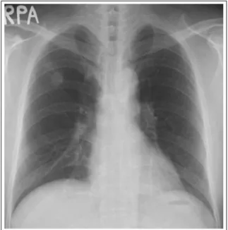
Figure 5. Chest PA (4 months later). Chest PA shows disappeared pleural effusion after anti-Tbc medi- cation.
A B
Figure 6. Chest CT (4 months after anti-tbc medication). Chest CT shows no interval change of previous SPN. But pleural effusion and consolidative lesions around the nodule were disappeared (A, B).
인다. 방사선학적으로 폐실질 결핵병변은 결핵성 흉수환자의 약 20%에서 발견된다. 환자의 42%에서 흉수배양 양성이며, 흉막조직검사의 64%에서 결핵에 합당한 소견을 보인다. ADA 값이 46에서 60 이상일 때는 결핵성 흉수 진단에95-97%의 예민도를 가진다7. 결핵성 흉수는 방사선학적으로 명백한 폐결핵의 증 거가 없이 나타날 때, 초 감염 후 6-12주 후에 발생하 는 원발성 폐결핵의 합병증이거나 재발성 결핵을 의 미할 수 있다1. 결핵성 흉수는 흉막강 아래 폐실질의
건락성 괴사 부위의 흉막강 내로의 파열로 인해 생기 는 것으로 생각된다. Stead 등은 결핵성 흉막염을 가 진 환자 15명을 수술하였을 때 12명에서 흉막염과 인 접한 폐실질에서 폐결핵병소를 발견하였다8. 이는 결 핵균항원에 대한 지연성 과민반응이 결핵성 흉수 발 생기전이라는 하나의 증거가 될 수 있다.
동물실험에서 결핵단백에 감작된 기니픽에 흉막강 내 튜버큘린 단백을 주입하였을 경우 삼출성 흉수가 48시간 내 발생하였고, 항림프구 혈청을 투여하였을 경우 흉수발생이 억제 되는 것으로 나타났다4,5. BCG 모형에서 대식세포는 2-5일째에 흉수 내에 풍부하게 되고6, 이 시기 후에는 림프구가 우세하게 되는데9, 이 때 결핵단백에 감작된 T-임파구는 흉수 내에 존재하 게 된다10. 결핵성 흉수 내에는 면역반응 세포와 세포 매개성 면역반응을 유도하는 물질이 풍부하며11. CD4, CD8 이 말초혈액에는 1:7 의 비율로 존재 하는데 반 해서 흉수 내에는 3:4의 비율로 존재한다11. 결핵성 흉 수에서 T-helper type cell은 지연성 과민반응을 일으 키고 대식세포를 활성화 시키는데, 결핵균을 억제하 기 위해서는 강력한 Th-1(IFN-γ 우위)가 필요하고, Th-2 cytokine인 IL-4에 의해 길항된다.
Cazzadori 등은 본 증례처럼 폐결절에 대한 경피적 바늘생검 17일 뒤 흉수가 발생한 1예를 보고하였다12. 저자들은 생검바늘을 제거하는 과정에서 감염된 폐조 직부위가 흉막에 노출되어 흉수가 발생했을 가능성을 언급하고 있다. 그러나 이 증례에서는 흉부X-선이나 전산화단층촬영을 제시하지 않고 있어 그 연관관계를
입증하기 어렵다고 생각된다. 본 증례에서는 검사 이 전 전산화단층촬영에서 결절주변 결핵의심병변이 있 었고, 경피생검 후 생검바늘이 지나간 자리 좌우로 흉 수가 발생한 것으로 생검바늘에 의한 결핵조직의 흉 막노출과 흉수발생의 연관성을 보다 강력히 의심해 볼 수 있겠다. 따라서 본 증례는 생검바늘에 의한 폐 실질 결핵병변의 흉막강 내로의 노출 후, 결핵성 흉수 가 발생 하는 과정을 보여주는 한 예라고 생각된다.
요 약
결핵성 흉수에는 결핵 단백에 감작된 T-림프구 가 존재하며 결핵 단백이 흉수에 노출되는 경우 지연성 과민 반응이 일어나 결핵성 흉막염이 발생하게 되는 것으로 알려져 있다. 저자들은 고립성 폐결절이 우연 히 발견된 52세 남자에서 흉부전산화단층촬영 유도 하 경피적 바늘생검 후 결핵성 흉막염이 발생한 예를 경험하였다. 지연성 과민반응이라는 결핵성 흉수 발 생기전에 부합하는 임상상황으로 생각하여 문헌고찰 과 함께 보고한다.
참 고 문 헌
1. Berger HW, Mejia E. Tuberculous pleurisy. Chest 1973;63:88-92.
2. Allen JC, Apicella MA. Experimental pleural effusion as a manifestation of delayed hypersensitivity to tuberculin PPD. J Immunol 1968;101:481-7.
3. Apicella MA, Allen JC. A physiologic differentiation between delayed and immediate hypersensitivity. J
Clin Invest 1969;48:250-9.
4. Leibowitz S, Kennedy L, Lessof MH. The tuberculin reaction in the pleural cavity and its suppression by antilymphocyte serum. Br J Exp Pathol 1973;54:
152-62.
5. Yamamoto S, Dunn CJ, Willoughby DA. Studies on delayed hypersensitivity pleural exudates in guinea pigs. Demonstration of substances in the cell-free exudate which cause inhibition of mononuclear cell migration in vitro. Immunology 1976;30:505-11.
6. Antony VB, sahn SA, Antony AC, Repine JE. Bacillus Calmette-Guerin-stimulated neutrophils release chemotaxins for monocytes in rabbit pleural space in vitro. J Clin Invest 1985;76:1514-21.
7. Riantawan P, Chaowalit P, Wongsangiem M, Rojanaraweewong P. Diagnostic value of pleural fluid adenosine deaminase in tuberculous pleuritis with reference to HIV coinfection and a bayesian analysis.
Chest 1999;116:97-103.
8. Stead WW, Eichenholz A, Stauss HK. Operative and pathologic findings in twenty-four patients with syndrome of idiopathic pleurisy with effusion, presu- mably tuberculous. Am Rev Tuberc 1955;71:473-502.
9. Widstrom O, Nilsson BS. Pleurisy induced by intrapleural BCG in immunized guinea pigs. Eur J Respir Dis 1982;63:425-34.
10. Fujiwara H, Tsuyuguchi I. Frequency of tuberculin reactive T-lymphocytes in pleural fluid and blood from patients with tuberculous pleurisy. Chest 1986;89:
530-2.
11. Ellner JJ, Barnes PF, Wallis RS, Modlin RL. The immunology of tuberculous pleurisy. Semin Respir Infect 1988;3:335-42.
12. Cazzadori A, Di Perri G, Marocco S, Carlotto A, Adami T, Concia E. Tuberculous pleurisy after percutaneous needle biopsy of a pulmonary nodule.
Respir Med 1994;88:477-8.