연구논문
1-Deoxynojirimycin 생산 균주 Bucillus subtilis MORI 3K-85의 단백질 분석
조용석1,3, 강경돈3, 박영식3, 이재연3, 김현수1, 육원정3, Shizuo George Kamita2, 황교열3, 성수일1,3*
Protein Analysis of Bacillus subtilis MORI 3K-85 with Reference to the Biosynthesis of 1-Deoxynojirimycin
Yong Seok Cho1,3, Kyung-Don Kang3, Young Shik Park3, Jae Yeon Lee3, Hyun Su Kim1, Won Jeong Yuk3, Shizuo George Kamita2, Kyo Yeol Hwang3, and Su-Il Seong1,3*
접수: 2011년 8월 31일 / 게재승인: 2011년 10월 22일
© 2011 The Korean Society for Biotechnology and Bioengineering
Abstract:1)In our previous study, we isolated and characterized a 1-deoxynojirimycin (DNJ)-producing bacterium, Bacillus subtilis MORI, from chungkookjang, a Korean traditional food. B. subtilis MORI was subjected to γ-irradiation and the resulting bacteria were screened for increased DNJ production.
A mutant was identified that produced 7.6 times more DNJ and named B. subtilis MORI 3K-85. In this study, the protein profiles of both strains were compared by one-dimensional and two-dimensional gel electrophoresis (1-DE and 2-DE, respectively) under both native and denaturing conditions.
The 1-DE native-PAGE and 1-DE SDS-PAGE analyses identified 5 and 7 bands, respectively, that were found at higher concentrations in B. subtilisMORI 3K-85 than in B.
subtilis MORI. Similarly, 2-DE analyses identified 20 protein spots which were found at higher concentrations in B. subtilis MORI 3K-85. The peptide mass profiles of these 20 proteins
1수원대학교 생명과학과
1Department of Life Science, The University of Suwon, Hwaseong-si, 445-743, Korea
Tel: +82-31-220-2483, Fax: +82-31-220-2483 e-mail: [email protected]
2캘리포니아주립대학교 (데이비스) 곤충학과
2Department of Entomology, University of California, Davis, CA 95616, USA
3(주)바이오토피아 부설생명과학연구소
3R&D center for Life Science, Biotopia Co., Ltd., Chuncheon 200-883, Korea
were analyzed by MALDI-TOF and compared with peptide sequences of B. subtilis and B. amyloliquefaciens in the MASCOT database. This screening suggested that three dehydrogenases, an aldolase, a synthetase, an isomerase, a reductase, and a peroxidase are elevated in B. subtilis MORI 3K-85. Based on this data, one or more of the elevated 8 enzymes might be related to the DNJ biosynthetic pathway.
Keywords: Bacillus subtilis, two-dimensional gelelectrophoresis, 1-deoxynojirimycin, α-glucosidase inhibitor, proteome
1. 서론
여러 종류의 식물과 미생물로부터 분리되고 있는 polyhydroxylated alkaloids는 당 내에 수개의 수산기를 갖고 산소가 질소로 치환되어 있는 구조적 특징을 갖는 물질로 지금까지 약 100여종이 알려져 있으며 이들은 다시 구조 상의 차이에 따라 piperidines, pyrrolidines, indolizidines, pyrrolizidines, nortropanes의 5계열로 분류된다 [1,2]. 이중 piperidine 계열은 monocycle의 6탄당 구조를 갖는 alkaloids 로 그의 대표적인 물질로는 1-deoxynojirimycin (DNJ)이 있 다. DNJ가 갖는 주요 생화학적 기능으로는 α-glucosidase 에 대한 효소 저해기능을 들 수 있는데 그 대표적인 사례 로는 체내 소장 내의 당 중간 소화산물인 maltose, sucrose, lactose 등 이당류의 glucose로의 전환 억제와 [1,3-5], 간염 바이러스 (HBV)나 AIDS 바이러스 (HIV) 및 송아지 설사
바이러스 (BVDV)와 같은 피막 바이러스의 glycoprotein 피막 형성에 필요한 glycosylation의 저해에 따른 바이러스의 증식 억제가 알려지고 있다 [6-11]. 이와 같은 DNJ의 α-glucosidase 저해작용을 이용하여 인슐린 비의존형 당뇨병 환자들을 위한 혈당강하제 개발 및 각종 인축의 바이러스 질병에 대한 항바 이러스제로의 이용 방안이 적극 검토되고 있다 [1,2,10,11].
이러한 DNJ의 산업적 이용에 대한 높은 기대에 부응하여 보다 경제적인 DNJ의 생산방법이 모색되고 있다. 현재까지 DNJ을 얻는 방법으로는 식물 추출법 [12], 화학적 합성법 [13], 미생물 발효법 [3,14-17] 등이 알려져 있다. 이중 식물 추출 법은 가장 오래 전부터 연구가 진행되어 왔으나 DNJ의 생산 성이 낮고 추출물 내에 유사 alkaloids 성분이 다량으로 함유 되어 있어 분리, 정제상의 어려움이 있으며 화학적 합성법은 합성 단계가 복잡하고 많은 이성질체들의 형성으로 단일 물 질로의 정제가 쉽지 않은 난점이 있다 [3]. 이에 비해 미생물 발효법은 생산공정이나 정제과정의 간소화가 가능하여 경제 적인 측면에서 가장 유리한 DNJ 생산방법이라 할 수 있다.
당 연구실은 한국전통식품에서 분리한 200개의 Bacillus sp.
균주들을 대상으로 α-glucosidase 저해활성이 우수하며 동시 에 DNJ 생산이 우수한 Bacillus subtilis MORI (KCCM- 10450)균주를 선발하였으며 [18], 나아가 이 균주에 감마 광선을 처리하여 DNJ 생산성이 7.6배 증가된 돌연변이 균주 B. subtilis MORI 3K-85의 획득에 성공하였다. 최근, 강 등 [19]은 MORI 3K-85 균주로부터 DNJ 생산에 관여 하는 3개의 유전자 gabT1, yktc1, gutB1을 확보하여 이 들 유전자의 putative transaminase, fructose phosphatase, dehydrogenase 기능을 토대로 glucose로부터 DNJ까지의 생합성 경로를 추정한 바 있다 [20]. 만약 이들 확보된 유전 자에 의한 DNJ 생합성 경로가 명확하게 밝혀진다면 유전 공학적 기법 등에 의해 large-scale의 DNJ overproduction이 가능할 수 있고 이는 결국 보다 효율적이며 경제적인 DNJ 의 생산도 가능하게 해줄 것으로 기대된다.
저자들은 Bacillus 균에서의 DNJ 생합성 경로를 밝히기 위한 연구의 일환으로 우선 균의 단백질 패턴을 조사하였 다. 즉, DNJ 생산 균주인 B. subtilis MORI와 이 균의 감마 선 처리에 의해 현저하게 DNJ 생산성이 증가된 돌연변이 균주 B. subtilis MORI 3K-85 간의 단백질 패턴의 차이를 1차원 및 2차원 전기영동으로 분석하고 두 균주 간 유의적 차이를 보이는 단백질들의 MALDI-TOF 분석 및 MASCOT 데이터베이스 검색을 통해 해당 단백질들을 동정하고 이들과 DNJ 생합성과의 관련성에 관해 고찰을 하였다.
2. 재료 및 방법
2.1. 균주 배양 및 시료조제
균의 단백질 분석을 위해 청국장에서 분리한 DNJ 생산 균주 B. subtilis MORI (KCCM-10450)와 이 균주에 감마광선 을 처리하여 DNJ 생산이 7.6배 증가된 돌연변이 균주 B.
subtilis MORI 3K-85를 사용하였다 [18]. 우선 두 균주를 각 각 YM broth 100 mL에 접종하고 37℃, 180 rpm으로 1일 간
배양하여 종균을 준비하였다. 다음 종균 배양액 10 mL을 YM broth 1 L에 접종하고 37℃, 180 rpm에서 1일 간 배양 후 원심분리 (4,500 × g, 4℃, 20분)하여 균체를 수집하였다.
이 균체에 PBS buffer (0.5 M sodium phosphate, 0.5 M NaCl, pH 6.8) 50 mL를 가하여 현탁하고 동일한 조건으로 원심분리하였다. 이 과정을 3회 반복한 후 얻어진 균체를 최종 PBS buffer 5 mL로 현탁하여 -20℃에 보관, 필요에 따라 꺼내어 전기영동 시료로 사용하였다.
2.2. 1차원 전기영동 (1-DE)
두 균주 간의 단백질 차이는 Bollag 등 [21]의 방법에 따라 Native-PAGE와 SDS-PAGE 분석에 의해 비교하였다. Native- PAGE를 실시하기 위해, 냉동보관 중인 시료 균주를 해동하 여 그의 현탁 균체액 1 mL를 sonicator (Sonics & Materials, USA)로 파쇄한 후 Bradford법으로 시료 내의 단백질을 정량 하였다. 시료의 제조는 동량의 균체 파쇄액 각 10 μL에 증 류수 70 μL와 5 × sample buffer (60 mM Tris-HCl pH 6.7, 25% glycerol, 0.1% bromophenol blue) 20 μL를 혼합한 후 3.4% polyacrylamide stacking gel과 7.5% polyacrylamide separation gel을 이용하여 전기영동을 실시하였다. 전기영동 이 완료된 gel은 0.1% coomassie blue R-250, 50% methanol, 10% glacial acetic acid의 혼합액에서 3시간 염색하고, 50% methanol과 10% glacial acetic acid의 혼합액에서 3회 탈색을 하였다. SDS-PAGE를 실시하기 위해, 냉동보관 중인 시료 균주를 해동하고 그의 현탁 균체액은 UV-VIS spectrophotometer (UV mini-1240, Shimadzu, Japan)를 이용하여 같은 농도가 되도록 조정하였다. 시료의 조제는 동량의 균체 시료 각 10 μL에 증류수 70 μL와 5 × sample buffer (2% SDS, 60 mM Tris-HCl pH 6.7, 25% glycerol, 14.4 mM 2-mercaptoethanol, 0.1% bromophenol blue) 20 μL 를 혼합한 다음 100℃에서 5분간 열처리하여 준비하였다.
SDS 전기영동은 4.5% polyacrylamide stacking gel과 12%
polyacrylamide separation gel을 이용하여 실시하였다.
2.3. 2차원 전기영동 (2-DE)
2-DE 시료의 조제를 위해 우선 PBS buffer 현탁 균체액 1 mL을 e-tube에 옮기고 원심분리 (5,900 × g, 4℃, 5분)하여 상등액을 제거하였다. 이 e-tube에 100 mM EDTA 5 μL, sample lysis buffer [8 M urea, 60 mM dithiothreitol, 2%
immobilized pH gradient (IPG) buffer 4-7, 4% (3-[(3- Cholamidopropyl)-Dimethylammonio]-1-Propane Sulfonate) (CHAPS)] 495 μL를 넣고 sonicator (Sonics & Materials, USA)를 이용하여 세포를 파쇄한 후 원심분리 (16,000 × g, 4℃, 60분)하여 얻어진 상등액을 -20℃에 보관하였다. 시료 내의 단백질 정량은 Bradford법을 이용하였다. 등전점전기 영동 (isoelectric focusing: IEF)은 Amersham Biosciences 의 2-DE principles and Methods [22]에 따라 다음과 같이 수행하였다. 우선 Immobiline Drystrip gel (18 cm, pH 4-7, Amersham Biosciences, Sweden)에 8 M urea, 0.2%
dithiothreitol (DTT), 0.5% IPG buffer, 0.5% CHAPS 및 0.002% bromophenol blue가 함유된 rehydration buffer
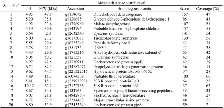
Table 1. Potential identity of protein spots from B. subtilis MORI and MORI 3K-85 by comparison with the B. subtilis database
Spot No.a Mascot database search result
pI MW (kDa) Accessionb Homologous protein Scorec Coverage (%)d
1 2 3 4 5 6 7 8 9 10 11 12 13 14 15 16 17 18 19 20
4.95 5.20 4.92 5.19 5.64 5.00 5.55 5.78 4.48 5.05 4.97 6.74 9.62 4.89 5.41 10.32
8.67 6.07 4.72 8.80
49.9 35.8 33.6 30.6 2.8 27.2 20.6 21.5 20.6 50.2 42.5 82.3 44.7 18.3 18.1 67.2 34.8 28.8 33.9 53.9
gi|118672 gi|120685 gi|1708969 gi|543796 gi|2632340 gi|6175067 gi|6225248 gi|551736 gi|1703216 gi|121359 gi|1730911 gi|44887474 gi|221322516 gi|6094509 gi|239938931 gi|3122736 gi|134763 gi|48428568 gi|3334468 gi|25453340
Dihydrolipoyl dehydrogenase
Glyceraldehyde-3-phosphate dehydrogenase 1 Malate dehydrogenase
Probable fructose-bisphosphate aldolase Cysteine synthase
Triosephosphate isomerase Peptide deformylase 2 ORF3C
Alkyl hydroperoxide reductase subunit C Glutamine synthetase
Uncharacterized protein ypgR
Exopolysaccharide polymerization protein Hypothetical protein BsubsS 06352 Probable thiol peroxidase
50S Ribosomal protein L10 50S Ribosomal protein L32
Sporulation sigma E factor processing peptidase Sirohydrochlorin ferrochelatase
Major intracellular serine protease Uncharacterized protein yjeA
157 85 107 157 141 130 43 43 53 42 42 36 37 100 64 37 35 46 48 59
47 48 53 55 54 56 39 35 42 34 29 19 28 66 37 62 32 27 25 21
aThe numbering corresponding to the 2-DE image in Fig. 2 bAccession number in NCBInr; cScore is -10 × Log (P), where P is the probability that the observed is a random event; Protein scores greater than 51 are significant (P < 0.05); dPercentage of the protein sequence covered by the matched peptides.
350 μL를 처리하여 16시간 동안 재수화를 하였다. 다음 1 mg 의 단백질이 함유된 sample lysis buffer (8 M urea, 60 mM DTT, 2% IPG buffer, 4% CHAPS) 100 μL를 재수화된 strip gel에 loading하고 Ettan IPGphor Ⅱ system (Amersham Biosciences, Sweden)을 이용하여 등전점전기영동을 실시 하였다. 등전점전기영동의 20℃를 유지하면서 100 voltage 에서 1시간, 200 voltage에서 1시간, 300 voltage에서 1시간, linear gradient로 1,000 voltage까지 1시간, linear gradient로 8,000 voltage까지 1시간, 8,000 voltage에서 8시간, 50 voltage 에서 5시간 동안 실시하였다. 영동이 완료된 strip gel들은 1% DTT가 함유된 평형화 buffer (50 mM Tris-HC1 pH 8.8, 6 M urea, 30% glycerol, 2% SDS, 0.002% bromophenol blue) 10 mL에 10분 간 침적 후 다시 2.5% iodoacetamide이 함유된 평형화 buffer 10 mL에 넣어 10분 간 부드럽게 교반 시켰다. pH 평형조절이 완료된 strip gel들은 이차증류수 로 세척한 다음12.5% SDS-polyacrylamide homogeneous gel (25.5 cm × 20 cm) 상에 놓고 그 위에 2 mL의 agarose sealing 용액 (0.5% agarose, 0.002% bromophenol blue, 25 mM Tris-HCl, pH 8.3, 192 mM glycine, 0.1% SDS)을 주입한 후 전기영동 하였다. 전기영동은 Ettan DALT six 이 차원 전기영동 시스템 (Amersham Biosciences, Sweden)을 이용하여 SDS electrophoresis buffer (25 mM Tris-HCl, pH 8.3, 192 mM glycine, 0.1% SDS)를 15℃로 유지하면서 30 mA/gel의 전류로 통전하였다. 이차원 전기영동이 끝난 gel은 40% methanol, 5% phosphoric acid 등의 혼합용액에 서 12시간 고정하고 17% ammonium sulfate, 3% phosphoric acid, 34% methanol, 0.1% coomassie brilliant blue G250 (Bio-Rad Laboratories, CA, USA)의 혼합용액에서 12시간
염색한 후 10% methanol, 10% glacial acetic acid 용액에 서 탈색하였다.
2.4. MALDI-TOF 분석
Coomassie brilliant blue G250으로 염색된 gel로부터 양 균주 간에 차이를 보이는 spot들을 회수한 후 연세대학교의 Yonsei Proteome Research Center에 의뢰하여 MALDI-TOF (Applied Biosystems 4700 proteomics analyzer, USA) 분 석을 하였다. 즉 회수된 spot들은 SDS, 염 및 염색약 등을 제거하기 위하여 60% acetonitrile에 50 mM ammonium bicarbonate 0.5 mL이 첨가된 용액으로 씻어내고, 유기용매 는 speed vacuum concentrator (Heto, Denmark)로 건조하 여 제거하였다. 건조된 gel 조각에 trypsin (12.5 ng/μL in 50 mM ammonium bicarbonate) 5 μL를 첨가하여 얼음에 45분 동안 방치한 후 다시 50 mM ammonium bicarbonate 10 μL를 첨가하여 37℃에서 12시간 동안 반응시키고 최종 0.5% trifluoracetic acid 5 μL를 첨가하여 반응을 정지시 켰다. 이 시료를 resin [Poros 10 R2 resin: Oligo R3 resin (2:1)]이 충진된 column에 통과시켜 염을 제거한 후 acetonitrile 5 μL로 추출하였다. 추출한 펩타이드에 matrix solution [10 mg/mL α-cyano-4-hydroxycinnamic acid (CHCA) in 70% (v/v) acetonitrile and 2% (v/v) formic acid] 1 μL를 넣고 혼합한 다음 MALDI plate (Opti-TOFTM 384-well Insert, Applied Biosystems, USA)에 넣고, MALDI-TOF (Applied Biosystems, USA) 분석을 하였다. MALDI-TOF 로 펩티드의 질량을 분석하고 MORI 균의 표준 균주인 B.
subtilis와 같은 Bacillus에 속하며 DNJ 생산 균주로 알려진 B. amyloliquefaciens에 대한 MASCOT 데이터베이스를 통해
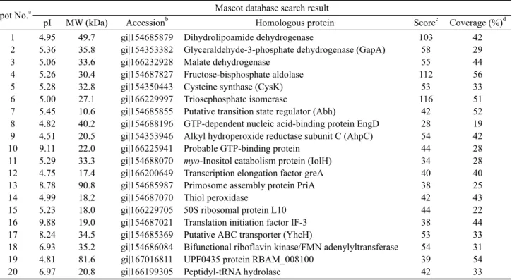
Table 2. Potential identity of protein spots from B. subtilis MORI and MORI 3K-85 by comparison with the B. amyloliquefaciens database
Spot No.a Mascot database search result
pI MW (kDa) Accessionb Homologous protein Scorec Coverage (%)d
1 2 3 4 5 6 7 8 9 10 11 12 13 14 15 16 17 18 19 20
4.95 5.36 5.06 5.26 5.28 5.00 5.45 4.82 4.51 9.11 5.29 4.75 8.78 4.99 5.23 9.88 8.24 6.93 4.81 6.97
49.7 35.8 33.6 30.4 32.8 27.1 10.6 40.2 20.5 22.0 33.3 17.4 90.8 18.2 18.0 19.0 34.5 35.2 81.6 20.8
gi|154685879 gi|154353382 gi|166232928 gi|154687827 gi|154350443 gi|166229997 gi|154685855 gi|154688196 gi|154353946 gi|166225941 gi|154688070 gi|166200649 gi|154685987 gi|154687070 gi|166229705 gi|154687021 gi|154685369 gi|154686084 gi|167016811 gi|166199305
Dihydrolipoamide dehydrogenase
Glyceraldehyde-3-phosphate dehydrogenase (GapA) Malate dehydrogenase
Fructose-bisphosphate aldolase Cysteine synthase (CysK) Triosephosphate isomerase
Putative transition state regulator (Abh)
GTP-dependent nucleic acid-binding protein EngD Alkyl hydroperoxide reductase subunit C (AhpC) Probable GTP-binding protein
myo-Inositol catabolism protein (IolH) Transcription elongation factor greA Primosome assembly protein PriA Thiol peroxidase
50S ribosomal protein L10 Translation initiation factor IF-3 Putative ABC transporter (YhcH)
Bifunctional riboflavin kinase/FMN adenylyltransferase UPF0435 protein RBAM_008100
Peptidyl-tRNA hydrolase
103 58 55 112
53 116
42 28 54 44 34 40 38 42 44 38 53 54 39 42
42 29 44 56 33 51 52 19 42 28 28 40 25 43 22 44 33 31 54 33
aThe numbering corresponding to the 2-DE image in Fig. 2 bAccession number in NCBInr; cScore is -10 × Log (P), where P is the probability that the observed is a random event; Protein scores greater than 51 are significant (P < 0.05); dPercentage of the protein sequence covered by the matched peptides.
단백질을 동정하였다.
3. 결과 및 고찰
DNJ 생산 균주인 MORI와 이 균에 감마선 처리에 의한 돌연 변이 유발로 DNJ 생산이 7.6배 증가한 MORI 3K-85 균주의 단백질 패턴을 Native-PAGE와 SDS-PAGE로 비교하였다.
먼저 Native-PAGE의 경우, 두 균주 간 전기영동 패턴에 정성 적인 차이는 발견할 수 없었고 다만 모균주에 비해 MORI 3K-85 균주에서 5개의 band가 다소 진하게 염색되어 이들 단백질의 농도가 증가하였음을 알 수 있었다 (Fig. 1(a)).
이러한 단백질 band의 농도 변화는 SDS-PAGE 분석에서도 재현되었다. 즉, SDS-PAGE의 경우, 역시 두 균주 간 기본 단백질 패턴은 동일하여 새로운 단백질의 출현이나 소멸과 같은 정성적 변화는 발견되지 않았으나 Native-PAGE의 경우 와 같이 두 균주 간 단백질 band의 농도에서 차이가 인정되었 는데 예를 들면 66.2 kDa 근처에서 1종, 32~45 kDa 부위에 서 4종, 그리고 21.5~32 kDa 부위에서 2종, 등 모두 7종의 단백질 band가 모균주에 비해 MORI 3K-85에서 진하게 염색 되었다 (Fig. 1(b)). 이와 같이 MORI 균주에 비해 DNJ 생산 이 크게 향상된 MORI 3K-85 균주에서 몇몇 단백질 band의 농도가 증가하였다는 사실은 이들 특정 단백질들이 DNJ의 생합성과 어느 정도 연관성이 있음을 시사하는 결과로 받아 들여진다. 그러나 1차원 전기영동 분석에 의한 이들 단백질
의 농도 변화만으로는 얻을 수 있는 정보에 한계가 있어 보 다 상세한 결과를 얻기 위해 2차원 전기영동과 MALDI-TOF 분석을 수행하였다.
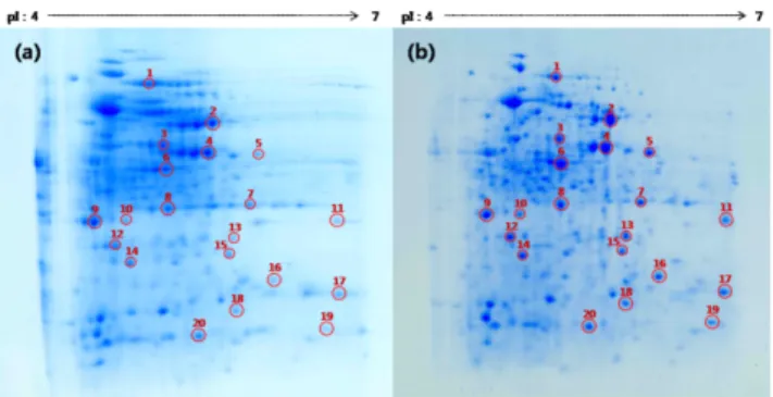
(a) (b)
Fig. 1. Native- and SDS-PAGE analyses of proteins isolated from B. subtilis strains MORI and MORI 3K-85. (a) Native-PAGE analysis (lane a: B. subtilis MORI, lane b: B. subtilis MORI 3K-85), (b) SDS- PAGE analysis (lane M: protein molecular weight markers, lane a:
B. subtilis MORI, lane b: B. subtilis MORI 3K-85).
2차원 전기영동의 결과 수 많은 단백질 spot들이 검출되 었으며 2-DE상에 나타난 두 균주의 spot들은 육안으로 1개 씩 정밀하게 대응 비교하면서 양자 간의 차이를 조사하였 다 (Fig. 2). 1-DE 경우에서와 같이 2-DE에서도 정성적 큰 차이는 없었으나 동일한 시료 양 (1 mg)의 loading 조건에서 일부의 spot들은 모균주에 비해 MORI 3K-85 균주에서 진한
염색성을 나타내 단백질의 농도가 상대적으로 증가함을 알 수 있었다. 이들 가운데 뚜렷하게 농도 증가를 보이는 20개 의 spot을 선정하여 MALDI-TOF로 펩티드의 질량을 분석 하고 MORI 균의 표준 균주인 B. subtilis와 같은 Bacillus에 속하며 DNJ 생산균주로 알려진 B. amyloliquefaciens에 대 한 MASCOT 데이터베이스를 통해 단백질을 동정하였다.
먼저 MORI 균의 표준 균주인 B. subtilis과의 단백질 동정 결과 20개의 spot들 가운데 13종 (spot no. 1, 2, 3, 4, 5, 6, 7, 9, 10, 14, 17, 18, 19)은 효소, 그리고 나머지 7종 (spot no. 8, 11, 12, 13, 15, 16 및 20)은 효소 이외의 기능을 보유 하는 단백질로 검색되었다. 동정된 13종의 효소는 3종의 dehydrogenase를 비롯하여 aldolase, synthase, isomerase, deformylase, reductase, synthetase, peroxidase, peptidase, ferrochelatase 및 protease 등으로 확인되었다 (Table 1). 한 편 DNJ 생산 균주로 알려진 B. amyloliquefaciens에 대한 단백질 검색 결과 이들 20개의 spot은 10종 (spot no. 1, 2, 3, 4, 5, 6, 9, 14, 18, 20)의 효소와 10종 (spot no.7, 8, 10, 11, 12, 13, 15, 16, 17, 19)의 효소 이외의 기능을 갖는 단백질 로 알려졌으며, 해당 효소로는 3종의 dehydrogenase외에 aldolase, synthase, isomerase, reductase, peroxidase, kinase/adenyltransferase, t-RNA hydrolase 등으로 B. subtilis 검색 결과와는 약간의 차이를 보였다 (Table 2).
Fig. 2. Two dimensional gel electrophoresis of crude protein extracts of B. subtilis MORI and B. subtilis MORI 3K-85 stained by Coomassie brilliant blue G250. (a) B. subtilis MORI, (b) B. subtilis MORI 3K-85.
이상의 MASCOT 데이터베이스를 통해 입수한 20개 spot 들의 단백질 정보와 DNJ의 생합성 관련성에 대해 고찰해 보기로 한다. 먼저 B. subtilis과의 단백질 검색결과 동정된 20종의 단백질들이 MORI 균주의 돌연변이 유발에 의해 DNJ 생산능력이 7.6배 증대된 MORI 3K-85 균주에서 상대 적으로 농도의 증가를 보였다고 하여 이들이 모두 DNJ 생 합성에 관여하는 단백질이라고 단정하기는 어렵다. 감마선 처리에 의한 유전자의 변이는 DNJ의 생합성과 무관하게 다양 한 형태의 단백질 발현으로 나타날 수 있기 때문이다. 다만 이들 20종의 단백질들이 유전적 기원은 같으나 돌연변이 유발 에 의해 DNJ 생산성에 차이를 갖는 두 균주를 통해 확인되 었다는 점에서 이들 단백질 특히 효소들의 DNJ 생합성과의 관련 가능성이 제기될 수 있다.
현재까지 알려진 DNJ 생합성 경로로는 Bacilli와
Streptomyces 균에서 제안된 glucose로부터 amination, oxidation, epimerization 등의 과정을 거치는 C2/C6 cyclization 경로 [23,24]와 닭의장풀 (Commelina communis)에서 보고된 C1/C5 cyclization 경로 [25]가 있으나 지금까지 식물이나 미생물에서 DNJ의 생합성 경로 해명을 위한 결정적인 유전 자나 효소에 관한 보고는 없었다. 최근 강 등 [19]은 MORI 3K-85 균주로부터 DNJ 생산에 관여하는 3개의 유전자 gabT1, yktc1, gutB1을 확보하고 나아가 이들 유전자의 putative transaminase, fructose phosphatase, dehydrogenase 의 기능을 토대로 glucose로부터 DNJ까지의 생합성 경로에 kinase, aminotransferase, phosphatase, dehydrogenase 등 4종의 효소와 cyclization, epimerization, deoxygenation 등 의 구조변화가 관여할 것이라는 일련의 DNJ 생합성 경로를 추정한 바 있다 [20]. 그러나 이들 DNJ 생합성 경로에 관여 할 것으로 추정한 4종의 효소들 가운데 본 연구의 2-DE 분석 에 의해 검출된 효소는 dehydrogenase 뿐이었으며 나머지 3종의 효소는 확인되지 않았다.
한편 dehydrogenase를 제외한 나머지 10종의 효소와 DNJ 생합성과의 관계에 대해서는 아직 어떠한 설명이나 결론을 내릴 수가 없다. 다만 본 연구에서 확인된 3종의 dehydrogenase에 의해 생성된 물질들이 매개체로 제공되어 직간접적으로 DNJ 생합성에 관여하는 것으로 추정된다.
그리고 같은 Bacillus속으로서 DNJ의 생산균주로 알려 진 B. amyloliquefaciens과의 단백질 비교 결과 3종의 dehydrogenase를 포함한 aldolase, synthetase, isomerase, reductase, peroxidase 등 8종의 효소가 B. subtilis와 동일한 기능의 효소로 밝혀진 사실은 매우 의미가 있다고 본다.
왜냐하면 최근 B. amyloliquefaciens로부터 보고된 DNJ 생산 관련 유전자 gabT1, yktc1, gutB1은 우리가 B. subtilis MORI 3K-85로부터 확인한 DNJ 유전자와 일치함으로써 [19,26], 두 균주의 DNJ 생합성 과정에 이들 효소가 공통으로 관련 될 가능성을 예상할 수 있기 때문이다. 현재까지 식물과 미생물 등에서 glucose을 전구물질로 하는 몇몇 DNJ 생합성 경로가 보고되고는 있으나 아직 추정 수준이며 따라서 앞 으로 DNJ에 대한 보다 다양하고 복잡한 생합성 경로들이 밝혀질 것을 예상하면서 금후 DNJ 생합성 경로 규명에 상기 효소들의 역할을 기대해 본다.
감사
이 논문은 2007년 정부 (교육인적자원부)의 재원으로 한국 학술진흥재단의 지원을 받아 수행된 연구임 (KRF-2007-313- C00645).
References
1. Asano, N., R. J. Nash, R. J. Molyneux, and G. W. J. Fleet (2000) Sugar-mimic glycosidase inhibitors: natural occurrence, biological activity and prospects for therapeutic application. Tetrahedron:
Asymmetry 53: 1645-1680.
2. Watson, A. A., G. W. G. Fleet, N. Asano, R. J Molyneux, and R.
J. Nash (2001) Polyhydroxylated alkaloids-natural occurrence and therapeutic applications. Phytochemistry 56: 265-295.
3. Schedel, M. (2008) Regioselective Oxidation of Aminosorbitol with Gluconobacter oxydans, Key Reaction in the Industrial 1-Deoxynojirimycin Synthesis, pp. 296-307. In: H.-J. Rehm and G. Reed (eds.), Biotechnology: Biotransformations II, Volume 8b, 2nd ed. Wiley-VCH Verlag GmbH, Weinheim, Germany.
4. Asano, N., K. Oseki, E. Tomioka, H. Kizu, and K. Matsui (1994) N-containing sugars from Morus alba and their glycosidase inhibitory activities. Carbohyr. Res. 259: 243-255.
5. Yoshikuni, Y. (1988) Inhibition of intestinal α-glycosidase activity and postprandial hyperglycemia by moranoline and its N-alkyl derivatives. Agric. Biol. Chem. 52: 121-128.
6. Gruters, R. A., J. J. Neefjes, M. Tersmette, R. E. Y. D. Goede, A. Tulp, H. G. Huisman, F. Miedema, and H. L. Ploegh (1987) Interference with HIV-induced syncytium formation and viral infectivity by inhibitors of trimming glucosidase. Nature 330: 74-77.
7. Fleet, G. W. J., A. Karpas, R. A. Dwek, L. E. Fellows, A. S. Tyms, S. Petursson, S. K. Namgoong, N. G. Ramsden, P. W. Smith, J. C.
Son, F. Wilson, D. R. Witty, G. S. Jacob, and T. W. Rademacher (1988) Inhibition of HIV replication by amino-sugar derivatives.
FEBS Lett. 237: 128-132.
8. Karpas, A., G. W. J. Fleet, R. A. Dwek, S. Petursson, S. K.
Namgoong, N. G. Ramsden, G. S. Jacob, and T. W. Rademacher (1988) Aminosugar derivatives as potential anti-human immunodeficiency virus agents. Proc. Natl. Acad. Sic. USA 85: 9229-9233.
9. Mehta, A., N. Zitzmann, P. M. Rudd, T. M. Block, and R. A.
Dwek (1998) α-Glucosidase inhibitors as potential broad anti-viral agents. FEBS Lett. 430: 17-22.
10. Dwek, R. A., T. D. Butters, F. M. Platt, and N. Zitzmann (2002) Targeting glycosylation as a therapeutic approach. Nat. Rev.
Drug. Discov. 1: 65-75.
11. Jacob, J. R., K. Mansfield, J. E. You, B. C. Tennant, and Y. H. Kim (2007) Natural iminosugar derivatives of 1-deoxynojirimycin inhibit glycosylation of hepatitis viral envelope proteins. J.
Microbiol. 45: 431-440.
12. Asano, N., T. Yamashita, K. Yasuda, K. Ikeda, H. Kizu, Y.
Kameda, A. Kato, R. J. Nash, H. S. Lee, and K. S. Ryu (2001) Polyhydroxylated alkaloids isolated from mulberry trees (Morus alba L.) and silkworms (Bombyx mori L.). J. Agric. Food Chem.
49: 4208-4213.
13. Afarinkia, K. and A. Bahar (2005) Recent advances in the chemistry of azapyranose sugars. Tetrahedron: Asymmetry 16:
1239-1287.
14. Cho, Y. S., Y. S. Park, J. Y. Lee, K.-D. Kang, K. Kim, K. Y. Hwang, and S. I. Seong (2008) Hypoglycemic effect of culture broth of Bacillus subtilis S10 producing 1-deoxynojirimycin. J. Korean Soc. Food Sci. Nutr. 37: 1401-1407.
15. Stein, D. C., L. K. Kopec, R. E. Yasbin, and F. E. Young (1984) Characterization of Bacillus subtilis DSM704 and its production of 1-deoxynojirimycin. Appl. Environ. Microbiol. 48: 280-284.
16. Ezure, Y., S. Maruo, K. Miyazaki, and M. Kawamata (1985) Moranoline (1-deoxynojirimycin) fermentation and its improvement. Agric. Biol. Chem. 49: 1119-1125.
17. Hardick, D. J., D. W. Hutchinson, S. J. Trew, and E. M. H.
Wellington (1991) The biosynthesis of deoxynojrimycin and deoxymannonojirimycin in Streptomyces subrutilus. J. Chem.
Soc. Chem. Commun. 10: 729-730.
18. Kim, H. S., J. Y. Lee, K. Y. Hwang, Y. S. Cho, Y. S. Park, K.-D.
Kang, and S. I. Seong (2011) Isolation and identification of a Bacillus sp. producing α-glucosidase inhibitor 1-deoxynojirimycin.
Korean J. Microbiol. Biotechnol. 39: 49-55.
19. Kang, K.-D., Y. S. Cho, J. H. Song, Y. S. Park, J. Y. Lee, K. Y.
Hwang, S. K. Rhee, J. H. Chung, O. Kwon, and S. I. Seong (2011) Identification of the gene involved in 1-deoxynojirimycin synthesis in Bacillus subtilis MORI 3K-85. J. Microbiol. 49:
431-440.
20. Cho, Y. S. (2011) Studies on 1-deoxynojirimycin biosynthesis genes in Bacillus subtilis MORI. Ph.D. Thesis. University of Suwon, Hwaseong-si, Gyeonggi-do, Korea.
21. Bollag, D. M., M. D. Rozycki, and S. J. Edelstein (1996) Protein Methods: Gel Electrophoresis Under Denaturing Conditions and Gel Electrophoresis Under Nondenaturing Conditions. 2nd ed., pp. 107-172. Wiley-Liss Inc., NY, USA.
22. Berkelman, T. and T. Stenstelt (2002) 2-D Electrophoresis Using Immobilized pH Gradie-nts, Principles and Methods.
2nd ed., pp. 17-93. Amersham Biosciences, Uppsala, Sweden.
23. Hardick, D. J. and D. W. Hutchinson (1993) The biosynthesis of 1-deoxynojirimycin in Bacillus subtilis var niger. Tetrahedron 49: 6707-6716.
24. Hardick, D. J., D. W. Hutchinson, S. J. Trew, and E. M. H.
Wellington (1992) Glucose is a precursor of 1-deoxynojirimycin and 1-deoxymannonojirimycin in Streptomyces subrutilus.
Tetrahedron 48: 6285-6296.
25. Shibano, M., Y. Fujimoto, K. Kushino, G. Kusano, and K. Baba (2004) Biosynthesis of 1-deoxynojirimycin in Commelina communis: a difference between the microorganisms and plants.
Phytochemstry 65: 2661-2665.
26. Clark, L. and N. Horenstein (2010) Biosynthesis of Azasugars.
First Southeast Enzyme Conference. April 10. Atlanta, GA, USA.