PGHN
Case Report
Tubular Colonic Duplication Presenting as Rectovestibular Fistula
Parag J. Karkera, Pradnya Bendre, Flavia D’souza, Mukunda Ramchandra, Amol Nage, and Nitin Palse
Department of Pediatric Surgery, Bai Jerbai Wadia Hospital for Children, Mumbai, India
Complete colonic duplication is a very rare congenital anomaly that may have different presentations according to its location and size. Complete colonic duplication can occur in about 15% of all gastrointestinal duplications. Double termination of tubular colonic duplication in the perineum is even more uncommon. We present a case of a Y-shaped tubular colonic duplication which presented with a rectovestibular fistula and a normal anus. Radiological evaluation and initial exploration for sigmoidostomy revealed duplicated colons with a common vascular supply. Endorectal mucosal resection of theduplicated distal segment till the colostomy site with division of the septum of the proximal segment and colostomy closure proved curative without compromise of the continence mechanism. Tubular colonic duplication should always be ruled out when a diagnosis of perineal canal is considered in cases of vestibular fistula alongwith a normal anus.
Key Words: Rectovestibular fistula, Colonic duplication, Tubular duplication
Received:December 23, 2014, Revised:February 1, 2015, Accepted:February 9, 2015
Corresponding author: Parag J. Karkera, Department of Pediatric Surgery, Bai Jerbai Wadia Hospital for Children, Acharya Donde Marg, Parel, Mumbai 400012, India. Tel: +91-22-4146963/64/65/66/67, E-mail: drpaggy@gmail.com
Copyright ⓒ 2015 by The Korean Society of Pediatric Gastroenterology, Hepatology and Nutrition
This is an openaccess article distributed under the terms of the Creative Commons Attribution NonCommercial License (http://creativecommons.org/licenses/by-nc/4.0/) which permits unrestricted noncommercial use, distribution, and reproduction in any medium, provided the original work is properly cited.
INTRODUCTION
Alimentary tract duplications are rare congenital malformations of the gastrointestinal tract, which may be cystic or tubular and occur anywhere from the mouth to the anus [1]. More than 80% of cases present before the age of 2 years as an acute abdo- men or bowel obstruction [2,3]. The commonest site for duplication is the ileum, while colonic duplica- tion is rare, accounting for 6-13% of all gastro- intestinal duplications, commonly located in the ce-
cum [1,4]. These lesions, if encountered incidentally, should be surgically managed to prevent any future complications. Although perineal canal or H-type congenital fistula is a relatively common anomaly in Asian countries, it’s association with Y-shaped tubu- lar colonic duplication is rare [5]. We report a case of colorectal duplication in a two month old female child passing stools from normal anal opening and a fistulous opening in the vestibule.
Fig. 1. Clinical picture showing 3 openings in the vestibule- urethra (foley catheter), vagina, vestibular fistula (infant feeding tube), in addition to the normal sited anus.
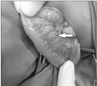
Fig. 2. Intra-operative picture showing duplicated colon (arrow points to the duplicated segment at the mesenteric border of native colon).
Fig. 3. Distal and proximal cologram showing duplicated colon with 2 separate lumens (arrow points to negative shadow of the intervening septum between the duplicated colons).
CASE REPORT
A two-month-old female child presented with the complaints of passing stools from normal anal open- ing and a fistulous opening in the vestibule. A provi- sional diagnosis of perineal canal was made. Abdo-
minal sonography, echocardiography and spinal ra- diography were normal, ruling out associated uri- nary tract, cardiac and vertebral anomalies. Exami- nation under anaesthesia revealed a normal anal opening. The vestibule had 3 openings, which were a normal urethral and vaginal opening and a rec- tovestibular fistula (Fig. 1) with no communication with the normal anorectum. Considering this to be a complex anomaly, a diverting stoma was planned.
On exploration of the abdomen for the stoma, tubu- lar duplication of the sigmoid colon was noted (Fig.
2). A high sigmoid loop colostomy was done which resulted in four stomal lumens. Later, for better de- lineation of the anatomy of the anomaly, a proximal and distal colostomogram was performed (Fig. 3). It revealed duplicated colon with separate lumens from the transverse colon downwards with two anal openings. The duplicated colonic segment towards the mesenteric side terminated as a rectovestibular fistula (Fig. 4). At 6 months of age, the patient was taken up for definitive surgical management. An ab- dominoperineal approach was used, in which ini- tially the colostomy was mobilized. For the distal du- plicated colonic segment, the mucosal cuff of the co- lon leading to the vestibular fistula was dissected like
Fig. 4. Diagrammatic presentation of the anatomy.
Fig. 5. Mucosal resection (arrow-mucosal cuff) of the distal duplicated colon.
Fig. 6. Septum (arrow) between the duplicated colons.
a Soave’s endorectal pullthrough from the distal sto- ma to the vestibular fistula and excised (Fig. 5). The remaining muscular cuff was plicated and closed.
The proximal duplicated colonic segment shared the vascular supply with the native colon, hence re- section was not possible. The duplicated segment ex- tended proximally till the ascending colon and com- mon wall comprised of two layers of mucosa. There-
fore, the intervening mucosal septum between the duplicated colon, upto the non-duplicated ascending colon was divided (Fig. 6). A colo-colic anastomosis between the proximal unified colon with the distal native colon leading to the normal anus was done. In view of the significant disparity in the proximal and distal colonic luminar diameter, a covering ileostomy was also done. The postoperative course was un- eventful and the ileostomy was closed after 3 months. At follow up of 6 months post-surgery, the opening in the vestibule had closed, with no bowel complaints.
DISCUSSION
Duplications of the colon are uncommon malfor- mations, which are tubular or cystic in nature. Early aberration in the formation of the primitive hindgut is hypothesized to cause a split or twinning process of the anlage, which results in terminal gut duplication with or without duplication of the genitourinary or- gans [6]. Tubular colonic duplications are double-bar- reled or Y-shaped. They possess a double muscular layer and epithelium similar to the rest of the colon [1,7]. Either both lumina may be unobstructed and function normally as two perineal ani, or terminate distally blindly as imperforate anus of one or both lumina. In some cases, the ventral colon may end as a rectourinary, rectovaginal or vestibular fistula [7].
The presenting features are constipation, vomiting, volvulus, perforation, and, most commonly, intestinal obstruction due to compression of the normal bowel by the blind end of the duplication [8]. Associated anomalies include genitourinary duplications, skel- etal anomalies bladder exstrophy, malrotation of gut, omphalocele, Meckel’s diverticulum [1,7,8]. In absence of other associated malformations or an ec- topic opening, tubular duplications of the colon re- main unnoticed, until it’s complications warrant surgical intervention.
Most authors recommend that once the diagnosis is made, an elective surgical procedure in an optimal state of the patient should be performed to avoid complications. The recommended surgical procedure is excision of the duplicated colon. Although malig- nant changes have been reported in adults [9], color- ectal duplications are essentially benign lesions and radical surgical excision is not required. Need for sur- gery when asymptomatic is debatable [8].
In most cases, resection of the duplicated colon may not be possible because of common blood supply with a single mesentery to the two colons [8]. In our case, separation of the two proximal colonic seg- ments was impossible because in addition to the common blood supply, the intervening wall com- prised of only mucosa. Also, excision of common wall resulted in gross disparity between proximal and distal loops.
The options to manage unresectable proximal du- plicated colons include dividing the septum to con- vert it into a common colonic channel; long side-to-side anastomosis of adjacent duplicated bowel; trans- ection of the rectum over the peritoneal reflection and anastomosis of the proximal end of the neigh- boring colon to the main colon, excision of the distal common septum with creation of a common channel should prevent accumulation of feces in the false lu- men or performing mucosectomy on one limb of the proximal duplication [10-13].
Excision of the distal duplicated segment is even more difficult, as although, all ectopic openings re- quire surgical intervention, it can compromise the continence mechanism of the normal sited anus.
Stephens and Smith [14] presented a series of dou- ble perineal ani and opined that, provided that there is no neurogenic element, and both recta appear to lie within the same puborectalis muscle sling and within a single external sphincter, it may be best to accept the 2 ani permanently. Attempt to excise one rectum from the other may jeopardize normal continence.
Hence, for the distal segment, if the colon proper ends as a fistula, posterior sagittal anorectoplasty ap- proach helps to prevent compromise of continence mechanism [15]. Management options for the distal duplicated segment include fistula closure with ex- cision of distal few centimeters of septum to create a common distal channel; and mucosal stripping of the duplicated colon like a soave’s procedure [15-17].
Also, in cases in whom hindgut duplication is dis- covered unexpectedly at initial laparotomy for stoma formation for anorectal malformations, sigmoidos- tomy has proven to be well tolerated as it does not compromise subsequent reconstruction, and facili- tates easier radiological evaluation of the complex anatomy [15]. To prevent unexpected findings on surgical exploration, radiological evaluation is re- commended. Contrast enema and a fistulogram per- formed concomitantly, preferably with contrast me- dia of differing densities, helps to delineate both colons. Occasionally, upper gastrointestinal contrast follow-through studies may help to locate the prox- imal extent of duplication [10,18].
In our case, the child was asymptomatic for the proximal colonic duplication and the duplicated co- lon could not be resected without resection of almost the entire colon in view of the shared vascular supply.
Hence, dividing the septum between the two lumens of the proximal colonic segment with mucosectomy and closure of fistula treated the problem without hindering continence.
In conclusion, tubular colorectal duplication should be considered as one of the differential diagnosis with perineal canal in cases of vestibular fistula along with a normal anus. Resection of the dupli- cated colon may not be possible due to shared blood supply between the duplicated segments. Sigmoi-
dostomy at initial laparotomy is useful for future re- construction and radiographic evaluation. Endorectal mucosal resection of the duplicated segment effec- tively manages the condition without compromise of the continence mechanism.
REFERENCES
1. Macpherson RI. Gastrointestinal tract duplications:
clinical, pathologic, etiologic, and radiologic conside- rations. Radiographics 1993;13:1063-80.
2. Kekez T, Augustin G, Hrstic I, Smud D, Majerovic M, Jelincic Z, et al. Colonic duplication in an adult who pre- sented with chronic constipation attributed to hypo- thyroidism. World J Gastroenterol 2008;14:644-6.
3. Merrot T, Anastasescu R, Pankevych T, Tercier S, Garcia S, Alessandrini P, et al. Duodenal duplications.
Clinical characteristics, embryological hypotheses, histological findings, treatment. Eur J Pediatr Surg 2006;16:18-23.
4. Liu R, Adler DG. Duplication cysts: diagnosis, manage- ment, and the role of endoscopic ultrasound. Endosc Ultrasound 2014;3:152-60.
5. Bryndorf J, Madsen CM. Ectopic anus in the female.
Acta Chir Scand 1960;118:466-78.
6. Ravitch MM. Hind gut duplication; doubling of colon and genital urinary tracts. Ann Surg 1953;137:588- 601.
7. Kaur N, Nagpal K, Sodhi P, Minocha VR. Hindgut du- plication--case report and literature review. Pediatr Surg Int 2004;20:640-2.
8. Jellali MA, Mekki M, Saad J, Zrig A, Elanes I, Mnari W, et al. Perinatally discovered complete tubular colon-
ic duplication associated with anal atresia. J Pediatr Surg 2012;47:e19-23.
9. Inoue Y, Nakamura H. Adenocarcinoma arising in co- lonic duplication cysts with calcification: CT findings of two cases. Abdom Imaging 1998;23:135-7.
10. Aworanti O, Twomey E, Awadalla S. Terminal ileum and total colonic duplication associated with a rec- tovestibular fistula in a child. Ir Med J 2014;107:241-2.
11. Yucesan S, Zorludemir U, Olcay I. Complete duplica- tion of the colon. J Pediatr Surg 1986;21:962-3.
12. Ravitch MM. Duplication of the gastrointestinal tract.
In: Ravitch MM, Welch KJ, Bandolph JG, eds. Pediatric surgery. 4th ed. Chicago: Year Book Medical, 1986:91l- 20.
13. Fuchs JR, Clark K, Breckler FD, Rescorla FJ. Complete colonic duplication--a case report. J Pediatr Surg 2008;
43:E11-3.
14. Stephens FD, Smith ED. Duplications and vesicoin- testinal fissure. In: Anorectal malformations in chil- dren: update 1988 (birth defects, original article series).
New York: Alan R Liss, 1988:554-5.
15. Craigie RJ, Abbaraju JS, Ba'ath ME, Turnock RR, Baillie CT. Anorectal malformation with tubular hindgut duplication. J Pediatr Surg 2006;41:e31-4.
16. Khaleghnejad Tabari A, Mirshemirani A, Khaleghnejad Tabari N. Complete colonic duplication in children.
Caspian J Intern Med 2012;3:436-9.
17. Zhang Z, Huang Y, Wang D, Su P. Rectosigmoid tubular duplication presenting as perineal sepsis in a neonate.
J Pediatr Surg 2010;45:627-9.
18. Rathi V, Singh S, Bhargava SK, Kaur N, Seth N.
Diagnosis of tubular colonic duplication by barium fol- low-through study. Australas Radiol 2005;49:157-9.