Breast cancer is one of the most common malignan- cies in women and the main cause of cancer-related deaths (1). Breast cancer usually metastasizes to the lymph nodes, lung, bone, liver or brain. However, metastases to the gastrointestinal (GI) tract are rare. The most frequent sites of the GI tract involved are the stom- ach and the small intestine, while a colonic metastases is extremely rare (2, 3). Because of its rarity and its non- specific clinical presentation, a colonic metastasis may be easily mistaken for a double primary colonic carcino- ma in patients with history of breast cancer.
Nevertheless, early recognition of this metastatic dis-
ease is important because the treatment for these two diseases could vary greatly. We herein report the case of a patient with breast cancer metastases to the colon and small bowel along with a review of literature.
J Korean Soc Radiol 2010;62:551-554
─ 551 ─
Localized Metastasis to Small and Large Bowel from Breast Cancer: A Case Report1
Jae Ho Shim, M.D., Eun Ju Son, M.D., Beom-Jin Lim, M.D.2, Ji Hyun Youk, M.D., Jeong-Ah Kim, M.D., Joon Jeong, M.D.3
1Department of Radiology, Gangnam Severance Hospital, Yonsei University College of Medicine
2Department of Diagnostic Pathology, Gangnam Severance Hospital, Yonsei University College of Medicine
3Department of General Surgery, Gangnam Severance Hospital, Yonsei University College of Medicine
Received November 25, 2009 ; Accepted February 16, 2010
Address reprint requests to : Eun Ju Son, M.D., Department of Radiology, Gangnam Severance Hospital, Yonsei University College of Medicine, 146-92, Dogok-dong, Gangnam-gu, Seoul 135-720, Korea.
Tel. 82-2-2019-3510 Fax. 82-2-3462-5472 E-mail: [email protected]
Breast cancer is one of the most common malignancies in women and the main lead- ing cause of cancer death. Breast cancer frequently metastasizes to the bones, lungs, and liver; however, gastrointestinal involvement is rare. The most frequent sites of the GI tract involved are the stomach and the small intestine, while colonic metastasis is extremely rare with the presentation being nonspecific. Early diagnosis is important for proper treatment. We present a case of metastatic breast cancer to the small and large bowel.
Index words :Breast Neoplasms Neoplasm Metastasis Colon
Intestine, Small
Fig. 1. A 70-year-old woman with locally advanced cancer in the left breast. US image demonstrates a 3 cm-sized irregular hypoechoic mass at 1 o’clock position.
Case Report
A 70-year-old woman had been diagnosed with locally advanced breast cancer in her left breast in 2007 (Fig. 1).
After six cycles of neoadjuvant chemotherapy (cy- clophosphamide, methotrexate, and 5-fluorouracil), she had underwent a left modified radical mastectomy and ipsilateral axillary lymph node dissection. A histopatho- logical examination revealed a 3.5 cm sized invasive ductal carcinoma (IDC) of nuclear grade 3, histologic grade III, and metastatic carcinoma in 10 out of 28 lymph nodes with perinodal soft tissue extension.
Immunohistochemistry for estrogen and progesterone receptors showed negative staining for both receptors.
Jae Ho Shim, et al: Localized Metastasis to Small and Large Bowel from Breast Cancer
─ 552 ─ Fig. 2. Colonoscopy shows an ulcerofungating mass at the dis- tal transverse colon with lobulation on the surface.
A B
C D
Fig. 3. A 70-year-old woman followed after undergoing a left mastectomy due to locally advanced breast cancer in her left breast.
A. Abdominal CT shows an enhancing mass (arrow) in the distal portion of transverse colon, corresponding to the finding on a colonoscopy.
B. There is another enhancing mass (arrow) in the distal ileum causing the intussusceptions (arrowhead).
C, D. Strong F-18 FDG uptakes are noted for each lesion (SUV 8.9 (C) and 10.4 (D), respectively).
There was no evidence of distant metastases at the time of surgery. Consequently, the breast cancer was consis- tent with stage IIIA. After the operation, she received one cycle of radiotherapy at 3,600 cGy dose and a 10th adjuvant cycle of oral chemotherapy (Capecitabine) for 7 months.
Although, there was neither evidence of recurrence, nor metastatic lesions during the two follow-up exami- nations (six monthly ultrasound, mammography, and biochemical investigations) over the course of two years after surgery. However, a hemoglobin drop of 3 g/L (10.7 to 7.7) was noted in the last follow-up study. An esophagogastroduodenoscopy (EGD) and colonoscopy was recommended for the further study.
The patient underwent an EGD and colonoscopy, which revealed an ulcerofungating mass occupying 30%
of circumference of the bowel within the distal trans- verse colon (Fig. 2). A biopsy was performed, in addition to pathological examination including special immuno- histochemical stains, confirmed the diagnosis of a metastatic breast cancer to the transverse colon; patho- logic findings were similar to those of the prior breast cancer specimen. Abdominal CT and FDG-PET/CT (Figs. 3A-D) for staging workup revealed an additional 3
× 2.5 cm-sized enhancing mass in the distal ileum caus- ing the intussusception. However, following a small
bowel series, there was no evidence of a small bowel mass or lead point. The patient underwent left hemi- colectomy and segmental resection of the small bowel with the tentative diagnosis of two concomitant metasta- tic lesions in the small and large intestine.
A histological examination of the surgical specimens confirmed the diagnosis of metastatic breast cancer in the colon and small intestine (Figs. 4A, B). The postoper- ative course was uneventful and adjuvant chemothera- py (taxol) was performed.
Discussion
Although common locations of metastases from breast cancer are the bones, lungs, liver, pleura, adrenals and the central nervous system, GI metastases of breast can- cer are rare and usually associated with disseminated disease (4). The stomach is the more frequently involved GI organ, while colonic and rectal metastases are ex- tremely rare (2, 3). Asch et al. (5) reported GI tract metastasis secondary to breast cancer in 1968, which was the largest case series in the medical literature. The sites of involvement of the GI tract included the esopha- gus (25%), stomach (25%), small intestine (28%), colon (19%), and rectum (4%). There was no mention of the primary histological subtype in this series.
J Korean Soc Radiol 2010;62:551-554
─ 553 ─
A B
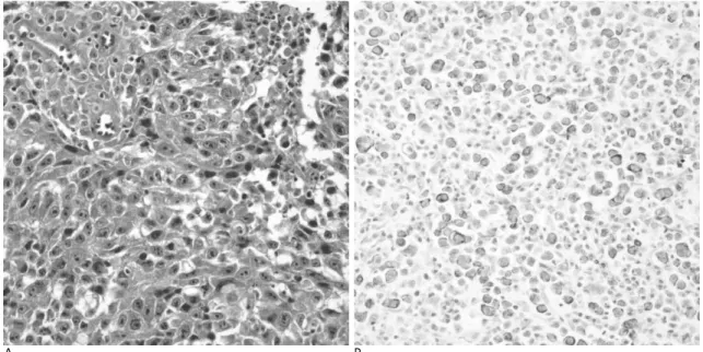
Fig. 4. Histopathologic findings of initial breast cancer and colonic metastasis.
A. Histologic finding of the initial breast mass. Infiltrating ductal carcinoma showing marked nuclear atypia with prominent nucle- oli (Original magnification: ×400).
B. Histologic finding of the colon. The tumor is composed of non-cohesive, pleomorphic tumor cells with prominent nucleoli. The immunohistochemical staining of CK7 (+) and CK20 (-) favors the diagnosis of metastatic carcinoma over primary gastrointestinal carcinoma.
In our case, the histological subtype of metastatic breast cancer was IDC. However, infiltrating lobular carcinoma was found to metastasize more frequently to the GI tract, peritoneum and retroperitoneum than the IDC (6). Taal and colleagues retrospectively identified 17 patients with metastatic breast cancer to the colon or rectum over a 15-year-period. Fifteen of these patients (88%) had lobular carcinoma and only one (6%) had IDC. Therefore, lobular carcinoma represents the most common subtype of breast cancer metastasizing to the colon and rectum (3, 7), although it is not the most com- mon type of primary breast malignancy, accounting for less than 10% (2). The reasons for this have not been clarified.
In our case, metastatic lesions were confined to the small and large intestine without disseminated metasta- sis. Metastases in multiple sites of the GI tract like our case were reported in the literature only as autopsy studies (8, 9). Our case is the first in non-autopsied case.
Although the therapeutic management is still controver- sial, there is general agreement on the need for palliative surgery (10). Especially, when the leading point of the intussusception is suspected as malignancy like in our case, surgical resection of the involved bowel without attempted reduction is required.
Isolated GI metastases without disseminating metasta- sis to other organs are extremely rare and are less com- mon than benign disease processes or second primaries of the intestinal tract in patients with a history of breast cancer. However, in patients with a history of breast
cancer, especially, a histological subtype of lobular car- cinoma and a newly identified GI neoplasm, the radiolo- gist should be concerned about the possibility of metastatic breast cancer. In conclusion, a detailed pathological analysis and repetition of endoscopy are necessary for early diagnosis.
References
1. Jemal A, Siegel R, Ward E, Hao Y, Xu J, Murray T, et al. Cancer statistics, 2008. CA: Cancer J Clin 2008;58:71-96
2. Schwarz RE, Klimstra DS, Turnbull AD. Metastatic breast cancer masquerading as gastrointestinal primary. Am J Gastroenterol 1998;93:111-114
3. Taal BG, den Hartog Jager FC, Steinmetz R, Peterse H. The spec- trum of gastrointestinal metastases of breast carcinoma: II. The colon and rectum. Gastrointest Endosc 1992;38:136-141
4. Rabau MY, Alon RJ, Werbin N, Yossipov Y. Colonic metastases from lobular carcinoma of the breast. Dis Colon Rectum 1988;31:
401-402
5. Asch MJ, Wiedel PD, Habif DV. Gastrointestinal metastases from carcinoma of the breast. Arch Surg 1968;96:840-843
6. Borst MJ, Ingold JA. Metastatic patterns of invasive lobular versus invasive ductal carcinoma of the breast. Surgery 1993;114:637-641 7. Washington K, McDonagh D. Secondary tumors of the gastroin-
testinal tract: surgical pathologic findings and comparison with au- topsy survey. Mod Pathol 1995;8:427-433
8. Cifuentes N, Pickren JW. Metastases from carcinoma of mamma- ry gland: an autopsy study. J Surg Oncol 1979;11:193-205
9. Graham WP 3rd, Goldman L. Gastro-intestinal metastases from carcinoma of the breast. Ann Surg 1964;159:477-480
10. Clavien PA, Laffer U, Torhost J, Harder F. Gastro-intestinal metas- tases as first clinical manifestation of the dissemination of a breast cancer. Eur J Surg Oncol 1990;16:121-126
Jae Ho Shim, et al: Localized Metastasis to Small and Large Bowel from Breast Cancer
─ 554 ─
대한영상의학회지 2010;62:551-554
소장과 대장에만 전이를 보인 유방암: 증례 보고1
1연세대학교 의과대학 강남세브란스병원 영상의학과
2연세대학교 의과대학 강남세브란스병원 해부병리과
3연세대학교 의과대학 강남세브란스병원 외과
심재호∙손은주∙임범진2∙육지현∙김정아∙정 준3
유방암은 여성들에 있어서 유병률이 가장 높은 암 중의 하나이자 암으로 인한 사망의 주요 원인이다. 유방암은 주 로 뼈, 폐, 그리고 간으로 전이를 보인다. 그러나 위장관 전이는 상대적으로 드물다. 위장관 전이에서 상대적으로 흔 한 곳은 위와 소장이지만 대장으로의 전이는 매우 드물고 그 임상증세는 비특이적이다. 적절한 치료를 위해서는 조 기에 정확한 진단이 중요하다. 저자는 유방암의 전이가 소장과 대장에만 국한된 증례를 보고 하고자 한다.