ABSTRACT
Background: Unenhanced computed tomography (UCT) may be useful for evaluating acute pyelonephritis; however, no study has compared UCT with enhanced computed tomography (ECT) as a diagnostic tool. We evaluated a clinical usefulness of UCT versus ECT in acute pyelonephritis (APN).
Methods: We reviewed the clinical and radiological data from 183 APN-suspected patients who underwent UCT and ECT simultaneously at emergency room (ER) over a two-year period. Demographic, clinical parameters and computed tomography (CT) parameters of 149 patients were compared.
Results: The average patient age was 61.2 (± 10) years: 31 patients were men. Ninety-nine (66.4%) patients showed stones (18.7%), perinephric infiltration (56%), swelling (21%), and hydronephrosis (6.7%) on UCT. Seventeen patients (11.4%) had an atypical clinical course, requiring additional tests for accurate diagnosis. In 7 patients UCT and ECT results did not differ; in 10 patients, the diagnosis changed on ECT. On ECT, 112/149 (75.2%) patients had stones (16.7%), perinephric infiltrations (57%), swelling (21%), and hydronephrosis (6.7%); 62.5% showed parenchymal involvement: 34 (22.8%) patients had no abnormal ECT findings. APN CT findings are similar on stone, perinephric infiltration, swelling and hydronephrosis on both CTs. Twelve patients (8.0%) had an abnormal ECT finding, i.e., low-grade (1 and 2) parenchymal involvement. Six (4%) patients developed contrast-induced acute kidney injury within 2 days after ECT.
Conclusion: We demonstrate that UCT is not inferior to ECT as an initial tool for evaluating APN for screening nephrolithiasis and hydronephrosis without the risk of contrast-induced acute kidney injury (CIAKI). However, patients with an atypical clinical course may still need ECT.
Keywords: Acute Pyelonephritis; Unenhanced Computed Tomography; Parenchymal Involvement; Perinephric Infiltration; Contrast-induced Nephropathy
INTRODUCTION
Acute pyelonephritis (APN) is the most common form of upper urinary tract infection (UTI), and approximately 20%–35% of women experience an episode of APN in their lifetime.1 APN usually occurs secondary to an ascending infection of gram-negative bacteria in women, and
Original Article
Received: Mar 21, 2018 Accepted: Jun 14, 2018 Address for Correspondence:
Sejoong Kim, MD, PhD
Department of Internal Medicine, Seoul National University Bundang Hospital, 82 Gumi-ro 173-beon-gil, Bundang-gu, Seongnam 13620, Republic of Korea.
E-mail: [email protected]
© 2018 The Korean Academy of Medical Sciences.
This is an Open Access article distributed under the terms of the Creative Commons Attribution Non-Commercial License (https://
creativecommons.org/licenses/by-nc/4.0/) which permits unrestricted non-commercial use, distribution, and reproduction in any medium, provided the original work is properly cited.
ORCID iDs Anna Lee
https://orcid.org/0000-0002-0823-4069 Hyo-Cheol Kim
https://orcid.org/0000-0002-6016-247X Sung Il Hwang
https://orcid.org/0000-0001-7516-5369 Ho Jun Chin
https://orcid.org/0000-0003-1185-2631 Ki Young Na
https://orcid.org/0000-0002-8872-8236 Dong-Wan Chae
https://orcid.org/0000-0001-9401-892X Sejoong Kim
https://orcid.org/0000-0002-7238-9962 Funding
This research was supported by a grant of the Korea Health Technology R&D Project through
Anna Lee ,1 Hyo-Cheol Kim ,2 Sung Il Hwang ,3 Ho Jun Chin ,1 Ki Young Na ,1 Dong-Wan Chae ,1 and Sejoong Kim 1
1Department of Internal Medicine, Seoul National University Bundang Hospital, Seongnam, Korea
2Department of Radiology, Seoul National University Hospital, Seoul, Korea
3Department of Radiology, Seoul National University Bundang Hospital, Seongnam, Korea
Clinical Usefulness of Unenhanced Computed Tomography in Patients with Acute Pyelonephritis
Nephrology
the diagnosis is made clinically.2 Incidences of nephrolithiasis related UTI have been on the rise.3
Enhanced computed tomography (ECT) is being widely used for diagnosis of APN.1,2,4-6 ECT shows both the intrarenal and extrarenal areas as precise images and provides comprehensive anatomical information.7 It also enables the detection of complications such as renal or perinephric abscess.8 However, the administration of iodinated contrast agents used during ECT increases the risk of contrast-induced acute kidney injury (CIAKI).9 specially in patients with poor renal function.10 Patients with APN are more likely to have an acute kidney injury (AKI).11 Nonetheless, ECT is widely used as a primary tool in the ERs, and there is growing concern about the damage it can cause to the kidney.
Unenhanced computed tomography (UCT), on the other hand, avoids both CIAKI and delays in testing due to fasting and can detect nephrolithiasis; parenchymal involvement, such as strings; and other anatomical disorders. However, to our knowledge, no studies yet have investigated the usability of UCT as a diagnostic tool or compared it with ECT for evaluating APN. This study aims to assess the correlations between the clinical and biochemical parameters of APN diagnosed using UCT and ECT as well as compare the clinical outcomes following the use of these techniques. Further, we will the define clinical predictors to help identify patients who can be diagnosed with UCT alone. The principal purpose of this study will be to compare the clinical outcome between UCT and ECT in patients with APN.
METHODS
Study design and measurements
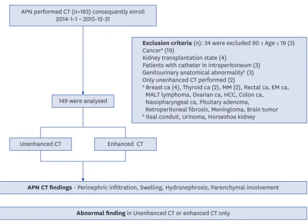
We conducted a retrospective review and analysis of the medical records of adult patients with clinically suggested APN who visited the ER at the Seoul National University Bundang Hospital and underwent both unenhanced- and enhanced-phase abdominal computed tomography (CT) scanning between January 2014 and December 2015. We excluded patients who had a history of cancer, kidney transplantation, intraperitoneal catheter implantation, and known genitourinary anatomical abnormalities (Fig. 1). We finally analyzed 149 patients. We collected demographic data and information on clinical parameters including sex, diabetes mellitus (DM), immunosuppressed state, recurrent urinary tract infection, neurogenic bladder, vesicoureteral reflux (VUR), and neurological diseases.12-14
We conducted the following biochemical and laboratory analyses: urine analysis with microscopy, urine culture and antibiotic sensitivity, blood culture, serum creatinine level assessment before and within 2 days after CT, determined the duration of antibiotic therapy and afebrile time, and reviewed the patient data. We also evaluated undetermined lesions in case of UCT and risks for missed diagnosis and incidence of CIAKI.
Clinical diagnosis of APN was made in patients fulfilling more than three of the following four diagnostic criteria: 1) costovertebral angle tenderness with or without lower urinary tract symptoms, including dysuria, urinary frequency, urinary urgency, nocturia, suprapubic discomfort, and, occasionally, gross hematuria, 2) fever of more than 37.5°C, 3) leukocytosis in the complete blood count (> 10,000/μL), and 4) abnormal urine test results (pyuria: white blood cells [WBC] of > 10/high-power field [HPF] or positive urine culture of > 105 colony- forming U/mL).2,4,8
the Korea Health Industry Development Institute (KHIDI), funded by the Ministry of Health & Welfare, Republic of Korea (grant number: HI17C1827).
Disclosure
The authors have no potential conflicts of interest to disclose.
Author Contributions
Conceptualization: Hwang SI, Chin HJ, Na KY, Chae DW, Kim S. Data curation: Lee A, Hwang SI. Validation: Hwang SI, Kim S. Methodology:
Kim HC. Investigation: Chin HJ. Writing - original draft: Lee A. Writing - review & editing:
Lee A.
CIAKI was defined as a rise in serum creatinine (Cr) of ≥ 0.5 mg/dL (≥ 44 mmol/L) or a 25% increase from the baseline value, assessed at 48 hours after a radiological procedure according to the guidelines of the Kidney Disease Improving Global Outcomes 2012.
CT analysis
Two radiologists who were unaware of the clinical outcomes recorded and graded the parenchymal involvement, perinephric infiltration, kidney swelling, hydronephrosis, cysts, abscesses,15 and stones on both UCT and ECT.16 Parenchymal abnormalities appeared as hypoenhanced regions that were ill-defined wedge-shaped lesions of decreased attenuation and that radiated from the papilla in the medulla to the cortical surface after contrast injection.16-19 Infiltration of perinephric fat represents APN with infection and obstruction.17 CT findings were divided into 5 grades based on the renal parenchymal involvement in this study, as follows: no renal parenchyma involvement (grade 0), less than 25% involvement
Exclusion criteria (n): 34 were excluded 90 ≤ Age ≤ 19 (3) Cancera (19)
Kidney transplantation state (4)
Patients with catheter in intraperitoneum (3) Genitourinary anatomical abnormalityb (3) Only unenhanced CT performed (2)
a Breast ca (4), Thyroid ca (2), MM (2), Rectal ca, EM ca, MALT lymphoma, Ovarian ca, HCC, Colon ca,
Nasopharyngeal ca, Pituitary adenoma,
Retroperitoneal fibrosis, Meningioma, Brain tumor
b Ileal conduit, Urinoma, Horseshoe kidney 149 were analysed
Unenhanced CT
APN CT findings - Perinephric infiltration, Swelling, Hydronephrosis, Parenchymal involvement Abnormal finding in Unenhanced CT or enhanced CT only
Undetermind renal lesion CT findings - Stone, Cyst, Abscess, Parenchymal involvement Missed diagnosis - Liver abscess, Appendicitis, Cholecystitis
CIAKI, Urologic procedure, Anatomical abnormality VUR, Febrile time, Microbiology Enhanced CT
APN performed CT (n=183) consequently enroll 2014-1-1 ~ 2015-12-31
Fig. 1. Study algorithm.
APN = acute pyelonephritis, CT = computed tomography, MM = multiple myeloma, EM = endometrial, MALT = mucosa-associated lymphoid tissue, HCC = hepatocellular carcinoma, UCT = unenhanced computed tomography, ECT = enhanced computed tomography, CIAKI = contrast induced acute kidney injury, VUR = vesicoureteral reflux.
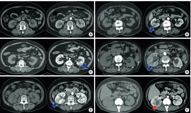
(grade 1), 25%–50% involvement (grade 2), 50%–75% involvement (grade 3), and greater than 75% (grade 4).19 Representative types of parenchymal involvement on ECT are shown in Fig. 2.
Statistical analysis
The baseline characteristics and laboratory data are presented as means and standard deviations for continuous variables and as frequencies and percentages for categorical variables. The accuracy of the two diagnostic tests performed simultaneously on an
individual subject was compared using the McNemar test. A value of P < 0.05 was considered significant. All analyses were performed using the SPSS Statistics software ver.22 (SPSS Inc., Chicago, IL, USA).
Ethics statement
This study was approved by the Institutional Review Board of Seoul National University Bundang Hospital (B-1604/344-108). All participants provided their written informed consent.
RESULTS
Baseline characteristics
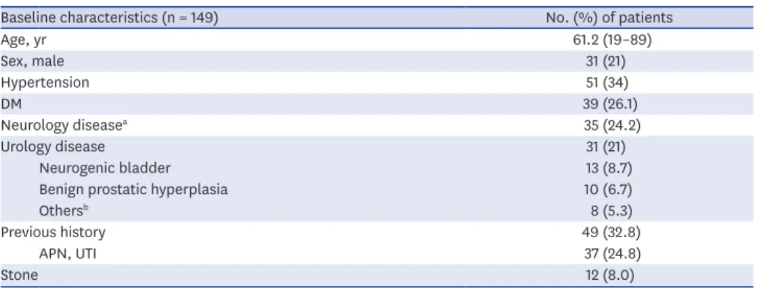
A total of 149 patients were finally enrolled in the study. Comparison of the baseline characteristics between the ECT and UCT groups are shown in Table 1.
B A
F E
D C
Fig. 2. Grade of parenchymal involvement. (A) grade 0: no renal parenchyma involvement, (B) grade 1: less than 25% involvement, (C) grade 2: 25%–50% involvement, (D) grade 3: 50%–75% involvement, (E) grade 4: greater than 75% involvement, and (F) Abcess. Left side present are UCT and right side present are ECT.
UCT = unenhanced computed tomography, ECT = enhanced computed tomography.
Of the 149 patients, 118 (79%) were women. Their mean age was 61.2 (± 10) years. A quarter of the patients had a history of DM and a neurological disease. Previous histories of UTI and nephrolithiasis were found in 37 (25%) and 12 (8%) patients, respectively. The average of Cr is 0.94 mg/dL at the time of ER visit, The mean of Cr at discharge is 0.87 mg/dL. The results of the APN clinical diagnosis criteria presented in this study are as follows: 1) costovertebral angle tenderness, 132 (88%); 2) fever of more than 37.5°C, 129 (86%); 3) leukocytosis in the complete blood count (> 10,000/μL), 98 (65%); 4) pyuria, WBC of > 10/high-power, 131 (87%).
Representative CT-APN CT findings
Table 2 summarizes the number of patients with and without abnormal findings.
UCT detected abnormal findings in 99(66.4%) patients, which included stone (18.7%), perinephric infiltration (56%), swelling (21%), hydronephrosis (6.7%), and parenchymal involvement (1.4%). The remaining 50 (33.6%) patients had no abnormal findings in UCT.
Seventeen patients (11.4%) had an atypical clinical course characterized by abdominal Table 1. Baseline characteristics
Baseline characteristics (n = 149) No. (%) of patients
Age, yr 61.2 (19–89)
Sex, male 31 (21)
Hypertension 51 (34)
DM 39 (26.1)
Neurology diseasea 35 (24.2)
Urology disease 31 (21)
Neurogenic bladder 13 (8.7)
Benign prostatic hyperplasia 10 (6.7)
Othersb 8 (5.3)
Previous history 49 (32.8)
APN, UTI 37 (24.8)
Stone 12 (8.0)
DM = diabetes mellitus, APN = acute pyelonephritis, UTI = urinary tract infection.
aCerebrovascular disease 15 (10), dementia 13 (8.7), Parkinson disease 4 (2.6), Alzheimer's disease 2 (1.3), cerebral palsy 1 (0.6); bOveractive bladder 3 (2.0), foley insertion state 3 (2.0), vesicoureteral reflux 2 (1.3).
Table 2. Representative CT findings
Characteristics No. (%) of patients
UCT ECT
Representative CT
Perinephric infiltration 84 (56) 85 (57)
Swelling 32 (21) 32 (21)
Hydronephrosis 10 (6.7) 10 (6.7)
Parenchymal involvement - bilateral involvement
0: no renal parenchyma involvement 147 (98.6) 56 (37.5)
1: less than 25% involvement 2 (1.4) 61 (40.9)–10 (6.7)
2: 25% to 50% involvement 0 (0) 21 (14)–4 (2.6)
3: 50% to 75% involvement 0 (0) 6 (4)
4: greater than 75% 0 (0) 5 (3.3)
Associated abnormality
Cyst 18 (12) 28 (18.7)
Abscess 0 (0) 7 (4.6)
Stone 28 (18.7) 25 (16.7)
Renal 13 (8.7) 10 (6.7)
UPJ 8 (5.3) 8 (5.3)
UVJ 7 (4.6) 7 (4.6)
CT = computed tomography, UCT = unenhanced computed tomography, ECT = enhanced computed tomography, UPJ = ureteropelvic junction, UVJ = ureterovesical junction.
pain without costovertebral angle tenderness, sustained fever regardless of an antibiotics treatment and a sustained shock state, requiring additional tests to confirm the diagnosis by UCT. Of these 17 patients, 7 received a comparable diagnosis by both UCT and ECT, whereas the remaining 10 patients had a different diagnosis including renal abscess by ECT. Evaluation by ECT revealed abnormal findings in 112 (75.2%) of the 149 patients, while the remaining 34 (22.8%) had no abnormal findings. These findings included stone (16.7%), perinephric infiltration (57%), swelling (21%), hydronephrosis (6.7%), and parenchymal involvement (62.5%). Of the 93 patients with parenchymal involvement, 14 presented bilateral parenchymal involvement. There were some differences in the clinical manifestations between the unilateral and bilateral parenchymal involvement groups. Of the 14 patients with bilateral parenchymal involvement, 21.4% were men, 28.5% had DM, 21.4% had shock (systolic blood pressure < 90 mmHg), 71.4% had bacteriuria, and 35.7%
had bacteremia. APN CT findings were comparable with regards to stone, perinephric infiltration, swelling and hydronephrosis on both UCT and ECT. In 12 patients (8.0%) ECT detected only a low grade (1 and 2) parenchymal involvement and no perinephric infiltration or other complications such as stone or abscess. These cases were diagnosed as simple APN and had a good prognosis.
Representative CT-stone, cyst, abscess
Table 2 summarizes the number of patients with stones, cysts, and/or abscesses.
While UCT detected various levels of stones in 28 (18.7%) patients, ECT detected the same in 25 (16.7%) patients. The stones were detected in the following anatomical locations:
renal (8.7%), ureteropelvic junction (UPJ) (5.3%), and ureterovesical junction (UVJ) (4.6%).
Although UCT detected more stones in the renal part, both CTs detected a comparable number of stones in the UPJ and UVJ, thereby suggesting that UCT is overall better than ECT in diagnosing stones.
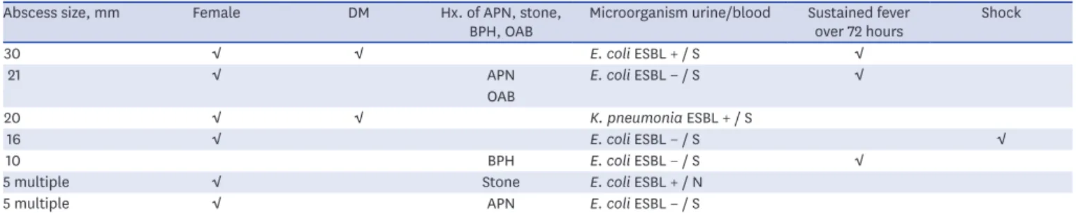
UCT detected renal cysts of multiple sizes in 18 (12%) of the patients, whereas ECT did the same in 28 (18%) patients. A renal abscess was detected in 7 (4.6%) patients with ECT (Table 3). Four of the 149 patients continued having fever 72 hours after starting antibiotics.
Three of these 4 patients showed renal abscess by ECT, which was not seen by UCT. The other 3 patients of 7 patients with renal abscess tested positive for extended-spectrum beta- lactamases (ESBL) and had a recurrence of APN respectively.
Missed diagnosis
UCT missed the diagnosis of cholecystitis, liver abscess, and appendicitis, in three men who were then diagnosed with ECT. The patient with cholecystitis had a delayed laparoscopic Table 3. Renal abscess
Abscess size, mm Female DM Hx. of APN, stone,
BPH, OAB Microorganism urine/blood Sustained fever
over 72 hours Shock
30 √ √ E. coli ESBL + / S √
21 √ APN E. coli ESBL − / S √
OAB
20 √ √ K. pneumonia ESBL + / S
16 √ E. coli ESBL − / S √
10 BPH E. coli ESBL − / S √
5 multiple √ Stone E. coli ESBL + / N
5 multiple √ APN E. coli ESBL − / S
DM = diabetes mellitus, Hx = history, APN = acute pyelonephritis, BPH = benign prostatic hyperplasia, OAB = overactive bladder, ESBL = extended spectrum beta- lactamase, S = same, N = no growth.
cholecystectomy following percutaneous transhepatic gallbladder drainage. The patient with appendicitis underwent a percutaneous abscess drainage on the perforated peripheral abscess. The third patient with the liver abscess, who was identified by aspiration and Klebsiella pneumoniae ESBL testing, improved after 6 weeks of antibiotics use. In all of these three cases, there was no imaging evidence for APN and therefore the treatment was completely different from that for APN.
CIAKI
Six patients were diagnosed with CIAKI within 2 days after ECT. CIAKI occurred despite hydration before ECT, but with supportive care, it improved by the time of discharge in all 6 cases. The incidence of CIAKI was related to age and DM in this study. The 6 patients were 89, 82, 80, 48, 42, and 33 years old (mean: 62.3 years). Three of these 6 patients have DM and were women.
Microorganisms
Comparison of identification strains between the urine culture and blood culture groups are shown in Table 4. Blood cultures tested positive in 21 (14%) patients, with Escherichia coli being the most common causative organism (n = 16; 10.7%) followed by K. pneumoniae and Pseudmonas aeruginosa. Only three patients had bacteremia caused by ESBL-producing E coli. Bacteriuria was seen in 75 (50.3%) patients, and E. coli was the most common causative organism, followed by K. pneumoniae and P. aeruginosa. All 7 patients with renal abscess had bacteriuria, and all 11 patients with more than 50% parenchymal involvement had bacteriuria.
DISCUSSION
APN is a clinical diagnosis, and recent studies have emphasized the importance of early imaging in patients with APN for cost-effective management.16,20-22 No studies to date have reported the use of UCT as a diagnostic tool for APN or compared it to other imaging techniques especially, ECT. So, we investigated the clinical usefulness of UCT for evaluating upper urinary tract infections and compared it to the ECT.
Recently, perirenal fat infiltration has been considered as important as parenchymal
involvement for the diagnosis of APN by UCT.22 Perirenal fat infiltration is observed when the inflammation of the renal parenchyma spreads beyond the renal capsule into the perirenal fat.7 The stranding likely represents fluid that collects within the bridging septa of the Table 4. Results of blood and urine culture
Microorganism No. (%) of patients
Bacteriuria Bacteraemia
E. coli ESBL (−) 49 (32.8) 13 (8.7)
E. coli ESBL (+) 14 (9.3) 3 (2.0)
K. pneumoniae ESBL (−) 2 (1.3) 1 (0.6)
K. pneumoniae ESBL (+) 1 (0.6) 1 (0.6)
Enterococcus faecium 2 (1.3) 0
P. aeruginosa 3 (2.0) 2 (1.3)
Citrobacter braakii 1 (0.6) 0
Enterobacter aerogenes 1 (0.6) 0
Coagulase negative Staphylococcus 2 (1.3) 1 (0.6)
Total 75 (50.3) 21 (14.0)
ESBL = extended spectrum beta-lactamase.
perinephric fat as a result of increased lymphatic pressure. Fluid collections may become asymmetric in the presence of unilateral obstruction or pyelonephritis.22
In this study, UCT was able to detect major APN findings. Whereas UCT detected perinephric infiltration for diagnosis of APN in 84 (56%) patients, ECT detected the same in 85 (57%) patients. A longer duration of fever, higher C-reactive protein levels, and grade of pyuria were related with perirenal fat infiltration,7 suggesting that the diagnostic accuracy of perinephric fat infiltration with UCT is almost identical to that with ECT for patients with APN. UCT may, therefore, be a good initial tool for assessing typical cases with low to moderate risk APN, with the advantage of avoiding CIAKI. Based on our findings we consider the following as risk factors for APN: being a woman (79%), DM (26.1%), previous recurrent APN/UTI (24.8%), neurogenic bladder (8.7%), and previous history of stone (8%). These factors are also risk factors for obstructive nephropathy. As a result of this study, perinephric infiltration was the most relevant finding in UCT for the diagnosis of APN. It is thought that perinephric infiltration is not inferior to parenchymal involvement for the diagnosis of APN. Although parenchymal involvement was absent from ECT, non-contrast CT showed 8 cases with perinephric infiltration. Four cases were secondary due to stone, and 4 cases were APN.
Six (4%) patients were diagnosed with CIAKI within 2 days after ECT. Potential benefits and disadvantages of administering contrast and its impact on patient management should be evaluated by physicians based on the complete clinical history. Customized CT examinations can be performed, to improve the diagnostic accuracy. Patients who get only the UCT due to fear of CIAKI, should keep undetermined lesions and missed diagnosis in mind. This study reveals that the high-risk group of patients with renal abscesses and missed diagnosis predominantly included men with DM, recurrence of APN, urologic disorders, and identification of a pathogenic strain in the urine culture.
In patients who did not show any APN findings by UCT, ECT detected parenchymal involvement in. Thirty-four patients with no abnormalities by ECT were treated for simple UTI with oral antibiotics. Sixteen patients who had only parenchymal involvement in ECT continued with the same treatment for APN.
Undetermined lesions visible only with contrast and not visible by UCT included renal abscesses in 7 (4.6%), cysts in 10 (1.4%) and missed diagnosis in 3 cases. These patients underwent additional procedures and different treatments. ECT can detect a renal abscess or other combined intra-abdominal infections. It is not the initial procedure for diagnosis but is used for confirmative imaging. ECT is recommended for patients with high-risk factors mentioned above and for the differential diagnosis of undetermined lesions such as renal abscesses.
Although no abnormalities found in ECT, UCT were detected stones. Stones were well detected in 28 (18.7%) and 25 (16.7%) patients, with UCT and ECT, respectively. In 3 cases, stones were visible only with UCT, all of which were renal stones. This suggests that a renal stone can be missed with ECT alone. Eleven of the 28 patients with stones were treated with urologic procedures. During the analysis, two patients were found to have VUR.
In typical APN patients, the rate of treatment using empirical antibiotics is very high.13 Patients who have unusually severe symptoms and do not respond to antibiotic therapy within 72 hours.23-27 need ECT despite the side effects of radiation defining the extent of the disease.28-30 This study recommends the use of UCT first, and then ECT to detect any missed diagnosis
only if the patient belongs to the high-risk group has a persistent fever after 72 hours of antibiotic use and shows no improvement in the symptoms. This two-step CT strategy may prove useful and cost-effective in diagnosing patients with typical low to moderate risk APN.
The limitations of this study are as follows: 1) clinical and laboratory parameters were used as the standard of reference to evaluate APN, 2) we could not identify the sensitivity of the different modalities to diagnosis, and 3) data from patients who were discharged from the ER with no follow-up could not be analyzed. Further studies are needed for prospective and other population settings for suggesting diagnostic guidelines for APN with UCT.
This study is the first one to evaluate the usefulness, efficacy, and validity of UCT as a diagnostic tool for APN. Our findings suggest that UCT is not inferior to ECT as an initial tool for evaluating typical APN patients with low to moderate risk as well as patients with an atypical clinical course, in which additional tests are required for accurate diagnosis.
REFERENCES
1. Jung YH, Cho IR, Lee SE, Lee KC, Kim JG, Jeon JS, et al. Comparative analysis of clinical parameters in acute pyelonephritis. Korean J Urol 2007;48(1):29-34.
CROSSREF
2. Ki M, Park T, Choi B, Foxman B. The epidemiology of acute pyelonephritis in South Korea, 1997–1999. Am J Epidemiol 2004;160(10):985-93.
PUBMED | CROSSREF
3. Johri N, Cooper B, Robertson W, Choong S, Rickards D, Unwin R. An update and practical guide to renal stone management. Nephron Clin Pract 2010;116(3):c159-71.
PUBMED | CROSSREF
4. Goldman SM. Acute and chronic urinary infection: present concepts and controversies. Urol Radiol 1988;10(1):17-24.
PUBMED | CROSSREF
5. Goldman SM, Fishman EK. Upper urinary tract infection: the current role of CT, ultrasound, and MRI.
Semin Ultrasound CT MR 1991;12(4):335-60.
PUBMED
6. Rabushka LS, Fishman EK, Goldman SM. Pictorial review: computed tomography of renal inflammatory disease. Urology 1994;44(4):473-80.
PUBMED | CROSSREF
7. Kim JS, Lee S, Lee KW, Kim JM, Kim YH, Kim ME. Relationship between uncommon computed tomography findings and clinical aspects in patients with acute pyelonephritis. Korean J Urol 2014;55(7):482-6.
PUBMED | CROSSREF
8. Akpinar E, Turkbey B, Eldem G, Karcaaltincaba M, Akhan O. When do we need contrast-enhanced CT in patients with vague urinary system findings on unenhanced CT? Emerg Radiol 2009;16(2):97-103.
PUBMED | CROSSREF
9. Mamoulakis C, Tsarouhas K, Fragkiadoulaki I, Heretis I, Wilks MF, Spandidos DA, et al. Contrast-induced nephropathy: Basic concepts, pathophysiological implications and prevention strategies. Pharmacol Ther 2017;180(1):99-112.
PUBMED | CROSSREF
10. Rathod SB, Kumbhar SS, Nanivadekar A, Aman K. Role of diffusion-weighted MRI in acute pyelonephritis: a prospective study. Acta Radiol 2015;56(2):244-9.
PUBMED | CROSSREF
11. Jang SH, Lee CS, Lee MY, Hwang WM, Yun SR. Clinical differences in acute kidney injury between unilateral acute pyelonephritis and bilateral acute pyelonephritis. Korean J Med 2012;82(6):696-703.
CROSSREF
12. Warren JW, Muncie HL Jr, Hall-Craggs M. Acute pyelonephritis associated with bacteriuria during long- term catheterization: a prospective clinicopathological study. J Infect Dis 1988;158(6):1341-6.
PUBMED | CROSSREF
13. The Korean Society of Infectious Diseases; The Korean Society for Chemotherapy; Korean Association of Urogenital Tract Infection and Inflammation; The Korean Society of Clinical Microbiology. Clinical guideline for the diagnosis and treatment of urinary tract infections: asymptomatic bacteriuria, uncomplicated & complicated urinary tract infections, bacterial prostatitis. Infect Chemother 2011;43(1):1-25.
CROSSREF
14. Chung VY, Tai CK, Fan CW, Tang CN. Severe acute pyelonephritis: a review of clinical outcome and risk factors for mortality. Hong Kong Med J 2014;20(4):285-9.
PUBMED | CROSSREF
15. Lim SK, Ng FC. Acute pyelonephritis and renal abscesses in adults--correlating clinical parameters with radiological (computer tomography) severity. Ann Acad Med Singapore 2011;40(9):407-13.
PUBMED
16. Yoo JM, Koh JS, Han CH, Lee SL, Ha US, Kang SH, et al. Diagnosing acute pyelonephritis with CT, Tc- DMSA SPECT, and Doppler ultrasound: a comparative study. Korean J Urol 2010;51(4):260-5.
PUBMED | CROSSREF
17. Kim SH, Kim YW, Lee HJ. Serious acute pyelonephritis: a predictive score for evaluation of deterioration of treatment based on clinical and radiologic findings using CT. Acta Radiol 2012;53(2):233-8.
PUBMED | CROSSREF
18. Mitterberger M, Pinggera GM, Colleselli D, Bartsch G, Strasser H, Steppan I, et al. Acute pyelonephritis:
comparison of diagnosis with computed tomography and contrast-enhanced ultrasonography. BJU Int 2008;101(3):341-4.
PUBMED | CROSSREF
19. Paick SH, Choo GY, Baek M, Bae SR, Kim HG, Lho YS, et al. Clinical value of acute pyelonephritis grade based on computed tomography in predicting severity and course of acute pyelonephritis. J Comput Assist Tomogr 2013;37(3):440-2.
PUBMED | CROSSREF
20. Johansen TE. The role of imaging in urinary tract infections. World J Urol 2004;22(5):392-8.
PUBMED | CROSSREF
21. Shen Y, Brown MA. Renal imaging in pyelonephritis. Nephrology (Carlton) 2004;9(1):22-5.
PUBMED | CROSSREF
22. Dalrymple NC, Casford B, Raiken DP, Elsass KD, Pagan RA. Pearls and pitfalls in the diagnosis of ureterolithiasis with unenhanced helical CT. Radiographics 2000;20(2):439-47.
PUBMED | CROSSREF
23. Stunell H, Buckley O, Feeney J, Geoghegan T, Browne RF, Torreggiani WC. Imaging of acute pyelonephritis in the adult. Eur Radiol 2007;17(7):1820-8.
PUBMED | CROSSREF
24. Browne RF, Zwirewich C, Torreggiani WC. Imaging of urinary tract infection in the adult. Eur Radiol 2004;14 Suppl 3:E168-83.
PUBMED | CROSSREF
25. Bjerklund Johansen TE. Diagnosis and imaging in urinary tract infections. Curr Opin Urol 2002;12(1):39-43.
PUBMED | CROSSREF
26. Scholes D, Hooton TM, Roberts PL, Gupta K, Stapleton AE, Stamm WE. Risk factors associated with acute pyelonephritis in healthy women. Ann Intern Med 2005;142(1):20-7.
PUBMED | CROSSREF
27. Warren JW, Abrutyn E, Hebel JR, Johnson JR, Schaeffer AJ, Stamm WE. Guidelines for antimicrobial treatment of uncomplicated acute bacterial cystitis and acute pyelonephritis in women. Clin Infect Dis 1999;29(4):745-58.
PUBMED | CROSSREF
28. Kim SH, Kim YW, Lee HJ. Serious acute pyelonephritis: a predictive score for evaluation of deterioration of treatment based on clinical and radiologic findings using CT. Acta Radiol 2012;53(2):233-8.
PUBMED | CROSSREF
29. Rollino C, Boero R, Ferro M, Anglesio A, Vaudano GP, Cametti A, et al. Acute pyelonephritis: analysis of 52 cases. Ren Fail 2002;24(5):601-8.
PUBMED | CROSSREF
30. Tseng CC, Wu JJ, Wang MC, Hor LI, Ko YH, Huang JJ. Host and bacterial virulence factors predisposing to emphysematous pyelonephritis. Am J Kidney Dis 2005;46(3):432-9.
PUBMED | CROSSREF