Objective Assessment of Surgical Restaging after Concurrent Chemoradiation for Locally Advanced Pancreatic Cancer
The role of neoadjuvant chemoradiation therapy in locally advanced pancreatic cancer (LAPC) is still controversial. The aim of this study was to evaluate surgical downstaging after concurrent chemoradiation therapy (CCRT) for LAPC by measuring the objective changes after treatment. From January 2003 through July 2011, 54 patients with LAPC underwent neoadjuvant CCRT. Computed tomography findings of the tumor size, including major vessel invasion, were analyzed before and after CCRT. Among the total recruited patients, 14 had borderline resectable malignancy and another 40 were unresectable before CCRT. After CCRT, a partial response was achieved in four patients.
Stable disease and further disease progression were achieved in 36 and 14 patients, respectively. Tumor size showed no significant difference before and after CCRT (3.6 ± 1.1 vs. 3.6 ± 1.0 cm, P = 0.61). Vessel invasion showed improvement in two patients, while 13 other patients showed further tumor progression. Thirty-nine patients with unresectable malignancy and 11 patients with borderline resectable malignancy at time of initial diagnosis remained unchanged after CCRT. Four patients with borderline pancreatic malignancy progressed to an unresectable stage, whereas one unresectable pancreatic malignancy improved to a borderline resectable stage. Only one patient with borderline resectable disease underwent operation after CCRT; however, curative resection failed due to celiac artery invasion and peritoneal seeding. The adverse events associated with CCRT were tolerable. In conclusion, preoperative CCRT in LAPC rarely leads to surgical downstaging, and it could lower resectability rates.
Keywords: Pancreatic Neoplasms; Neoadjuvant Therapy; Chemotherapy; Radiotherapy;
Locally Advanced Pancreatic Cancer Woo Hyun Paik,1* Sang Hyub Lee,2*
Yong-Tae Kim,2 Jin Myung Park,2 Byeong Jun Song,2 and Ji Kon Ryu2
1Department of Internal Medicine, Inje University Ilsan Paik Hospital, Goyang; 2Department of Internal Medicine and Liver Research Institute, Seoul National University College of Medicine, Seoul, Korea
* Woo Hyun Paik and Sang Hyub Lee contributed equally to this work.
Received: 13 November 2014 Accepted: 1 April 2015 Address for Correspondence:
Yong-Tae Kim, MD
Division of Gastroenterology, Department of Internal Medicine, Seoul National University College of Medicine, 101 Daehak-ro, Jongno-gu, Seoul 110-744, Korea
Tel: +82.2-2072-2944, Fax: +82.2-762-9662 E-mail: [email protected]
http://dx.doi.org/10.3346/jkms.2015.30.7.917 • J Korean Med Sci 2015; 30: 917-923
INTRODUCTION
Despite recent advances in medicine, attempts at improving survival rates in pancreatic cancers have failed to show any sig- nificant difference (1). Surgery is the only potentially curative treatment for pancreatic cancer, but only 10%-15% of patients are operable (2, 3). Most patients with pancreatic cancer are not candidates for surgical resection due to distant metastasis or lo- cally advanced malignancy (4). Locally advanced malignancy refers to tumor extension that involves the adjacent organs and results in failure to achieve complete tumor free margins after surgery. In pancreatic cancer, local extension is found in about 40% of patients at the time of presentation and most commonly includes invasion of vascular structures, such as the superior mesenteric vessels or the celiac trunk (5, 6).
Interest has been increasing in preoperative treatment for pan- creatic cancer, especially in locally advanced pancreatic cancer (LAPC) (7). Local treatments such as radiotherapy can attenu- ate the locoregional extent of the tumor. Advanced stages with vascular invasion have a higher probability of tumor microme-
tastasis, which can provoke early recurrence even after radical resection (8). Therefore, in these cases, systemic chemotherapy is equally important for prevention of early dissemination.
Many studies have attempted to determine the efficacy of preoperative chemoradiotherapy; however, the use of chemo- radiotherapy for downstaging LAPC and conversion from an unresectable or borderline resectable state to a resectable dis- ease remains controversial (9). Recent systematic reviews show- ed marginal survival benefit and little surgical downstaging in response to preoperative chemoradiotherapy in LAPC (4, 10).
Nevertheless, most of the previous studies did not provide ob- jective changes after neoadjuvant treatment (11-14). In addi- tion, some of these studies have pitfalls, as the resectability post CRRT was assessed by the response evaluation criteria in solid tumors (RECIST) without commenting on the extent of major vessel invasion (15-17).
The aim of the present study was to assess the objective chang- es occurring after neoadjuvant CCRT for locally advanced pan- creatic cancer and to reevaluate surgical resectability after CCRT based on the extent of vascular invasion.
MATERIALS AND METHODS Patients and clinical data
A total of 1,299 patients with pancreatic cancer from January 2003 through July 2011 were initially screened. Computed to- mography (CT) indicated that 328 of these patients (25%) had suspicious major vessel invasion [superior mesenteric artery and vein (SMA/SMV), portal vein (PV), celiac artery (CA), and common hepatic artery (CHA)] without distant metastasis, which represented LAPC at the time of diagnosis. We ultimately re- cruited 54 patients who had pathologically confirmed pancre- atic ductal adenocarcinoma and had undergone CCRT for the purpose of surgical downstaging of the tumor. The clinical data of enrolled patients were collected retrospectively based on electronic medical records.
Resectability criteria
LAPC was classified into borderline resectable or unresectable disease, based on the National Comprehensive Cancer Network (NCCN) Guidelines 2011 criteria (18). Borderline resectable disease was defined as pancreatic cancer with an abutment of the SMV/PV and of the SMA, CA, or CHA. Short-segment en- casement/occlusion of the SMV/PV that allowed a safe vascu- lar resection and reconstruction was also considered as border- line resectable. Unresectable disease was defined as pancreatic cancer with an encasement of the SMA, CA, or CHA or unre- constructible SMV/PV occlusion.
Chemotherapy and radiotherapy protocol
The CCRT regimen was based on gemcitabine, 5-fluorouracil or capecitabine with concurrent 50.4 Gy of external-beam ra- diotherapy given in 28 fractions. If the patients tolerated this ra- diotherapy, additional doses were given. The dose of each che- motherapeutic agent during CCRT was as follows: gemcitabine iv 400 mg/m2 weekly, 5-fluorouracil 500 mg/m2/day iv bolus for 3 days at 1st and 5th week, and capecitabine 800 mg/m2 per oral- ly twice daily.
Tumor response assessment and toxicity
Resectability after CCRT was assessed by changes in vascular invasion seen by dual-phase spiral CT as well as based on the RECIST criteria. The CT images of all 54 patients were reviewed on a Picture Archiving and Communications System. CT find- ings regarding tumor size, major vessel invasion, and distant metastases were analyzed before and after CCRT. CT studies were assessed within 4 weeks prior to CCRT and 8 weeks after CCRT. Tumor size was measured at its longest diameter. Vessel invasion was measured by the degree of tumor contact with ad- jacent vasculature. Tumor involvement of vessels was graded on a scale from 0 to 4 (0 :not involved; 1:1°-90°; 2: 91°-180°; 3:
181°-270°; 4: 271°-360°) (19). Scores of 1 and 2 represented ves-
sel abutment, and scores of 3 and 4 represented vessel encase- ment (20).
Toxicities during CCRT were graded by the Estern Coopera- tive Oncology Group (ECOG) common toxicity criteria grading system. Treatment for toxicities, including gastrointestinal symp- toms, constitutional symptoms, and hematologic toxicity, were recorded.
Statistical analysis
Descriptive statistical analysis was performed using SPSS v.18.0 (IBM Corp., Armonk, NY, USA). Differences in tumor size and vessel invasion before and after CCRT were compared using a paired t-test. The relationship between chemotherapeutic agents and surgical downstaging was assessed using the chi-square test. The Kaplan-Meier method was used to analyze time-de- pendent variables. A P value of < 0.05 was considered to be sta- tistically significant.
Ethics statement
The protocol was reviewed and approved by the institutional review board of Seoul National University Hospital (No. 1110- 099-382). In light of the retrospective nature of the study, inform- ed consent was waived by the board.
RESULTS
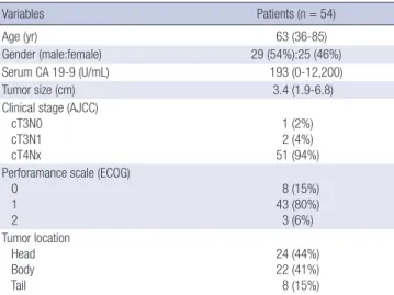
Baseline demographics and clinical characteristics The median age of study patients was 63 yr (range 36 to 85) and there were 29 males and 25 females (Table 1). The median se- rum CA 19-9 level before CCRT was 193 U/mL (range 0 to 12,200).
Almost all patients (51 patients, 94%) had stage III disease at baseline CT, according to the 7th edition American Joint Com- mittee on Cancer (AJCC) Cancer Staging Manual. Of the 54 pa- tients, 14 (26%) were found to have borderline resectable and
Table 1. Baseline demographics and clinical characteristics
Variables Patients (n = 54)
Age (yr) 63 (36-85)
Gender (male:female) 29 (54%):25 (46%)
Serum CA 19-9 (U/mL) 193 (0-12,200)
Tumor size (cm) 3.4 (1.9-6.8)
Clinical stage (AJCC) cT3N0
cT3N1 cT4Nx
1 (2%) 2 (4%) 51 (94%) Perforamance scale (ECOG)
0 1 2
8 (15%) 43 (80%) 3 (6%) Tumor location
Head Body Tail
24 (44%) 22 (41%) 8 (15%) Data are expressed as the median (range) or number (%).
40 (74%) were found to have unresectable pancreatic cancers prior to CCRT, according to NCCN criteria. Invasions into the SMA, SMV, PV, CA, and CHA were detected before treatment in 38, 30, 19, 28, and 20 patients, respectively.
Objective changes in the tumor after chemoradiotherapy The CCRT treatment consisted of administration of gemcitabine, 5-fluorouracil, and capecitabine to 17, 9, and 28 patients, respec- tively. The median radiation dose was 54 (range 32.4-59.4) Gy. No complete response after CCRT was observed in this study. A par- tial response was achieved in four patients (7%). Stable disease
and progressive disease were determined in 36 (67%) and 14 pa- tients (26%), respectively. Among the 14 patients with disease progression, four patients had local progression and the other 10 patients had distant metastasis. Tumor size showed no sig- nificant difference before and after neoadjuvant treatment (3.6
± 1.1 vs. 3.6 ± 1.0 cm, P = 0.61). Vascular invasion was improved in two patients (4%), while further disease progression was seen in 13 patients (24%). No significant differences were noted for PV (P > 0.99), CA (P = 0.53) and CHA (P = 0.20) invasion follow- ing neoadjuvant treatment. SMA (P = 0.008) and SMV (P = 0.04) invasions were rather aggravated after neoadjuvant treatment.
A
B
C A
B
C A
B
C A
B
C A
B
C A
B
C
Before After
A
B
C
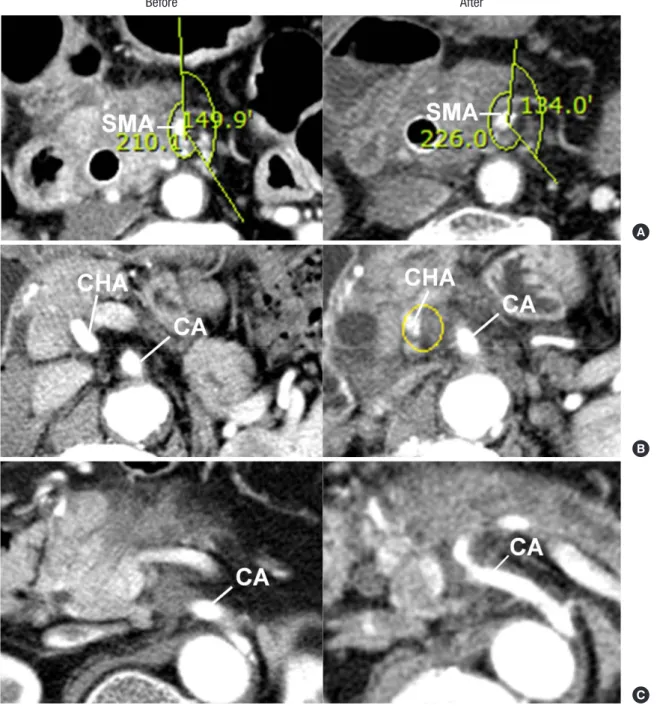
Fig. 1. Objective measurements of vascular invasion before and after chemoradiotherapy. (A) Computed tomography (CT) images at the level of superior mesenteric artery (SMA).
Post-chemoradiotherapy image shows similar vascular involvement with pre-chemoradiotherapy image. (B) CT images at the level of celiac artery (CA) and common hepatic ar- tery (CHA). Post-chemoradiotherapy image shows increased CHA involvement. (C) CT images at the level of CA. Post-chemoraidotherapy image shows decreased CA involvement.
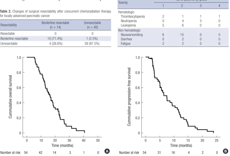
Table 2. Changes of surgical resectability after concurrent chemoradiation therapy for locally advanced pancreatic cancer
Resectability Borderline resectable
(n = 14) Unresectable (n = 40)
Resectable 0 0
Borderline resectable 10 (71.4%) 1 (2.5%)
Unresectable 4 (28.6%) 39 (97.5%)
Table 3. Toxicities during chemoradiation therapy
Toxicity No. of patients by grade
1 2 3 4
Hematologic Thrombocytopenia Neutropenia Leukopenia
2 0 5
1 4 6
1 3 0
1 0 0 Non-hematologic
Nausea/vomiting Diarrhea Fatigue
6 0 2
15 2 2
0 0 0
0 0 0
Fig. 2. Kaplan-Meier curves showing overall survival and progression-free survival of patients. (A) The median overall survival was 16.5 (95% CI 13.2-19.9) months. (B) The median progression-free survival was 6.4 (95% CI 4.0-8.8) months.
Cummulative overall survival
Time (months)
Number at risk 54 42 14 3 1 0
0 10 20 30 40 50 1.0
0.8
0.6
0.4
0.2
0
A
Cummulative progression-free survival
Time (months)
Number at risk 54 31 16 4 2 0
0 5 10 15 20 25 1.0
0.8
0.6
0.4
0.2
0
B Both patients who showed vascular improvement after CCRT
had unresectable disease initially: One had grade 4 CA encase- ment along with grade 2 CHA abutment, and the other had grade 4 CA encasement along with grade 2 CHA abutment. The first patient showed a radiologic response at CA (grade 2) and was surgically downstaged to borderline resectable disease. How- ever, surgical resection was abandoned due to poor general con- dition. The second patient showed a radiologic response at CHA;
however, the tumor was still unresectable as CA encasement showed no improvement. Thirty-nine patients with unresect- able and 11 patients with borderline resectable pancreatic can- cer at initial diagnosis still had unresectable and borderline re- sectable pancreatic cancers, respectively, after CCRT (Fig. 1A).
Four borderline pancreatic cancers progressed further to an unresectable stage (Fig. 1B), whereas one unresectable pancre- atic cancer improved to borderline resectability after CCRT (Fig.
1C). No patient improved to resectable disease staging after CCRT (Table 2). Only one patient with borderline pancreatic cancer (a 75-yr-old man with CA abutment) underwent explor- ative laparotomy although the radiologic response was minimal.
Curative resection failed, however, due to tumor infiltration to the CA and peritoneal metastasis detected during surgery.
We investigated the relationship between chemotherapeutic
agents used for CCRT and tumor progression. Tumor progres- sion after CCRT was observed in two patients (12%) with gem- citabine, one patient (11%) with 5-fluorouracil, and one patient (4%) with capecitabine. No significant difference was seen in tumor progression after CCRT based on the choice of chemo- therapeutic agent (P = 0.54).
Toxicity
A total of 36 patients (67%) experienced CCRT-related adverse events (Table 3). The most common adverse event reported was hematology toxicity (23 patients, 43%). No CCRT-related mortality occurred. Severe toxicities (grade 3-4) arose in five patients (9%). Grade 3 toxicities developed in four patients (one developed thrombocytopenia and three developed neutrope- nia). One patient developed Grade 4 thrombocytopenia and treatment was withheld.
Survival analysis
During follow-up, 46 patients (85%) died and the median over- all survival was 16.2 (95% CI 12.7-19.7) months (Fig. 2A). Dis-
ease progression was observed in 47 patients (87%) and the me- dian progression-free survival was 6.4 months (95% CI 4.0-8.8) (Fig. 2B). Among the patients with disease progression, system- ic presentations (36 patients, 77%) were more frequent than lo- cal progression (11 patients, 23%).
DISCUSSION
Many studies have evaluated the efficacy of neoadjuvant CCRT for LAPC; however, the ambiguous definition of the criteria for determining tumor resectability in these reports complicates judgment of the effectiveness of CCRT. Recent meta-analysis of neoadjuvant treatment in pancreatic cancer showed that the criteria for surgical resectability were either not clearly defined or not stated in more than half of the published studies (4). Our primary interest was to measure the objective changes of vas- cular invasion after CCRT in LAPC. Unfortunately, we found that the objective improvements of vascular invasion after neo- adjuvant treatment were negligible, as none of the enrolled pa- tients were eligible for a curative resection.
Previous studies reported various curative resection rates for pancreatic cancer after neoadjuvant treatment ranging from 1%
to 71% (9, 12, 17, 20-22). The proportion of resectable, border- line resectable, and unresectable patients could significantly affect the curative resection rate after neoadjuvant treatment.
In addition, the heterogeneity of radical resection rates could be explained by the different resectability criteria adopted in these studies. No patients underwent radical resections after CCRT in the present study because most patients were at an unresectable stage at the time of initial diagnosis. About 30%- 40% of patients with pancreatic cancer are diagnosed as LAPC (4), but the proportion of LAPC in this study was only 25%. This showed that, at our institution, LAPC was defined when defi- nite vascular invasion was present on CT imaging, which could have given rise to the low radical resectability rate after neoad- juvant CCRT compared to previous studies. Surgeons have dif- ferent strategies for resection of pancreatic cancers and a sur- geon’s propensity for resection, especially in borderline resect- able disease, could significantly affect the resection rate.
Surgical exploration is the most accurate method for tumor staging. One report diagnosed LAPC by intraoperative staging (either laparoscopy and/or laparotomy) (9). Of a total of 87 pa- tients with LAPC who underwent neoadjuvant chemoradiation, only three patients had a sufficient clinical response on restag- ing to warrant a re-exploration. Of these three patients, only one patient had a potentially curative resection. This result is con- sistent with our study.
The strength of our study is that it quantitatively investigates the extent of tumor invasion in each major vessel. Some studies have assessed the effect of neoadjuvant treatment by RECIST criteria (15-17), which mainly assess the response based on tu-
mor size and distant metastasis (23). However, the RECIST cri- teria do not reflect changes in vascular involvement, which is a most important determinant for the evaluation of surgical re- sectability. A recent study that evaluated the radiological response of patients with locally advanced pancreatic cancer found that neoadjuvant therapy did not induce radiological tumor regres- sion (23). They evaluated the invasion of major vessels based on pre-and post-treatment CT scans. We classified vascular in- volvement from grade 0 to 4 and assessed the change after CCRT.
These objective findings allowed accurate surgical restaging af- ter CCRT.
The development of dual phase, thin-sectioned, and multi- detector CT imaging has improved the accuracy of preoperative staging in pancreatic cancer (24-26). However, radiotherapy causes local inflammation, resulting in over-diagnosis of vascu- lar involvement after CCRT. This could result in misdiagnosis of patients who were candidates for surgery after CCRT as having an unresectable disease. Nevertheless, the possibility of over- diagnosis is assumed to be low, as no improvement of vascular involvement was evident on consecutive follow-up CT imaging.
As this was a retrospective study, we limited the enrollment criteria to patients with LAPC who received CCRT. This minimiz- ed the potential bias that could result from various treatments.
The small number of enrolled patients precludes any recom- mendation that neoadjuvant CCRT should be contraindicated in pancreatic cancer. Palliative chemotherapy with FOLFIRI- NOX (5-fluorouracil, oxaliplatin, irinotecan, and leucovorin) has proven efficacy in metastatic pancreatic cancers (27), and FOLFIRINOX as a neoadjuvant therapy in LAPC recently showed promising results (28, 29). Future research should therefore eval- uate the efficacy of preoperative treatment with FOLFIRINOX for LAPC, with a clear definition of the resectability criteria and presentation of the objective changes after therapy.
We would like to note that the present study is a hallmark study in LAPC, since the objective changes after neoadjuvant treatment were described and the surgical restaging assessment was based on these findings. The objective improvements after neoadjuvant treatment were minimal and the real efficacy of neoadjuvant chemoradiation therapy in LAPC was disappoint- ing. Prospective studies implicating precise resectability criteria and objective changes after treatment are required to clarify conflicting results regarding the efficacy of neoadjuvant treat- ment for pancreatic cancer.
DISCLOSURE
The authors declare no conflicts of interest.
AUTHOR CONTRIBUTION
Conceived and designed the study: Paik WH, Kim YT. Data col-
lection and statistical analysis: Paik WH, Lee SH, Kim YT. Wrote the initial draft: Paik WH, Lee SH, Kim YT. Contributed to the critical aspects of the research: Lee SH, Kim YT, Park JM, Song BJ, Ryu JK. All the authors approved final version of this manu- script to be published.
ORCID
Woo Hyun Paik http://orcid.org/0000-0001-8708-3280 Sang Hyub Lee http://orcid.org/0000-0003-2174-9726 Yong-Tae Kim http://orcid.org/0000-0002-4842-6874 Jin Myung Park http://orcid.org/0000-0002-8798-0587 Byeong Jun Song http://orcid.org/0000-0001-6478-9838 Ji Kon Ryu http://orcid.org/0000-0001-8798-0491
REFERENCES
1. Hidalgo M. Pancreatic cancer. N Engl J Med 2010; 362: 1605-17.
2. Conlon KC, Klimstra DS, Brennan MF. Long-term survival after curative resection for pancreatic ductal adenocarcinoma. Clinicopathologic anal- ysis of 5-year survivors. Ann Surg 1996; 223: 273-9.
3. Chua YJ, Cunningham D. Adjuvant treatment for resectable pancreatic cancer. J Clin Oncol 2005; 23: 4532-7.
4. Gillen S, Schuster T, Meyer Zum Büschenfelde C, Friess H, Kleeff J. Pre- operative/neoadjuvant therapy in pancreatic cancer: a systematic re- view and meta-analysis of response and resection percentages. PLoS Med 2010; 7: e1000267.
5. Jemal A, Murray T, Ward E, Samuels A, Tiwari RC, Ghafoor A, Feuer EJ, Thun MJ. Cancer statistics, 2005. CA Cancer J Clin 2005; 55: 10-30.
6. Mancuso A, Calabrò F, Sternberg CN. Current therapies and advances in the treatment of pancreatic cancer. Crit Rev Oncol Hematol 2006; 58:
231-41.
7. Lowy AM. Neoadjuvant therapy for pancreatic cancer. J Gastrointest Surg 2008; 12: 1600-8.
8. Park DI, Lee JK, Kim JE, Hyun JG, Shim SG, Lee KT, Palk SW, Rhee JC, Choi KW, Lim JH, et al. The analysis of resectability and survival in pan- creatic cancer patients with vascular invasion. J Clin Gastroenterol 2001;
32: 231-4.
9. Kim HJ, Czischke K, Brennan MF, Conlon KC. Does neoadjuvant chemo- radiation downstage locally advanced pancreatic cancer? J Gastrointest Surg 2002; 6: 763-9.
10. Morganti AG, Massaccesi M, La Torre G, Caravatta L, Piscopo A, Tam- baro R, Sofo L, Sallustio G, Ingrosso M, Macchia G, et al. A systematic review of resectability and survival after concurrent chemoradiation in primarily unresectable pancreatic cancer. Ann Surg Oncol 2010; 17: 194- 205.
11. Epelbaum R, Rosenblatt E, Nasrallah S, Faraggi D, Gaitini D, Mizrahi S, Kuten A. Phase II study of gemcitabine combined with radiation therapy in patients with localized, unresectable pancreatic cancer. J Surg Oncol 2002; 81: 138-43.
12. Joensuu TK, Kiviluoto T, Kärkkäinen P, Vento P, Kivisaari L, Tenhunen M, Westberg R, Elomaa I. Phase I-II trial of twice-weekly gemcitabine and concomitant irradiation in patients undergoing pancreaticoduode-
nectomy with extended lymphadenectomy for locally advanced pancre- atic cancer. Int J Radiat Oncol Biol Phys 2004; 60: 444-52.
13. Laurent S, Monsaert E, Boterberg T, Demols A, Borbath I, Polus M, Hen- dlisz A, de Hemptinne B, Mahin C, Scalliet P, et al. Feasibility of radio- therapy with concomitant gemcitabine and oxaliplatin in locally advanc- ed pancreatic cancer and distal cholangiocarcinoma: a prospective dose finding phase I-II study. Ann Oncol 2009; 20: 1369-74.
14. Andriulli A, Festa V, Botteri E, Valvano MR, Koch M, Bassi C, Maison- neuve P, Sebastiano PD. Neoadjuvant/preoperative gemcitabine for pa- tients with localized pancreatic cancer: a meta-analysis of prospective studies. Ann Surg Oncol 2012; 19: 1644-62.
15. Talamonti MS, Small W Jr, Mulcahy MF, Wayne JD, Attaluri V, Colletti LM, Zalupski MM, Hoffman JP, Freedman GM, Kinsella TJ, et al. A multi- institutional phase II trial of preoperative full-dose gemcitabine and con- current radiation for patients with potentially resectable pancreatic car- cinoma. Ann Surg Oncol 2006; 13: 150-8.
16. Cardenes HR, Moore AM, Johnson CS, Yu M, Helft P, Chiorean EG, Vin- son J, Howard TJ, Stephens AW, Tai DF, et al. A phase II study of gem- citabine in combination with radiation therapy in patients with local- ized, unresectable, pancreatic cancer: a Hoosier Oncology Group study.
Am J Clin Oncol 2011; 34: 460-5.
17. Pipas JM, Barth RJ Jr, Zaki B, Tsapakos MJ, Suriawinata AA, Bettmann MA, Cates JM, Ripple GH, Sutton JE, Gordon SR, et al. Docetaxel/Gem- citabine followed by gemcitabine and external beam radiotherapy in patients with pancreatic adenocarcinoma. Ann Surg Oncol 2005; 12:
995-1004.
18. National Comprehensive Cancer Network (NCCN). Pancreatic adeno- carcinoma. Practice Guidelines in Oncology. v.1. Available at http://www.
nccn.org/professionals/physician_gls/PDF/ pancreatic.pdf [accessed on 14 Jul 2014].
19. Saldinger PF, Reilly M, Reynolds K, Raptopoulos V, Chuttani R, Steer ML, Matthews JB. Is CT angiography sufficient for prediction of resect- ability of periampullary neoplasms? J Gastrointest Surg 2000; 4: 233-7;
discussion 8-9.
20. Varadhachary GR, Tamm EP, Abbruzzese JL, Xiong HQ, Crane CH, Wang H, Lee JE, Pisters PW, Evans DB, Wolff RA. Borderline resectable pan- creatic cancer: definitions, management, and role of preoperative thera- py. Ann Surg Oncol 2006; 13: 1035-46.
21. Kamthan AG, Morris JC, Dalton J, Mandeli JP, Chesser MR, Leben D, Cooperman A, Bruckner HW. Combined modality therapy for stage II and stage III pancreatic carcinoma. J Clin Oncol 1997; 15: 2920-7.
22. McFarland EG, Kaufman JA, Saini S, Halpern EF, Lu DS, Waltman AC, Warshaw AL. Preoperative staging of cancer of the pancreas: value of MR angiography versus conventional angiography in detecting portal venous invasion. AJR Am J Roentgenol 1996; 166: 37-43.
23. Dudeja V, Greeno EW, Walker SP, Jensen EH. Neoadjuvant chemora- diotherapy for locally advanced pancreas cancer rarely leads to radio- logical evidence of tumour regression. HPB (Oxford) 2013; 15: 661-7.
24. Valls C, Andia E, Sanchez A, Fabregat J, Pozuelo O, Quintero JC, Serra- no T, Garcia-Borobia F, Jorba R. Dual-phase helical CT of pancreatic ad- enocarcinoma: assessment of resectability before surgery. AJR Am J Roent- genol 2002; 178: 821-6.
25. Tamm EP, Loyer EM, Faria S, Raut CP, Evans DB, Wolff RA, Crane CH, Dubrow RA, Charnsangavej C. Staging of pancreatic cancer with multi- detector CT in the setting of preoperative chemoradiation therapy. Ab-
dom Imaging 2006; 31: 568-74.
26. Wong JC, Lu DS. Staging of pancreatic adenocarcinoma by imaging studies. Clin Gastroenterol Hepatol 2008; 6: 1301-8.
27. Conroy T, Desseigne F, Ychou M, Bouché O, Guimbaud R, Bécouarn Y, Adenis A, Raoul JL, Gourgou-Bourgade S, de la Fouchardière C, et al.;
Groupe Tumeurs Digestives of Unicancer; PRODIGE Intergroup. FOL- FIRINOX versus gemcitabine for metastatic pancreatic cancer. N Engl J Med 2011; 364: 1817-25.
28. Christians KK, Tsai S, Mahmoud A, Ritch P, Thomas JP, Wiebe L, Kelly T,
Erickson B, Wang H, Evans DB, et al. Neoadjuvant FOLFIRINOX for borderline resectable pancreas cancer: a new treatment paradigm? On- cologist 2014; 19: 266-74.
29. Ferrone CR, Marchegiani G, Hong TS, Ryan DP, Deshpande V, McDon- nell EI, Sabbatino F, Santos DD, Allen JN, Blaszkowsky LS, et al. Radio- logical and surgical implications of neoadjuvant treatment with FOL- FIRINOX for locally advanced and borderline resectable pancreatic can- cer. Ann Surg 2015; 261: 12-7.