The Effect of Preceding Biopsy on Complete Endoscopic Resection in Rectal Carcinoid Tumor
Biopsy of rectal carcinoid tumor is commonly taken before endoscopic resection. However the preceding biopsy can inhibit complete resection by causing blurred tumor border and fibrosis of the tissue. The objective of the study was to investigate the effect of preceding biopsy on complete endoscopic resection in rectal carcinoid tumor. It was also determined if rectal carcinoid tumors can be macroscopically distinguished by endoscopy. We reviewed retrospectively the records of patients with rectal carcinoid tumor who had undergone an endoscopic treatment at our hospital, during a 7-yr period. The resection margin was clear in 57 of 98 cases. The preceding biopsy was taken in 57 cases and the biopsy was
significantly associated with the risk of incomplete tumor resection (OR, 3.696; 95% CI, 1.528-8.938, P = 0.004). In 95.9% of the cases, it was possible to suspect a carcinoid tumor by macroscopic appearance during initial endoscopy. The preceding biopsy may disturb complete resection of rectal carcinoid tumor. In most cases, the carcinoid tumor could be suspected by macroscopic appearance. Therefore the preceding biopsy is not essential, and it may be avoided for the complete resection.
Keywords: Carcinoid Tumor; NETs; Rectal Carcinoid Tumor; Endoscopic Treatment; Biopsy Sang Pyo Lee, In-Kyung Sung,
Jeong Hwan Kim, Sun-Young Lee, Hyung Seok Park, and Chan Sup Shim Department of Internal Medicine, Konkuk University School of Medicine, Seoul, Korea
Received: 7 November 2013 Accepted: 14 February 2014 Address for Correspondence:
In-Kyung Sung, MD
Department of Internal Medicine, Digestive Disease Centre, Konkuk University School of Medicine, 120-1 Neungdong-ro, Gwangjin-gu, Seoul 143-729, Korea
Tel: +82.2-2030-5100, Fax: +82.2-2030-7748 E-mail: [email protected]
http://dx.doi.org/10.3346/jkms.2014.29.4.512 • J Korean Med Sci 2014; 29: 512-518
INTRODUCTION
Carcinoid tumors are rare, slow-growing malignancies arising from enterochromaffin cells (1, 2). They can occur in nearly any gastrointestinal tissue; and rectum is one of the most common- ly reported sites (3-5). According to a previous study, rectal car- cinoids comprised 18.5% of all carcinoid tumors, and 27.4% of all gastrointestinal carcinoids (1). Carcinoid tumors of the rec- tum are likely increasing in incidence (1, 6).
Most rectal carcinoid tumors are small and movable subepi- thelial lesions located between 4 and 20 cm above the dentate line and are mainly discovered incidentally on routine lower endoscopy (7-10). Such a small tumor rarely metastasizes and endoscopic resection is curative. Metastases to bone, lymph nodes and liver are only likely in tumors that are 2 cm and larg- er and they invade the muscularis propria (6, 7, 11). Overall dis- tant metastases from rectal carcinoids occur in only 2.3%-3.9%
(1, 3).
It is enough to remove a small lesion with the endoscopic treatment only, but it is important to resect completely (12-14).
Any unsuccessfully resected lesions may need frequent follow- up or further treatment such as additional endoscopic treatment and operation.
Biopsy of carcinoid tumor is commonly taken before endo- scopic resection. However most carcinoid tumors can be mac- roscopically distinguished by endoscopy, and a biopsy is not essential. In addition, biopsy can inhibit a complete resection
by causing blurred tumor border and fibrosis of the tissue. Pre- vious studies have revealed that the method of endoscopic treat- ment is an important factor in complete resection of rectal car- cinoid tumor (12, 15). However little has been known about the effect of preceding biopsy on complete resection. Only one pre- vious study suggested that the post-biopsy scar can interfere with successful endoscopic resection of rectal carcinoid tumor (16). However it had limited significance due to a small number of cases.
The primary objective of this study is to investigate the risk factors of incomplete tumor resection in rectal carcinoid tumor and to look into the effect of preceding biopsy on complete en- doscopic resection. The secondary objective is to determine if rectal carcinoid tumors can be macroscopically distinguished by endoscopy.
MATERIALS AND METHODS Patients and inclusion criteria
We reviewed retrospectively the records of patients with rectal subepithelial tumor who had undergone an endoscopic treat- ment at Konkuk University Hospital (Seoul, Korea), during a 7-yr period from July 2005 to July 2012. The data gathered on the patients and procedures were through endoscopic images, review of chart, and histologic results.
To be eligible for inclusion, the tumors had to be located in the rectum and be less than 20 mm in diameter. The endoscop-
ic treatments which were included in the study were endoscopic submucosal dissection (ESD), endoscopic mucosal resection- band ligation (EMR-L), and conventional snare polypectomy.
We excluded tumors resected by other methods, including hot biopsy, argon plasma coagulation (APC) and cold biopsy. De- pending on the histological results, well-differentiated neuro- endocrine tumors (grade I or II), namely carcinoid tumors, were included and other tumors were excluded. The tumors which involved muscularis propria, adjacent vessels, lymph nodes, or distant organ were additionally excluded. The cases with inad- equate medical records were excluded as well. For patients with multiple rectal carcinoid tumors, each tumor removed by en- doscopic treatment was categorized as a separate case.
Probable risk factors for incomplete tumor resection According to completeness of resection, each tumor was classi- fied into clear resection margin group (A group) and positive margin group (B group). When both the lateral and deep mar- gins were free of tumor cells, it was denominated as complete resection. If it was unclear whether resection margin was involv- ed, it was included in the positive margin group, as the subse- quent therapeutic plan would be similar.
Probable risk factors for incomplete tumor resection were as follows: preceding biopsy before endoscopic resection; size of the tumor measured by endoscopist; location of tumor; mor- phology of tumor; and method of endoscopic treatment. In ad- dition, we investigated the depth of invasion and whether en- doscopic ultrasonography (EUS) was performed.
The location of tumor was divided into proximal and distal part of the rectum. Distal rectum was defined as the rectum lo- cated below the second valve of Houston. We used the diame- ter as measured by endoscopist during endoscopy rather than the diameter of specimen measured by pathologist, as part of the mass may have been removed by preceding biopsy. Also, in the positive resection margin group, a small portion of the re- sidual tumor may have still remained on the resection site. There- fore the size of specimen as measured by pathologist may be smaller than the actual size.
The endoscopic morphology of tumor was divided into three types: protruded type; slightly elevated or flat type; and depress- ed or ulcerative type (Fig. 1). If biopsy was taken before the en- doscopic resection, morphology after the preceding biopsy was selected as a risk factor of incomplete resection, as morphology of tumor may be transformed by biopsy. Describing tumor ac- cording to the Parris-Japanese classification, protruded type belonged to Ip, Isp and Is lesions, slightly elevated or flat type belonged to IIa and IIb lesions and depressed or ulcerative type were IIc and III lesions.
The endoscopic treatments were classified into three meth- ods: ESD, EMR-L and conventional snare polypectomy. Simpli- fied ESD which performed submucosal dissection with snaring
was included in the ESD method.
Review of endoscopic records
In order to determine if carcinoid tumors were distinguished by endoscopy, we searched for the endoscopic records at the time of diagnosis. If the patient was referred from another hospital, we additionally reviewed the medical treatment request and endoscopic images of the previous hospital. Then we investi- gated whether “carcinoid” was written in the medical records before the patient was histologically confirmed to carcinoid tu- mor. In addition, we reviewed the records of patients who had rectal subepithelial tumor during the same study period and investigated the cases where the rectal tumor was recorded as
“carcinoid” on medical record, but in fact were not.
We also reviewed the endoscopic images to see whether the shape and color of tumor were typical and if it looked like a sub- epithelial lesion, macroscopically. Review of the images was performed by two experienced endoscopists. If there were dif- fering views, an agreement was reached through a discussion.
Endoscopic procedure
Standard colonoscope (CF-H260; Olympus, Tokyo, Japan) was used for the procedure. The colon was cleansed with 4 L poly- ethylene glycol electrolyte solution. Midazolam (Dormicum®) for conscious sedation and meperidine (Demerol®) for pain control were selectively given to patients. The procedure was performed in a standard fashion by one endoscopist and one assistant, and tumor removed following a saline injection mixed with indigocarmine and epinephrine. Conventional snare pol- ypectomy was performed using oval-shaped snares (Olympus, Tokyo, Japan). Band ligation method was performed by stan- dard upper endoscope (GF-260; Olympus) and pneumo-acti- vate ELV device (Sumitomo Bakelite Co., Ltd., Tokyo, Japan). ESD was mostly carried out using a dual knife (KD-650L; Olympus).
Statistical analysis
Continuous variables were summarized as mean ± standard deviation (SD), and categorical variables as frequency (%). To analyze the risk factors of incomplete tumor resection, categori- cal data were compared using chi-square or Fisher’s exact tests.
Then, we used logistic regression analysis to estimate the odds ratio (OR) and 95% confidence interval (CI) of risk factors that caused incomplete tumor resection. All analyses were conduct- ed using SPSS for Windows (versions 19.0). A P value of less than 0.05 was considered statistically significant.
Ethics statement
The study was approved by the institutional review board of Konkuk University School of Medicine which confirmed that the study was in accordance with the ethical guidelines of the Helsinki Declaration (KUH1010502). All patients provided writ-
A
C-1
B
C-2
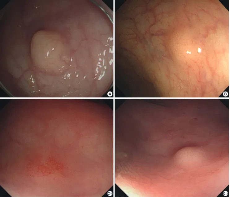
Fig. 1. Color and endoscopic morphology of rectal carcinoid tumor. Among the 98 cases, tumors were yellow in 85 cases, white and pale in 10 cases, and the other three cas- es were similar in color to the surrounding normal mucosa. Tumor morphology after biopsy was classified into protruded type in 51 cases, slightly elevated or flat type in 43 and depressed or ulcerative type in 4. (A) The tumor is white and pale and morphologically belonged to the protruded type. (B) The color of tumor is yellow and it is of the slightly el- evated type. (C) In the C-1 image, the tumor is a depressed type, and the color is neither yellow nor white. It could not be easily distinguished from the surrounding mucosa, but the tumor had definite capillary bed. C-2 image was the same patient’s endoscopic image before the biopsy was taken. It shows typical features of carcinoid tumor, which was yellow-colored and slightly elevated type.
ten consents to undergo endoscopic treatment and were in- formed of the risks and potential benefits of the procedures.
RESULTS
Characteristics of patients and tumors
Over the 7-yr period studied, 5,235 patients had undergone colonoscopic polypectomy, EMR or ESD in our unit. Among the patients, 113 patients were diagnosed with rectal subepi- thelial tumor and 16 of them were not carcinoid tumor. There- fore we identified 97 patients with rectal carcinoid who had un- dergone endoscopic tumor resection. There were three cases
with no tumor in the resected tissue by endoscopic resection, although preceding biopsy revealed a carcinoid tumor. It may be that the small tumor was almost entirely resected by biopsy forceps, when the biopsy was performed. These three cases were excluded from the study, as they were not treated by the prescribed methods and the resection margin status could not be identified. In five patients, carcinoid tumor was found si- multaneously at more than two locations. Four of these patients had two concurrent rectal carcinoids, and the other had one rectal carcinoid and one ampulla of Vater carcinoid. Thus, a to- tal of 98 cases (94 patients) were finally included for the study (Fig. 2). The overall mean age and tumor size were 48.04 ± 11.12
(range 29-75) yr and 7.40 ± 2.86 (range 3-15) mm, respectively.
Other characteristics of the tumors are shown in Table 1.
Comparison of group A and B
The clear resection margin group (A group) and the positive margin group (B group) had 57 and 41 cases, respectively (Table
2). The B group included six cases where the margin status was unclear. Thirty five cases with definitely positive resection mar- gin were composed of 18 deep margin positive cases, 1 lateral margin positive case, 2 both margin positive cases and 14 cases which were not marked separately.
The mean age of the A group was 49 ± 12 (range 29-75) yr, and the mean tumor size was 7.26 ± 2.83 (range 3-15) mm in diameter. The mean age of the B group was 47 ± 10 (range 29- 68), and the mean tumor size was 7.59 ± 2.90 mm (range 3-15).
A previous biopsy had been performed in 26 cases (45.6%) in group A and 31 cases (75.6%) in group B.
In group A, 21 (36.8%) tumors were removed by ESD, 28 (49.1%) tumors resected by snare polypectomy and 8 (14.0%) were EMR- L. In group B, ESD, snare polypectomy and EMR-L were perform- ed in 13 (31.7%), 25 (61.0%) and 3 (7.3%) cases, respectively.
Tumor morphology before biopsy was divided into 57 pro- truded types and 39 slightly elevated or flat types. There was no depressed or ulcerative type. Two cases were not evaluated due to poor endoscopic images. However, tumor morphology after biopsy was changed into 51 protruded types, 43 slightly elevat- ed or flat types, and 4 depressed or ulcerative types. Lesions con- fined to the mucosal layer were 2 in group A and 1 in group B.
The rest had submucosal invasion. In both A and B groups, EUS was performed in 15 cases for each group. Of these several risk factors, only preceding biopsy was significantly associated with the risk of incomplete tumor resection (P = 0.004, chi-square tests, Table 2). Logistic regression analysis revealed that patients who were performed preceding biopsy were about 3.7 times more likely to fail to completely remove tumors as compared to 5,235 patients who underwent colonoscopic polypectomy,
EMR, or ESD during the study period
Subepithelial tumors on rectum: 113 patients
Rectal carcinoid tumor: 94 patients (98 cases) Inclusion criteria:
- Tumors located in rectum and below 20 mm in diameter - Tumors confined to the submucosal layer
- Tumors resected by conventional polypectomy, EMR-L, or ESD
- Well differentiated neuroendocrine tumors (grade I or II)
Excluded patients
Chronic nonspecific inflammation: 5 patients Inflammatory polyp: 2
Hyperplastic polyp: 2 Lymphoid polyp: 2 Leiomyoma: 2 Lipoma: 2
Malignant lymphoma, extranodal marginal zone B-cell lymphoma: 1 Carcinoid tumor which was removed by cold biopsy: 3
The A group (the clear resection margin group): 57 cases The B group (the positive margin group): 41 cases
Fig. 2. Selection of the cases. A total of 5,235 patients underwent endoscopic treatment of colonic tumor (polypectomy, EMR or ESD) at our hospital, during a 7-yr period from July 2005 to July 2012. Among them, we identified 113 patients with subepithelial tumor on rectum who had undergone endoscopic tumor resection. Finally, 98 rectal carci- noid cases (94 patients) were included, and the resection margin was clear in 57 cases.
Table 1. Baseline characteristics of the patients and tumors
Characteristics No. (%) or mean (SD)*
Age (yr) 48.04 (11.1)
Female sex 34 (34.7)
Size† (mm) 7.40 (2.9)
Preceding biopsy 57 (58.2)
Morphology Protruded
Slightly elevated or flat Depressed or ulcerative
51 (52.0) 43 (43.9) 4 (4.1) Treatment method
ESD
Snare polypectomy EMR-L
34 (34.7) 53 (54.1) 11 (11.2) Location
Proximal rectum
Distal rectum 16 (16.3)
82 (83.7) Color
Yellow
White or pale mucosa Normal colored mucosa
85 (86.7) 10 (10.2) 3 (3.1)
EUS 30 (30.6)
Clear resection margin 57 (58.2)
*Continuous variables were summarized as mean ± standard deviation (SD) and cate- gorical variables as frequency (%); †The tumor size was measured by endoscopist during endoscopy. ESD, endoscopic submucosal dissection; EMR-L, endoscopic mu- cosal resection-band ligation; EUS, endoscopic ultrasonography.
patients with naïve lesions (OR, 3.696; 95% CI, 1.528-8.938; P = 0.004).
Identification of carcinoid tumor by initial endoscopy All 98 carcinoid cases were observed as subepithelial lesion by the endoscopic images. Eighty five cases were yellow-colored tumor and ten cases were white and pale. Only three cases were similar in color to the surrounding normal mucosa. There were five cases which had definite capillary bed on tumor (Fig. 1). In all cases without biopsy and in 53 out of the 57 cases with pre- ceding biopsy, “carcinoid” was written in the initial medical re- cords before tumor was histologically confirmed as carcinoid.
There were no unevaluated cases due to lack of the initial endo- scopic record. Therefore, in 95.9% (94 cases) of all cases, carci- noid tumor could be suspected by macroscopic appearance at the time of the initial endoscopic examination.
There were five cases where the endoscopist attempted to re- move the tumor with biopsy forceps, carcinoid tumor was not
suspected at the time. In three of these cases, there was no re- sidual tumor in the specimen obtained by additional polypec- tomy, and they were excluded from our study as stated earlier.
The other two cases came under the four cases where “carcinoid”
was not written in the initial medical records.
In three cases, although a preceding biopsy did not reveal a carcinoid tumor, a polypectomy was done from a strongly sus- pected carcinoid tumor macroscopically. These three cases were all diagnosed with carcinoid tumor, finally.
There were 16 patients who had rectal subepithelial tumor excepting carcinoid tumor in the study period. The histologic results were as follows: chronic nonspecific inflammation, in- flammatory polyp, hyperplastic polyp, lymphoid polyp, leiomy- oma, lipoma and malignant lymphoma (Fig. 2). Among these, according to the initial endoscopic records, there were six pa- tients where the tumor looked like rectal carcinoid, but were not. None of the cases underwent EUS, and the final histologic results showed chronic nonspecific inflammation in three pa- tients, lymphoid polyp in one, lipoma in one, and hyperplastic polyp in one (Fig. 3).
Follow-up
In the B group, an evaluation was performed by patient’s medi- cal record from the timing of the polypectomy to October 2013.
There has been no recurrence within the follow-up period and one patient underwent a surgical operation (lower anterior re- section) due to the carcinoid tumor. Only 24 out of 41 patients were properly monitored. Their mean follow-up period was 33.29 ± 24.75 (range 12-95) months and a first follow-up endos- copy and imaging studies were performed at 7.25 ± 6.42 (range 2-24) months after the procedure. In one patient, follow-up study was performed at four months after the procedure, and then the patient did not visit the hospital for six years. The remaining 16 patients did not visit our outpatient gastroenterology clinic after the procedure. Two of them underwent health examina- tion in our hospital after two and seven years, respectively, and there was no relapse.
DISCUSSION
Rectal carcinoids are diagnosed in relatively young patients in their 40s to 60s, and the incidence of multicentric carcinoids of the colon is low (1, 17). In our study, the mean age was 48 yr old and multicentric carcinoids were found in 5 out of 94 patients.
These results were similar to the results of previous studies.
According to our results, preceding biopsy performed before endoscopic excision was the only factor of disturbing a com- plete resection of carcinoid tumor. The reasons for this are de- termined to be as follows. First, biopsy can make uncertain bor- ders of tumors. It is important to localize the lesion to remove a carcinoid tumor, because carcinoid tumor is located in deeper Table 2. Probable risk factors for incomplete resection of rectal carcinoid tumor and
comparison of group A and B Probable risk factors A group
(n = 57)
B group (n = 41)
P
value* OR (95% CI)*
Age, yr, mean (SD)†
< 40 yr, No. (%) 40-49 50-59 60 ≤
48.51 (11.6) 15 (26.3) 19 (33.3) 12 (21.1) 11 (19.3)
47.39 (10.4) 12 (29.3) 13 (31.7) 10 (24.4) 6 (14.6)
0.281
Female sex, No. (%) 24 (42.1) 10 (24.4) 0.139 Size‡, mm, mean (SD)†
1-9 mm, No. (%) 10-19 mm
7.26 (2.8) 41 (71.9) 16 (28.1)
7.59 (2.9) 27 (65.9) 14 (34.2)
0.657
Preceding biopsy, No. (%) 26 (45.6) 31 (75.6) 0.004 3.696 (1.53-8.94) Morphology, No. (%)
Protruded
Slightly elevated or flat Depressed or ulcerative
32 (56.1) 24 (42.1) 1 (1.75)
19 (46.3) 19 (46.3) 3 (7.3)
0.348
Treatment method, No. (%) ESD
Snare polypectomy EMR-L
21 (36.8) 28 (49.1) 8 (14.0)
13 (31.7) 25 (61.0) 3 (7.3)
0.443
Location, No. (%) Proximal rectum
Distal rectum 9 (15.8)
48 (84.2) 7 (17.1) 34 (82.9)
0.865
Color Yellow White or pale Normal colored
47 7 3
38 3 0
0.232
EUS 15 (26.3) 15 (36.6) 0.374
Depth of invasion
Confined to mucosa 2 (3.5) 1 (2.4)
*Data were analyzed by chi-square test or Fisher’s exact tests. Then, we used logistic regression analyses to estimate the odds ratio (OR) and 95% confidence intervals (CI).
The significant results are written in bold; †Continuous variables were summarized as mean ± standard deviation (SD) and categorical variables as frequency (%); ‡The tu- mor size was measured by endoscopist during endoscopy. The A and B group indicate the clear resection margin group and the positive margin group, respectively. ESD, en- doscopic submucosal dissection; EMR-L, endoscopic mucosal resection-band ligation.
Fig. 3. Rectal subepithelial tumors mimicking carcinoid tumor. There were six patients with tumor which looked to be rectal carcinoid, but were not. The final histologic results showed chronic nonspecific inflammation in 3 patients (A-C), lymphoid polyp (D), lipoma (E) and hyperplastic polyp (F). All of these patients did not undergo EUS. The retrospec- tive review of the images showed that image C and F did not look like a subepithelial lesion.
A
D
B
E
C
F
layer than other polypoid lesions and the tumor is commonly small and subtle. If the lesion is flattened and margins blurred after biopsy, snaring and targeting of the lesion may be difficult.
The second reason is tissue fibrosis after biopsy. Tissue fibrosis may disturb dissection in ESD procedure and suction in EMR-L.
Previous studies revealed that pathologically complete resec- tion of small rectal carcinoid tumors was more likely to be achi- eved using advanced endoscopic techniques rather than con- ventional polypectomy (12, 15, 18). The complete resection rates were 30.9%, 72.0%, and 81.8% for conventional polypecto- my, advanced endoscopic techniques and surgical local exci- sion, respectively (15). Another study showed that the complete resection rates were 20.0% by conventional polypectomy, 84.6%
by two-channel endoscopic mucosal resection and 77.8% by ESD (12). In our study, such tendency was also observed, but the result of ESD method was not as good as expected. The rea- sons for this may be that it is very difficult to dissect the submu- cosal layer of small lesions below 1 cm in size. It is also techni- cally difficult to resect a tumor which is located in distal rectum.
In addition, previous results of ESD method were not adjusted for the effect of preceding biopsy. In general, preceding biopsy tends to be taken less often before ESD than other methods due to the concern for tissue fibrosis. However biopsy was frequent- ly done before ESD in our study. This may be one reason for low complete resection rate of rectal carcinoid tumor treated by
ESD in our study.
Rectal carcinoid tumor has many distinguishable endoscopic features. Endoscopic findings of carcinoid tumor are character- ized by smooth, round and sessile elevations covered with yel- low and pale colored mucosa (19). The tumor is located in deep mucosa and submucosa, and is usually movable and hard (3, 20). In our study, almost every carcinoid lesion was also observ- ed as a yellowish or whitish subepithelial tumor, except in three cases. To add to these findings, it is imperative to make accurate diagnosis by checking for hardness and movability of tumor us- ing forceps. In 95.9% of all our cases, carcinoid tumor could be suspected by macroscopic appearance during initial endosco- py. If diagnosis is difficult by these findings only, EUS may be helpful in the diagnosis and treatment plan. Among the six pa- tients who were misdiagnosed, there was no one who under- went EUS. In addition, the retrospective review of the endoscop- ic images showed that two of six tumors did not look like a sub- epithelial lesion (Fig. 3C and F). Therefore careful observation by experienced endoscopist may lower the rate of mis diagnosis.
Our study had some limitations. First, if a detailed record was omitted by the endoscopist, it would have caused spurious re- sults, as our study was based on the medical records. Over the 7-yr study period, there were six cases where the tumors looked like rectal carcinoid, but were not. However we assume that there may have been more cases lacking formal records of en-
doscopic findings. In addition, among the cases where “carci- noid” was not written in the initial medical records, it was un- clear whether the endoscopists had suspected carcinoid tumor or not. Second, there may be some misdiagnoses, as the initial endoscopic impressions are founded on the interpretations of individual endoscopist. According to review of the endoscopic images, some of the lesions which were classified subepithelial tumor seemed to not be such (Fig. 3). Third, in many cases, we could not sort the resection margin status into lateral and deep margin positive due to poor pathologic record and fragmenta- tion of the specimen. However categorization of the lateral or deep margin involvement may be important, as lateral and deep margin involvement can occur for different reasons. Therefore, in the future, well-designed prospective studies are required for separate analysis of the lateral and deep margin involvement.
Forth limitation is a lack of data on the prognosis of the B group.
There was no recurrence within the follow-up period, however the period of follow-up was relatively short and there were a lot of losses to follow-up. Rectal carcinoid tumor is slow-growing tumor. Therefore long-term follow-up study is needed to over- come this limitation.
In conclusion, biopsy of rectal carcinoid tumor may have to be avoided. Most carcinoid tumors can be macroscopically dis- tinguished by endoscopy, and biopsy can inhibit a complete resection. Incompletely resected lesions may need frequent fol- low-up endoscopy and/or further treatments. As a result, it can increase additional medical costs and cause needless suffering.
At this time, biopsy of rectal carcinoid tumor is commonly tak- en before endoscopic resection. According to our study, a pre- ceding biopsy was taken in 41.8% of the cases. Therefore we cau- tiously suggest that biopsy of rectal carcinoid tumor should not be taken in the field for the complete resection of tumor, espe- cially if the tumor has macroscopically distinguishable features.
However, in proof of this, more research must be done.
DISCLOSURE
The authors have no conflicts of interest to disclose.
ORCID
Sang Pyo Lee http://orcid.org/0000-0002-4495-3714 In-Kyung Sung http://orcid.org/0000-0002-3848-5571
REFERENCES
1. Modlin IM, Lye KD, Kidd M. A 5-decade analysis of 13,715 carcinoid tu- mors. Cancer 2003; 97: 934-59.
2. Klimstra DS, Modlin IR, Coppola D, Lloyd RV, Suster S. The pathologic classification of neuroendocrine tumors: a review of nomenclature, grad- ing, and staging systems. Pancreas 2010; 39: 707-12.
3. Pinchot SN, Holen K, Sippel RS, Chen H. Carcinoid tumors. Oncologist
2008; 13: 1255-69.
4. Pascarella MR, McCloskey D, Jenab-Wolcott J, Vala M, Rovito M, McHugh J. Large cell neuroendocrine carcinoma of the colon: a rare and aggres- sive tumor. J Gastrointest Oncol 2011; 2: 250-3.
5. Kang H, O’Connell JB, Leonardi MJ, Maggard MA, McGory ML, Ko CY.
Rare tumors of the colon and rectum: a national review. Int J Colorectal Dis 2007; 22: 183-9.
6. Shebani KO, Souba WW, Finkelstein DM, Stark PC, Elgadi KM, Tanabe KK, Ott MJ. Prognosis and survival in patients with gastrointestinal tract carcinoid tumors. Ann Surg 1999; 229: 815-21.
7. Ramage JK, Goretzki PE, Manfredi R, Komminoth P, Ferone D, Hyrdel R, Kaltsas G, Kelestimur F, Kvols L, Scoazec JY, et al. Consensus guidelines for the management of patients with digestive neuroendocrine tumours:
well-differentiated colon and rectum tumour/carcinoma. Neuroendo- crinology 2008; 87: 31-9.
8. Klöppel G, Perren A, Heitz PU. The gastroenteropancreatic neuroendo- crine cell system and its tumors: the WHO classification. Ann N Y Acad Sci 2004; 1014: 13-27.
9. Vilallonga R, Espín Basany E, López Cano M, Landolfi S, Armengol Car- rasco M. Neuroendocrine carcinomas of the colon and rectum: a unit’s experience over six years. Rev Esp Enferm Dig 2008; 100: 11-6.
10. Cesar D, Zanatto RM, da Silva MV, Golçalves R, de Mello EL, de Jesus JP. Colon and rectum neuroendocrine tumors: experience of the Nation- al Cancer Institute in Brazil. Arq Bras Cir Dig 2013; 26: 36-9.
11. Kim MS, Hur H, Min BS, Baik SH, Lee KY, Kim NK. Clinical outcomes for rectal carcinoid tumors according to a new (AJCC 7th edition) TNM staging system: a single institutional analysis of 122 patients. J Surg On- col 2013; 107: 835-41.
12. Onozato Y, Kakizaki S, Iizuka H, Sohara N, Mori M, Itoh H. Endoscopic treatment of rectal carcinoid tumors. Dis Colon Rectum 2010; 53: 169-76.
13. Chagpar R, Chiang YJ, Xing Y, Cormier JN, Feig BW, Rashid A, Chang GJ, You YN. Neuroendocrine tumors of the colon and rectum: prognostic relevance and comparative performance of current staging systems. Ann Surg Oncol 2013; 20: 1170-8.
14. Ishikawa H, Imanishi K, Otani T, Okuda S, Tatsuta M, Ishiguro S. Effec- tiveness of endoscopic treatment of carcinoid tumors of the rectum. En- doscopy 1989; 21: 133-5.
15. Son HJ, Sohn DK, Hong CW, Han KS, Kim BC, Park JW, Choi HS, Chang HJ, Oh JH. Factors associated with complete local excision of small rectal carcinoid tumor. Int J Colorectal Dis 2013; 28: 57-61.
16. Cho SB, Park SY, Yoon KW, Lee S, Lee WS, Joo YE, Kim HS, Choi SK, Rew JS. The effect of post-biopsy scar on the submucosal elevation for endosco- pic resection of rectal carcinoids. Korean J Gastroenterol 2009; 53: 36-42.
17. Tichansky DS, Cagir B, Borrazzo E, Topham A, Palazzo J, Weaver EJ, Lan- ge A, Fry RD. Risk of second cancers in patients with colorectal carcinoids.
Dis Colon Rectum 2002; 45: 91-7.
18. Choi CW, Kang DH, Kim HW, Park SB, Jo WS, Song GA, Cho M. Com- parison of endoscopic resection therapies for rectal carcinoid tumor: en- doscopic submucosal dissection versus endoscopic mucosal resection us- ing band ligation. J Clin Gastroenterol 2013; 47: 432-6.
19. Shim KN, Yang SK, Myung SJ, Chang HS, Jung SA, Choe JW, Lee YJ, By- eon JS, Lee JH, Jung HY, et al. Atypical endoscopic features of rectal car- cinoids. Endoscopy 2004; 36: 313-6.
20. Eggenberger JC. Carcinoid and other neuroendocrine tumors of the co- lon and rectum. Clin Colon Rectal Surg 2011; 24: 129-34.