대한임상신경생리학회지 18(1):18-20, 2016 pISSN 1229-6414 eISSN 2288-1026 http://dx.doi.org/10.14253/kjcn.2016.18.1.18
Copyright 2016 by The Korean Society of Clinical Neurophysiology
This is an Open Access article distributed under the terms of the Creative Commons Attribution Non-Commercial License (http://creativecommons.org/licenses/by-nc/3.0) which permits unrestricted non-commercial use, distribution, and reproduction in any medium, provided the original work is properly cited.
Address for correspondence;
Dong Hoon Shin
Department of Neurology, Gachon University, Gil Medical Center 21, Namdong-daero 774beon-gil, Namdong-gu, Incheon 21565, Korea Tel: +82-32-460-3346 Fax: +83-32-460-3344
E-mail: [email protected]
Case Report
근위축성측삭경화증 환자에서의 myelin water fraction MRI 1예
가천대학교 길병원 신경과1, 서울대학교 전자전기공학과 바이오메디컬 영상과학연구실2, 가천대학교 길병원 영상의학과3
양지원
1․이종호
2․김응엽
3․신동훈
1Myelin Water Fraction MRI in a Case of Clinically Probable Amyotrophic Lateral Sclerosis
Jiwon Yang
1, Jongho Lee
2, EungYeop Kim
3, Dong Hoon Shin
11Department of Neurology, Gachon University Gil Medical Center, Incheon, Korea
2Laboratory for Imaging Science and Technology (LIST), Department of Electrical and Computer Engineering, Seoul National University, Seoul, Korea
3Department of Radiology, Gachon University Gil Medical Center, Incheon, Korea
Amyotrophic lateral sclerosis (ALS) is a progressive motor neuron degenerative disease that clinically manifests both upper and lower motor neuron signs. However, it is unknown where and how the motor neuron degeneration begins, and conflicting hypotheses have been suggested. Recent advanced radiological techniques enable us to look into ALS neuropathology in vivo. Herein, we report a case with upper motor neuron-predominant ALS in whom the results of brain magnetic resonance imaging (MRI) and myelin water fraction MRI suggest axonal degeneration. (Korean J Clin Neurophysiol 2016;18:18-20)
Key Words: Amyotrophic lateral sclerosis, Magnetic resonance imaging, Pathology
Received 7 January 2016; received in revised form 20 February 2016; accepted 9 May 2016.
Amyotrophic lateral sclerosis (ALS) is a relentlessly pro- gressive neurodegenerative disease with heterogeneous clinical manifestations and both upper and lower motor neuron signs.
Pathologically, ALS is characterized by motor neuron degener- ation and death with gliosis replacing lost neurons.1 It has not been established where ALS begins. Three hypotheses have been developed pertaining to the site of disease onset in ALS.
The ‘dying-forward’ hypothesis proposes that ALS is mainly a disorder of corticomotoneurons, which connect monosynaptically with anterior horn cells, mediating anterograde degeneration of anterior horn cells. The ‘dying-backward’ hypothesis proposes that ALS begins in the muscle defect or neuromuscular junc- tional synaptic denervation and is retrograde transported up the presynaptic axon to the cell body. In contrast with two hy- potheses mentioned above, some investigators have proposed that the upper and lower motor neuron degeneration occur independently.1
Herein, we report a case with upper motor neuron-predom- inant ALS supporting the dying-forward hypothesis of ALS path- ology, through the corticospinal tract degeneration of brain mag- netic resonance imaging (MRI) and myelin water imaging (MWI).
MWF MRI in One ALS Patient
Korean J Clin Neurophysiol / Volume 18 / June 2016 19
A B
C D
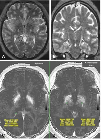
Figure 1. (A, B) Axial (A) and coronal (B) T2-weighted MRI of the brain display bilateral high signal intensity along the corticospinal tract (CST) (white arrows). (C, D) The mean myelin quantity was larger in the CST area than in the adjacent splenium of corpus callosum (right CST = 8,812, left CST = 8,677, splenium = 8,193) (green circles). MRI; magnetic reso- nance image.
Case Report
A 48-year-old woman complaining of dysarthria of seven months’ standing was hospitalized in the neurology department.
Examination revealed spastic dysarthria and minimal right side weakness; however, no fasciculation or muscle wasting was noted. She was hypertonic and hyperreflexic in the ipsilateral limbs. Hoffman’s sign and Babinski sign were observed in all limbs. Based on the upper motor neuron-dominant unilateral symptom and signs, we speculated that her condition might arise from chronic stroke. Brain MRI was performed for de- tecting vascular etiopathogenesis. MRI showed bilateral hyper- intensity along the corticospinal tract (CST), which was more prominent at the contralateral side to the symptomatic limb, on T2-weighted and FLAIR images (Fig. 1A, B). No additional
lesion was seen that might be provoking her symptoms. From those findings, we suspected the possibility of motor neuron disease. To reveal the lower motor neuron involvement, we performed electromyography. There were signs of acute and chronic denervation with reinnervation in two of the four re- gions (cervical, lumbosacral), even in the asymptomatic con- tralateral limbs muscles. Laboratory results including complete blood count, chemistry, electrolytes, and thyroid function tests were all within normal ranges. According to the revised El Escorial criteria,2 she was diagnosed with clinically probable ALS. On MWI, which is specific for myelin content, the mean myelin quantity was outnumbered in the abnormal CST area than the normal splenium of corpus callosum (Fig. 1C, D).
The result of brain MRI and MWI indicated intact myelin with pure axonal degeneration in CST on both sides.
Discussion
ALS involves in both upper and lower motor neurons.
Therefore, patients of ALS present spasticity, pathologic re- flexes with muscle atrophy, fasciculation, and paralysis at the same myotome.
The pathophysiology of ALS, where it begins from, is not established: whether the motor neuron degeneration begins in the neuronal cell body and proceeds anterograde, or from the muscle and neuromuscular junction and proceeds retrograde.1 Recent advances in neuroimaging such as diffusion tensor im- age (DTI), magnetic resonance spectroscopy (MRS) and func- tional MRI are utilized to reveal upper motor neuron-pathol- ogy in ALS.3 Furthermore, these tools could provide the sup- portive evidences for dying-forward hypothesis.
A small percentage of ALS patients displayed bilateral CST hyperintensity in the brain MRI.4 It was thought that cortical motor cells disappear, leading to anterograde axonal loss and gliosis in the CST, and this gliosis results in the bilateral white matter changes sometimes seen in the brain MRI. Some dis- pute over the clinical meaning of CST hyperintensity exists, because it has been described in healthy subjects,5 and in pa- tients with other conditions, such as liver cirrhosis.6 However, several studies suggested that CST hyperintensity was seen more frequent in ALS patients and it reflected the upper motor neuron dysfunction due to pyramidal tract degeneration.7 Non- conventional studies supporting the ‘dying-forward axonal de-
양지원․이종호․김응엽․신동훈
Korean J Clin Neurophysiol / Volume 18 / June 2016 20
generation’ hypothesis include: 1) transmagnetic stimulation studies documenting cortical hyperexcitability in patients with early sporadic ALS,8 2) DTI studies identifying abnormal dif- fusion properties involving CST at the multiple level,4 3) 11C- flumazenil PET which indicate reduced inhibitory GABAergic cortical effects, and 4) MRS showed a reduced primary motor cortex N-acetylaspartate to creatine ratio, implicating upper motor neuron dysfunction in the patients of ALS.4
MWI shows specificity for myelin content and integrity in both abnormal and normal appearing regions of white matter tissue. It has been used to evaluate myelin content in patients with multiple sclerosis, phenylketonuria, schizophrenia and chronic stroke.9 However, it has been rarely studied in ALS as a tool exploring white matter alteration. Only one study found that MWI can be used to distinguish primary lateral sclerosis from ALS, and may have value as a marker of extra- motor involvement.10 Our patient showed a preserved myelin signal in the corresponding CST hyperintensity. Although it is single case, we thought these images resulted from the ante- rograde primary axonal degeneration, not secondarily due to demyelination, suggesting the possible ALS mechanism of the dying-forward hypothesis. Because of prominent upper motor neuron signs, absence of muscle atrophy and fasciculation and minimal muscle weakness, we did not consider the dy- ing-backward hypothesis. We thought MWI can be comple- mentary to DTI for the evaluation of brain microstructure, al- though the relationship between white matter status and upper motor neuron dysfunction needs to be further examined.
In conclusion, this case implies the pathophysiology of ALS as a primary ‘dying-forward’ axonal degeneration by combina- tion of two MRI results, CST degeneration with preserved mye- lin water content. We suppose to conduct further study with larger ALS subjects to evaluate a role of MWI in ALS re- search in the future.
REFERENCES
1. Kiernan MC, Vucic S, Cheah BC, Turner MR, Eisen A, Hardiman O, et al. Amyotrophic lateral sclerosis. Lancet 2011;377:942-955.
2. Brooks BR, Miller RG, Swash M, Munsat TL; World Federation of Neurology Research Group on Motor Neuron Diseases. El Escorial revisited: revised criteria for the diagnosis of amyotrophic lateral sclerosis. Amyotroph Lateral Scler Other Motor Neuron Disord 2000;1:293-299.
3. Turner MR, Agosta F, Bede P, Govind V, Lule D, Verstraete E.
Neuroimaging in amyotrophic lateral sclerosis. Biomark Med 2012;6:319-337.
4. Zhang L, Ulug AM, Zimmerman RD, Lin MT, Rubin M, Beal MF.
The diagnostic utility of FLAIR imaging in clinically verified amyotrophic lateral sclerosis. J Magn Reson Imaging 2003;17:
521-527.
5. Mirowitz S, Sartor K, Gado M, Torack R. Focal signal-intensity variations in the posterior internal capsule: normal MR findings and distinction from pathologic findings. Radiology 1989;172:
535-539.
6. Córdoba J, Raguer N, Flavià M, Vargas V, Jacas C, Alonso J, et al.
T2 hyperintensity along the cortico-spinal tract in cirrhosis relates to functional abnormalities. Hepatology 2003;38:1026-1033.
7. Waragai M. MRI and clinical features in amyotrophic lateral sclerosis. Neuroradiology 1997;39:847-851.
8. Vucic S, Ziemann U, Eisen A, Hallett M, Kiernan MC. Transcranial magnetic stimulation and amyotrophic lateral sclerosis: pathophy- siological insights. J Neurol Neurosurg Psychiatry 2013;84:1161- 1170.
9. Borich MR, Mackay AL, Vavasour IM, Rauscher A, Boyd LA.
Evaluation of white matter myelin water fraction in chronic stroke.
Neuroimage Clin 2013;2:569-580.
10. Kolind S, Sharma R, Knight S, Johansen-Berg H, Talbot K, Turner MR. Myelin imaging in amyotrophic and primary lateral sclerosis.
Amyotroph Lateral Scler Frontotemporal Degener 2013;14:562- 573.