Influence of Propofol and Fentanyl on Deep Brain Stimulation of the Subthalamic Nucleus
We investigated the effect of propofol and fentanyl on microelectrode recording (MER) and its clinical applicability during subthalamic nucleus (STN) deep brain stimulation (DBS) surgery. We analyzed 8 patients with Parkinson’s disease, underwent bilateral STN DBS with MER. Their left sides were done under awake and then their right sides were done with a continuous infusion of propofol and fentanyl under local anesthesia. The electrode position was evaluated by preoperative MRI and postoperative CT. The clinical outcomes were assessed at six months after surgery. We isolated single unit activities from the left and the right side MERs. There was no significant difference in the mean firing rate between the left side MERs (38.7 ± 16.8 spikes/sec, n = 78) and the right side MERs (35.5 ± 17.2 spikes/sec, n = 66). The bursting pattern of spikes was more frequently observed in the right STN than in the left STN. All the electrode positions were within the STNs on both sides and the off-time Unified Parkinson’s Disease Rating Scale part III scores at six months after surgery decreased by 67% of the preoperative level. In this study, a continuous infusion of propofol and fentanyl did not significantly interfere with the MER signals from the STN. The results of this study suggest that propofol and fentanyl can be used for STN DBS in patients with advanced Parkinson’s disease improving the overall experience of the patients.
Keywords: Parkinson Disease; Microelectrodes; Propofol; Fentanyl; Subthalamic Nucleus;
Deep Brain Stimulation Wonki Kim,1 In Ho Song,2 Yong Hoon Lim,1
Mi-Ryoung Kim,1 Young Eun Kim,3 Jae Ha Hwang,1 In Keyoung Kim,1 Sang Woo Song,1 Jin Wook Kim,1 Woong-Woo Lee,3 Han-Joon Kim,3 Cheolyoung Kim,4 Hee Chan Kim,5 In Young Kim,6 Hee Pyoung Park,7 Dong Gyu Kim,1 Beom Seok Jeon,3,8 and Sun Ha Paek1,8,9,10
1Department of Neurosurgery, Seoul National University College of Medicine, Seoul; 2Medical Device Development Center, Osong Medical Innovation Foundation, Cheongwon; 3Department of Neurology, Seoul National University College of Medicine, Seoul; 4Medical Imaging Laboratory and CyberMed, Inc., Seoul; 5Department of Medical Engineering, Seoul National University College of Medicine, Seoul; 6Department of Biomedical Engineering, Hanyang University, Seoul;
7Department of Anaesthesiology and Pain Medicine, Seoul National University College of Medicine, Seoul; 8Ischemic/Hypoxic Disease Institute, Seoul National University College of Medicine, Seoul;
9Cancer Research Institute, Seoul National University College of Medicine, Seoul; 10Clinical Research Institute, Seoul National University Hospital, Seoul, Korea
Received: 3 March 2014 Accepted: 26 June 2014 Address for Correspondence:
Sun Ha Paek, MD
Department of Neurosurgery, Seoul National University Hospital, 101 Daehak-ro, Jongno-gu, Seoul 110-744, Korea
Tel: +82.2-2072-3993, Fax: +82.2-744-8459 E-mail: [email protected]
Funding: This work was supported by a grant from the Korea Institute of Planning & Evaluation for Technology in Food, Agriculture, Forestry, and Fisheries, Republic of Korea (No.
311011-05-3-SB020) and a grant from the Korea Healthcare Technology R&D Project, Ministry of Health & Welfare, Republic of Korea (No. HI09C13540100, HI10C14110400 and HI12C02050101).
http://dx.doi.org/10.3346/jkms.2014.29.9.1278 • J Korean Med Sci 2014; 29: 1278-1286
INTRODUCTION
Deep brain stimulation (DBS) of the subthalamic nucleus (STN) reduces motor disabil- ity, motor fluctuations and levodopa-induced dyskinesias (1). Intraoperative micro- electrode recordings (MER) and clinical monitoring with electrical stimulation of the STN have been recommended for the accurate positioning of the electrodes in the STN (2, 3). These procedures have the STN DBS done usually under local anesthesia (LA).
It is reported that the intraoperative use of propofol can alleviate the patients’ dis- comfort but interfere with the electrophysiological signals (4-6). However, it has not been thoroughly evaluated regarding how much it influences the electrical signals of MER during the STN DBS and the accuracy of positioning the electrodes after surgery.
The purpose of this study was to investigate the influence of propofol and fentanyl on microelectrode recording and their applicability in STN DBS.
MATERIALS AND METHODS Patients and clinical evaluation
Seventeen patients received a bilateral STN DBS operation under LA between October 2010 and June 2011 as described elsewhere (7). Eight patients whose electrical signals for MER were recorded from both sides for the comparative analysis were included in this study. The patients were evaluated with the use of the Unified Parkinson Disease Rating Scale (UPDRS), Hoehn and Yahr (H&Y) Staging, Schwab and England Activities
of Daily Living (SEADL), the Short Form-36 Health Survey (SF- 36), and neuropsychological tests. Evaluations were performed before surgery and 6 months after surgery. The neurological evaluations were performed by two neurologists. Patients were assessed in two conditions (off medication when the patients had taken no medication for 8 to 12 hr and on medication when the patients had experienced maximal clinical benefit 1 to 3 hr after the usual morning dose of dopaminergic treatment) before and after surgery. The levo-dopa equivalent daily dose (LEDD) was computed as described elsewhere (7). The clinical infor- mation of the eight patients with advanced Parkinson’s disease (PD) who had undergone bilateral STN stimulation is summa- rized in Table 1.
Surgical procedure
Anti-Parkinsonian drugs were tapered over two days and stop- p ed overnight before the surgery. In all cases, a stereotactic Lek- sell®-G frame (Elekta Instruments AB, Stockholm, Sweden) was mounted on the head of the patients under LA. Brain images were acquired on a 1.5-T Signa system (General Electric Medi- cal System, Milwaukee, WI, USA). The STNs were localized by a combination of direct visualization with MRI, MER, and an in- traoperative stimulation technique as previously described else- where (7). The FSPGR 3-D sequence was used for anterior com- missure (AC) - posterior commissure (PC) calculations. To bet- ter define the STN, T2 spin-echo images were obtained. Plan- ning of STN targeting and selection of trajectories were accom- plished with SurgiPlan® (Eleckta, Stockholm, Sweden). The char- acteristic discharge of the left STN was identified using multi- channel MER by LeadPoint (Medtronic, Minneapolis, MN, USA).
Additionally, a trial stimulation to assess clinical improvement was carried out before the permanent quadripolar electrodes
(DBS 3389, Medtronic) were positioned in the middle of the area of discharge. Then, the same procedure was performed for right side, under the monitored anesthesia care (MAC) with continuous infusion of propofol (25 μg/kg/min) and fentanyl (25 ng/kg/min). The depth of sedation was estimated with the degree of the patients’ awakening response to a loud sound and shaking as well as the response to pinching. The permanent quadripolar electrodes were positioned in the middle of the area of discharge. Then, the stereotactic frame was removed and the implantable pulse generators (IPG) (Medtronic) were then implanted in a subcutaneous pocket below the clavicle under general anesthesia in a single session. Electrical stimula- tion was started one day after surgery. The stimulation parame- ters and medications were progressively adjusted using an N’vi- sion® programmer (Medtronic) as previously described else- where (7).
Analysis of microelectrode recordings
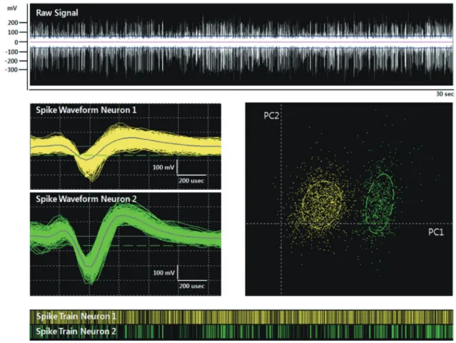
The MER signals along the finally selected trajectories were se- lected for the analysis. Each MER signal was band-pass filtered at 500-5,000 Hz with a gain 10,000 and was sampled at a rate of 24 kHz. Only an MER signal having stable single/multiunit ac- tivity that was clearly over the amplitude of the background noise baseline was selected and a threshold was applied to detect spikes. We isolated a total of 78 single unit activities under LA and 66 single unit activities under MAC. Spike sorting for a sin- gle unit was performed using the Offline Sorter software (Of- fline Sorter, Plexon, TX, USA). Principal components were cal- culated for unsorted waveforms and the waveforms were as- signed to the clusters using the expectation-maximization algo- rithm based on the T-distribution method (Fig. 1). Two statisti- cal parameters, the J3 statistic and the Davies-Bouldin (DB) va- lidity, were used to examine sorting quality statistics between classified clusters. A high value for the J3 statistic and a low val- ue for the DB validity indicate that the clusters are compact and well-separated.
Image fusion of preoperative MRI and postoperative CT For all patients 3-D spiral stereotactic CT scans (64-channel Bril- liance CT, Philips, Eindhoven, the Netherlands) with a 1-mm slice thickness were taken at an immediate postoperative peri- od and one month after bilateral STN stimulation to localize the electrodes by image fusion with the preoperative MRI by using mutual information techniques as previously described else- where (7). With CT-MRI image fusion, the electrode positions were plotted on the human brain atlas of Schaltenbrand and Wharen (8).
Statistical analysis
The data for the aforementioned variables were presented as the mean ± standard deviation. To examine the effect of anes- Table 1. Clinical findings of the 8 patients with advanced Parkinson’s disease
Parameters Findings
Number of patients by sex Male
Female
4 4
Age (yr) Mean ± SD
Range 57.1 ± 6.1 48-68
Symptom duration (yr) Mean ± SD
Range
11.6 ± 3.6 7-18 Duration of medication (yr) Mean ± SD
Range
9.5 ± 2.2 7-14
LEDD (mg/day) Mean ± SD 1,482.4 ± 360.9
Total scores of the UPDRS On-medication
Off-medication Mean ± SD
Mean ± SD 21.9 ± 13.2 53.9 ± 15.4 Scores of the UPDRS part III On-medication
Off-medication Mean ± SD
Mean ± SD 13.2 ± 8.3 41.4 ± 8.8 Hoehn & Yahr stage On-medication
Off-medication
Mean ± SD Mean ± SD
2.1 ± 0.2 3.1 ± 0.7 Schwab & England ADL On-medication
Off-medication Mean ± SD
Mean ± SD 81.3 ± 14.6 41.3 ± 24.7 ADL, Activities of Daily Life; LEDD, Levodopa equivalent daily dose; UPDRS, Unified Parkinson’s Disease Rating Scale.
thesia and the pattern on the mean firing rate and the effect of different combinations of anesthesia and the pattern on the mean firing rate, analysis of variance (ANOVA) was performed.
Single unit activity was classified as non-burst (tonic, irregu- lar), or as a burst discharge pattern using the method of the pre- vious study (9). The discharge density histogram was estimated from each 30-sec spike train containing at least 300 spikes. A tonic discharge pattern was characterized by a quasi-normal density distribution histogram. An irregular discharge pattern was characterized by a Poisson distribution, and a burst dis- charge pattern was tested with the chi-square test from a Pois- son distribution with a mean of 1.0 and characterized by a sig- nificant different distribution histogram (P < 0.05), a significant- ly positive skewness (P < 0.05) of the density distribution histo- gram and a minimum of four spikes per burst (Fig. 2).
Paired and unpaired t-tests were used for the comparison of means and Fisher’s exact test for the comparison of variables measured at the baseline before surgery and 6 months after sur- gery. P values of 0.05 were considered to indicate statistical sig- nificance. All statistical analyses were used in SAS statistical soft- ware (Version 9.0).
Ethics statement
This study was approved by the institutional review board of the Seoul National University Hospital (IRB No. H-1402-057-555).
Informed consent was exempted by the board.
RESULTS
The demographics in Table 1 show that the eight patients of this cohort study group were similar to those in our previous studies regarding age, sex ratio, age of onset of PD, duration of PD and preoperative levodopa equivalent dosage (7). There were no perioperative complications.
Results of MER analysis
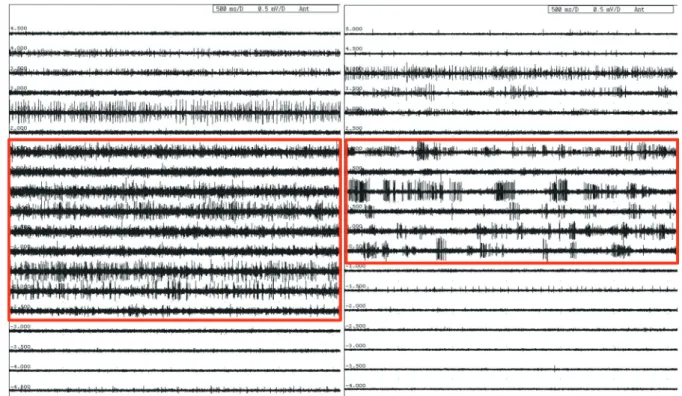
The typical STN bursting pattern and widening of the signal background noise baseline could be identified by the interaop- erative MER under LA. A typical bursting pattern in MAC could be also identified, whereas the signal background noise base- line seemed to be slightly reduced (Fig. 3). The mean firing rates were 38.7 ± 16.8 spikes/sec for LA, and 35.5 ± 17.2 spikes/sec for MAC. There was no significant difference in mean firing rate between the LA and MAC conditions (P = 0.256).
Fig. 1. A sorted single unit on microelectrode recording. This figure demonstrates a sorted single unit. The expectation-maximization algorithm was applied to raw signal (white) for getting isolated unit waveforms (green) from noise signal (yellow). Top, raw spiking activity; middle left, unit waveforms (green) and isolated form noise (yellow); middle right, isolated unit and noise on a principle component plot (x-axis, PC1;y-axis, PC2); bottom, raster trace of unit events (green) and noise events (yellow) over a selected time period.
Fig. 2. Burst discharge patterns on microelectrode recording. This figure shows results of burst discharge patterns under the MAC and the LA conditions. Spike discharge in left column and discharge density histogram in right column with/without burst discharge patterns in MAC and LA.
Raster plot MAC Non-burst
2.5 s
2.5 s
2.5 s
2.5 s MAC Burst
LA Non-burst
LA Burst
Density histogram
Fig. 3. Typical bursting patterns (see the boxes). The typical bursting patterns are demonstrated according to anesthetic methods. Left column, bursting pattern in LA; Right column, bursting pattern in MAC.
The recorded neuronal discharge exhibited two types of dis- charge pattern. The majority of the single unit STN neurons un- der LA had a burst discharge pattern (52.6%), and the remain- der exhibited a non-burst discharge pattern (47.4%). In the case of the MAC, the majority of the single unit STN neurons exhib- ited a burst discharge pattern (75.4%), and the non-bursting ac- tivity had an irregular discharge pattern (24.6%). The relative proportion of these discharge patterns differed significantly be- tween the LA and MAC (P = 0.006).
Since the mean firing rate between the LA and MAC condi- tions was different significantly, to examine whether the mean firing rate is affected by bursting and non-bursting patterns and whether there is the interaction effects of anesthesia and pattern on the mean firing rate, ANOVA was performed. ANOVA showed a significant difference in pattern (PATTERN, P < 0.001), but no significant differences in anesthesia and the combination of two factors (ANESTHESIA, P = 0.174: PATTERN × ANESTHESIA, P = 0.247, respectively).
Results of electrode position after bilateral STN DBS Based on the intraoperative MER findings, the microelectrodes positions were plotted in the sagittal and coronal planes aligned along the AC-PC line. All the electrode positions of 8 patients were within the STNs on both sides in the reformatted axial im- ages of the fused images between the preoperative MRI and post- operative CT taken one month after surgery (Fig. 4).
Results of Clinical Outcome 6 months after bilateral STN DBS
The clinical outcome was compared between the preoperative and postoperative status 6 months after bilateral STN stimula- tion (Tables 2 and 3). Significant improvement in the off-time scores for the total UPDRS, UPDRS III, H&Y scores, SEADL, and dyskinesia disability with decreased LEDD was observed 6 mon- ths after surgery. The LEDD was decreased 6 months after sur- gery (1,482.4 ± 360.9 mg/day at baseline and 513.6 ± 322.8 mg/
day at 6 months; P = 0.024). Regarding the eight sub-scales of the SF-36, the scores for bodily pain and summary scores for physical health and mental health were improved 6 months af- ter the surgery.
From the neuropsychological evaluation, the verbal memory test using the Rey-Kim memory battery showed a decline in rec- ognition 6 months after the surgery (P = 0.002), whereas non- verbal memory showed no meaningful change. In the frontal lobe function tests, the Stroop test (Stroop-a, P = 0.006; Stroop- b, P = 0.004; Stroop-c, P = 0.034) and the fluency test (P = 0.013) scores tended to be worse 6 months after the surgery (P = 0.046 for Stroop-a; P = 0.053 for fluency); however, they lacked statis- tical significance after a Bonferroni correction. Other tests in- cluding the Boston Naming test, Grooved Pegboard test, Mini- Mental state examination, Trail-Making test, Beck Depression
Inventory, and Wisconsin Card Sorting test, did not show any significant changes.
DISCUSSION
Since the introduction of DBS by Benabid and colleagues in 1987, this technique has become the preferred treatment for patients with advanced PD showing motor fluctuation and dys- kinesia after long-term medication (10). The clinical outcome of advanced PD patients after STN DBS depends on careful pa- tient selection and optimal targeting of the STN. Precise posi- tioning of the electrodes in the STN is considered the most im- portant factor to achieve good clinical outcome following STN DBS in appropriately selected surgical candidates. However, many unexpected factors, such as brain shift due to CSF leak- age, electrode artifacts in the MRI, errors in the image fusion, and manipulation errors from instruments make it difficult to precisely position the electrodes in the center of the STN using only the method of image-based targeting (11-13). Therefore, MER and intraoperative stimulation tests were adopted to im- prove the accuracy of the electrode location (2). Thus, STN DBS has been performed with awake and cooperative patients in most cases. For those reasons, anti-parkinsonian drugs should be stopped overnight before surgery and the patients fasted for approximately 12 hr on the day of surgery, which can be a pain- ful memory for most patients with advanced PD.
Sedatives have been used in some cases to alleviate severe anxiety, painful dystonia or respiratory difficulties experienced by the patients who were unable to tolerate the whole proce- dure of bilateral STN DBS (14). Propofol has been widely used alone or combined with remifentanyl. The mean infusion rate of propofol reported in the literature is approximately 50 μg/kg/
min (15, 16). The use of propofol alleviates intraoperative pa- tients’ discomfort to make the operating time shorter and frees the patients from a bad memory of the painful procedure of bi- lateral STN DBS. It can also allow neurosurgeons to concentrate on the surgical procedure for the accurate positioning of the electrodes because there is no need to communicate with the sleeping patients. However, there are two concerns regarding the use of propofol in STN DBS. One is about the selective de- pression of cerebral activity and the reduction of electrophysio- logical signals by propofol. The other one is that the patients could not be cooperative during the operation to observe the clinical benefits and adverse effects from the intraoperative stim- ulation tests of STN.
Regarding depression of the electrophysiological signals, there are few reports in the literature on the use of propofol for STN DBS (4-6, 17, 18). In addition, neuronal firing patterns are not well characterized and there are no prospective, randomized, blinded studies to compare their clinical outcome with that of an awaken technique. In this study, we found that the continu-
Fig. 4. Location of the electrodes plotted onto the human brain atlas. Based on the CT-MRI fusion images of the preoperative brain MRI and postoperative brain CT scan taken one month after surgery. All the electrode positions are mostly located to the middle one third part of the STNs on both sides in the fused images. (A) It shows location of the electrodes plotted onto the human brain atlas of Schaltenbrand and Wahren. (B) The microelectrodes positions were plotted in the sagittal and coronal planes aligned along an- terior commissure and posterior commissure line (AC-PC line).
Dorsal-ventral distance from AC-PC (mm)
Anterior-posterior distance from MCP (mm) -5 -4 -3 -2 -1 0 1 -2
-3 -4 -5 -6 -7 -8 -9 -10
Dorsal-ventral distance from AC-PC (mm)
Anterior-posterior distance from MCP (mm) 10 11 12 13 14 -2
-3 -4 -5 -6 -7 -8 -9 -10
LA bursting LA Non-bursting MAC bursting MAC Non-bursting
B Axial Hv -3.5
Sagittal SI 12.0 Sagittal SI 12.0
Coronal Fp 3.0 AC-PC
Rt electrode Lt electrode
Rt electrode
Rt electrode Lt electrode
Lt electrode mm
mm mm
A
ous infusion of propofol and fentanyl did not significantly inter- fere with the mean firing rates of the STN in comparison with awake monitoring in the same patient and showed good clini-
cal outcomes comparable to a previous report (7). The results of the mean firing rates in both awake and MAC conditions are similar to some previous reports ranging between 33.1 Hz and
Table 3. UPDRS III subscores of the 8 patients with advanced Parkinson’s disease
Scales Site Medication Subscores
P value Baseline 6 months
Resting tremor Lt Rt
On Off On Off
0.5 ± 1.0 1.1 ± 1.2 0.3 ± 0.6 0.6 ± 0.6
0.0 ± 0.0 0.1 ± 0.2 0.0 ± 0.0 0.0 ± 0.0
0.180 0.109 0.180 0.066 Action tremor Lt
Rt On Off On Off
0.4 ± 0.5 1.2 ± 0.6 0.2 ± 0.4 0.9 ± 0.9
0.0 ± 0.0 0.6 ± 0.8 0.0 ± 0.0 0.1 ± 0.2
0.059 0.066 0.180 0.072
Rigidity Lt
Rt On Off On Off
1.1 ± 1.0 3.3 ± 1.0 0.6 ± 0.8 3.1 ± 1.4
0.4 ± 0.5 0.6 ± 0.4 0.2 ± 0.3 1.3 ± 1.1
0.109 0.027 0.102 0.027 Bradykinesia Lt
Rt On Off On Off
3.3 ± 2.4 8.7 ± 2.6 3.0 ± 2.3 8.0 ± 2.5
1.8 ± 1.2 1.8 ± 1.1 2.0 ± 2.1 3.1 ± 2.1
0.102 0.028 0.026 0.027
Gait On
Off 0.6 ± 0.7
2.2 ± 0.9 0.5 ± 0.8
0.5 ± 0.8 0.786 0.026 Postural instability On
Off 0.3 ± 0.4
1.6 ± 1.2 0.2 ± 0.4
0.2 ± 0.4 1.000 0.026
Speech On
Off
0.8 ± 0.7 1.6 ± 1.0
0.6 ± 0.5 0.8 ± 0.6
0.564 0.066 UPDRSa-part III Lt
Rt On Off On Off
5.3 ± 4.1 14.4 ± 2.7 4.1 ± 3.6 12.5 ± 2.9
2.2 ± 1.2 4.9 ± 1.4 2.2 ± 2.3 4.5 ± 3.0
0.080 0.027 0.027 0.026 UPDRS, Unified Parkinson’s Disease Rating Scale.
Table 2. Clinical outcomes of the 8 patients with advanced Parkinson’s disease 6 months after bilateral subthalamic nucleus deep brain stimulation
Outcomes Medications Baseline 6 months P value
LEDD (mg/day) 1,482.4 ± 360.9 513.6 ± 322.8 0.024
Total scores of the UPDRS On
Off 21.9 ± 13.2
53.9 ± 15.4 12.9 ± 10.1
21.6 ± 9.6 0.028
0.027
Scores of the UPDRS part III On
Off 13.2 ± 8.3
41.4 ± 8.8 6.8 ± 5.4
13.8 ± 5.9 0.028
0.028
Hoehn & Yahr Stage On
Off
2.1 ± 0.2 3.1 ± 0.7
1.9 ± 0.5 2.1 ± 0.2
0.317 0.026
Schwab & England ADL On
Off 81.3 ± 14.6
41.3 ± 24.7 92.9 ± 7.6
72.9 ± 19.8 0.059
0.042
FOGQ On
Off 5.0 ± 4.6
16.4 ± 6.6 2.9 ± 3.3
7.1 ± 6.3 0.068
0.028
Dyskinesia disability 3.5 ± 0.9 0.9 ± 1.1 0.024
MMSE 26.8 ± 2.9 27.0 ± 3.2 0.705
BDI 20.4 ± 9.3 18.3 ± 10.4 0.463
SF36-Physical Health 131.7 ± 88.8 245.4 ± 90.3 0.018
SF36-Mental Health 168.0 ± 81.7 248.5 ± 91.0 0.128
ADL, Activities of Daily Life; BDI, Beck Depression Inventory; FOGQ, Freezing of Gait Questionnaire; LEDD, Levodopa equivalent daily dose; MMSE, Mini-mental state examina- tion; SF 36, Short Form-36; UPDRS, Unified Parkinson’s Disease Rating Scale.
42.3 Hz (3, 19-25). These results imply that the MAC has just mi- nimal effects on the firing rate of STN neurons in PD patients.
In this study, the typical STN bursting patterns in both awake and MAC conditions could be identified (Fig. 3). These results are consistent with the previous findings observed under vari- ous anesthetic conditions with propofol and remifentanyl in the literature (4, 9, 26). Most of the signal background noise base-
lines seemed to be slightly reduced under MAC compared with the awakened status. Meanwhile, they were significantly wider than the baseline outside the STN. Based on these observations, it is suggested that the MAC has little effect on the STN back- ground activity.
The prominent burst discharge pattern of the STN is consid- ered one of the characteristics in a parkinsonian state. We clas- sified the single unit activity of STN neurons into two discharge patterns with a method based on the discharge density histo- gram. We found that the burst discharge pattern was a predom- inant feature in both conditions. To assess whether MAC could change the single unit discharge pattern of the STN, we com- pared the relative proportion of two main patterns between the awake and MAC conditions. The proportion of burst discharge pattern was increased in the MAC (75.4%) compared to the awak- en status (52.6%). To assess the possibility of topographical re- cording bias within the STN, we present a topographical distri- bution of the burst and non-burst discharge patterns within the STN in each condition, respectively (Fig. 4). Because there was no particular different topographical distribution of the differ- ent discharge patterns, the recording bias cannot be regarded as a significant factor to influence the results. Therefore, the dif- ferent patterns of burst discharge observed may be a result of the difference between the awake and MAC conditions rather than the recording bias within the STN.
Regarding the different patterns of burst discharge, Rodriguez- Oroz et al. found burst-like activity in 60.5% of the single unit STN neurons in awake PD patients, using a different analysis method and named it ”irregular activity with long pause” (22).
Likewise, Steigerwald et al. classified 70% of the single unit dis- charge activities of the STN neurons in the awake status as burst- ing using a method based on ISI distribution (24). Although there was some difference in the relative proportion between the awake
and MAC conditions, our relative proportions are closely coin- cident with previous studies, supporting that our result is valid (3, 19-25).
The result of ANOVA showed a significant difference in the pattern (PATTERN, P < 0.001; ANESTHESIA, P = 0.174), con- firming the different types of pattern did affect the mean firing rate, whereas indicating two types of anesthesia states did not affect the mean firing rate. In addition, the types of anesthesia did not interact with the types of pattern each other (PATTERN
× ANESTHESIA, P = 0.247), indicating that interpretation of the main effects is complete, namely, the mean firing rate was af- fected by the different types of pattern not by the types of anes- thesia.
We conclude that the MAC does alter the single unit discharge pattern of the STN and it is the only type of pattern that affects the mean firing rate of the STN. Despite some differences in the MER signals between the LA and MAC conditions, there is no difficulty in identifying the MER signals characteristic of the STN under MAC with continuous infusion of propofol and fen- tanyl shown in Fig. 3.
It is very difficult for the patients to endure the whole proce- dure of bilateral STN DBS and properly respond to the intraop- erative stimulation tests in the awaken status, although sedation might disturb the assessment of clinical responses and compli- cations by DBS (27, 28). Fortunately, we found no difference in the electrode positions in all eight patients; all electrodes were located within STN on both sides (Fig. 4). The clinical outcomes of the eight patients were also comparable to the results of our previous reports in which both sides STN DBS were performed in the awaken status. There was no difference in the UDPRS to- tal scores and part III subscores six months after STN DBS when compared to our previous results (26).
This study has several limitations. First, we did not monitor the depth of anesthesia in this study. The depth of sedation was estimated only by the degree of the patients’ awakening response to loud sound and shaking as well as their response to pinching in this study. The monitoring of the depth of anesthesia to titrate sedation and the state of arousal during DBS insertion would be ideal. The Bispectral index (BIS) is used to monitor the depth of anesthesia (16, 29, 30). A BIS value of 65-85 is recommended for sedation and 40-65 for general anesthesia. Second, the num- ber of patients in this study was limited and the analysis was done retrospectively.
In conclusion, we compared MER signals between awake and MAC conditions in eight advanced PD patients treated with bi- lateral STN DBS as well as their clinical outcomes six months after surgery. We found that the continuous infusion of propo- fol and fentanyl did not significantly interfere with the MER sig- nals from the STN and with the clinical outcome after STN DBS surgery. The results of this study suggest that propofol and fen- tanyl can be used for STN DBS in patients with advanced PD
improving the overall experience of the patients.
DISCLOSURE
The authors declare no conflicts of interest to disclose.
ORCID
Wonki Kim http://orcid.org/0000-0001-7197-2366 In Ho Song http://orcid.org/0000-0001-6330-3453 Yong Hoon Lim http://orcid.org/0000-0002-0346-2345 Mi-Ryoung Kim http://orcid.org/0000-0003-0671-2263 Young Eun Kim http://orcid.org/0000-0002-7182-6569 Jae Ha Hwang http://orcid.org/0000-0002-6455-3590 In Keyoung Kim http://orcid.org/0000-0002-7761-9456 Sang Woo Song http://orcid.org/0000-0002-5523-3798 Jin Wook Kim http://orcid.org/0000-0002-3773-3940 Woong-Woo Lee http://orcid.org/0000-0002-8767-7967 Han-Joon Kim http://orcid.org/0000-0001-8219-9663 Cheolyoung Kim http://orcid.org/0000-0001-5586-9342 Hee Chan Kim http://orcid.org/0000-0002-2112-426X In Young Kim http://orcid.org/0000-0001-9580-7074 Hee Pyoung Park http://orcid.org/0000-0002-4772-0780 Dong Gyu Kim http://orcid.org/0000-0002-5740-6189 Beom Seok Jeon http://orcid.org/0000-0003-2491-3544 Sun Ha Paek http://orcid.org/0000-0003-3007-8653 REFERENCES
1. Deep-Brain Stimulation for Parkinson’s Disease Study Group. Deep- brain stimulation of the subthalamic nucleus or the pars interna of the globus pallidus in Parkinson’s disease. N Engl J Med 2001; 345: 956-63.
2. Benabid AL. Deep brain stimulation for Parkinson’s disease. Curr Opin Neurobiol 2003; 13: 696-706.
3. Hutchison WD, Allan RJ, Opitz H, Levy R, Dostrovsky JO, Lang AE, Loz- ano AM. Neurophysiological identification of the subthalamic nucleus in surgery for Parkinson’s disease. Ann Neurol 1998; 44: 622-8.
4. Hertel F, Züchner M, Weimar I, Gemmar P, Noll B, Bettag M, Decker C.
Implantation of electrodes for deep brain stimulation of the subthalamic nucleus in advanced Parkinson’s disease with the aid of intraoperative microrecording under general anesthesia. Neurosurgery 2006; 59: E1138.
5. Lefaucheur JP, Gurruchaga JM, Pollin B, von Raison F, Mohsen N, Shin M, Ménard-Lefaucheur I, Oshino S, Kishima H, Fénelon G, et al. Out- come of bilateral subthalamic nucleus stimulation in the treatment of Parkinson’s disease: correlation with intra-operative multi-unit record- ings but not with the type of anaesthesia. Eur Neurol 2008; 60: 186-99.
6. Maltête D, Navarro S, Welter ML, Roche S, Bonnet AM, Houeto JL, Mes- nage V, Pidoux B, Dormont D, Cornu P, et al. Subthalamic stimulation in Parkinson disease: with or without anesthesia? Arch Neurol 2004; 61:
390-2.
7. Paek SH, Kim HJ, Yoon JY, Heo JH, Kim C, Kim MR, Lim YH, Kim KR, Kim JW, Han JH, et al. Fusion image-based programming after subtha- lamic nucleus deep brain stimulation. World Neurosurg 2011; 75: 517-
24.
8. Schaltenbrand G, Wharen W. Atlas of stereotaxy of the human brain.
New York: Thieme, 1998.
9. Harries AM, Kausar J, Roberts SA, Mocroft AP, Hodson JA, Pall HS, Mit- chell RD. Deep brain stimulation of the subthalamic nucleus for advanc- ed Parkinson disease using general anesthesia: long-term results. J Neu- rosurg 2012; 116: 107-13.
10. Benabid AL, Pollak P, Louveau A, Henry S, de Rougemont J. Combined (thalamotomy and stimulation) stereotactic surgery of the VIM thalamic nucleus for bilateral Parkinson disease. Appl Neurophysiol 1987; 50: 344-6.
11. Khan MF, Mewes K, Gross RE, Skrinjar O. Assessment of brain shift re- lated to deep brain stimulation surgery. Stereotact Funct Neurosurg 2008;
86: 44-53.
12. Martinez-Santiesteban FM, Swanson SD, Noll DC, Anderson DJ. Mag- netic field perturbation of neural recording and stimulating microelec- trodes. Phys Med Biol 2007; 52: 2073-88.
13. Miyagi Y, Shima F, Sasaki T. Brain shift: an error factor during implanta- tion of deep brain stimulation electrodes. J Neurosurg 2007; 107: 989-97.
14. Benarroch EE. Subthalamic nucleus and its connections: anatomic sub- strate for the network effects of deep brain stimulation. Neurology 2008;
70: 1991-5.
15. Deiner S, Hagen J. Parkinson’s disease and deep brain stimulator place- ment. Anesthesiol Clin 2009; 27: 391-415.
16. Duque P, Mateo O, Ruiz F, de Viloria JG, Contreras A, Grandas F. Intra- operative microrecording under general anaesthesia with bispectral anal- ysis monitoring in a case of deep brain stimulation surgery for Parkin- son’s disease. Eur J Neurol 2008; 15: e76-7.
17. Lin SH, Chen TY, Lin SZ, Shyr MH, Chou YC, Hsieh WA, Tsai ST, Chen SY. Subthalamic deep brain stimulation after anesthetic inhalation in Parkinson disease: a preliminary study. J Neurosurg 2008; 109: 238-44.
18. Yamada K, Goto S, Kuratsu J, Matsuzaki K, Tamura T, Nagahiro S, Mu- rase N, Shimazu H, Kaji R. Stereotactic surgery for subthalamic nucleus stimulation under general anesthesia: a retrospective evaluation of Jap- anese patients with Parkinson’s disease. Parkinsonism Relat Disord 2007;
13: 101-7.
19. Benazzouz A, Breit S, Koudsie A, Pollak P, Krack P, Benabid AL. Intraop- erative microrecordings of the subthalamic nucleus in Parkinson’s dis-
ease. Mov Disord 2002; 17: S145-9.
20. Maciver MB, Bronte-Stewart HM, Henderson JM, Jaffe RA, Brock-Utne JG. Human subthalamic neuron spiking exhibits subtle responses to sed- atives. Anesthesiology 2011; 115: 254-64.
21. Magnin M, Morel A, Jeanmonod D. Single-unit analysis of the pallidum, thalamus and subthalamic nucleus in parkinsonian patients. Neurosci- ence 2000; 96: 549-64.
22. Rodriguez-Oroz MC, Rodriguez M, Guridi J, Mewes K, Chockkman V, Vitek J, DeLong MR, Obeso JA. The subthalamic nucleus in Parkinson’s disease: somatotopic organization and physiological characteristics. Brain 2001; 124: 1777-90.
23. Schrock LE, Ostrem JL, Turner RS, Shimamoto SA, Starr PA. The sub- thalamic nucleus in primary dystonia: single-unit discharge characteris- tics. J Neurophysiol 2009; 102: 3740-52.
24. Steigerwald F, Pötter M, Herzog J, Pinsker M, Kopper F, Mehdorn H, Deu- schl G, Volkmann J. Neuronal activity of the human subthalamic nucle- us in the parkinsonian and nonparkinsonian state. J Neurophysiol 2008;
100: 2515-24.
25. Weinberger M, Mahant N, Hutchison WD, Lozano AM, Moro E, Hodaie M, Lang AE, Dostrovsky JO. Beta oscillatory activity in the subthalamic nucleus and its relation to dopaminergic response in Parkinson’s disease.
J Neurophysiol 2006; 96: 3248-56.
26. Hutchison WD, Lang AE, Dostrovsky JO, Lozano AM. Pallidal neuronal activity: implications for models of dystonia. Ann Neurol 2003; 53: 480-8.
27. Anderson BJ, Marks PV, Futter ME. Propofol: contrasting effects in move- ment disorders. Br J Neurosurg 1994; 8: 387-8.
28. Böhmdorfer W, Schwarzinger P, Binder S, Sporn P. Temporary suppres- sion of tremor by remifentanil in a patient with Parkinson’s disease dur- ing cataract extraction under local anesthesia. Anaesthesist 2003; 52:
795-7.
29. Velly LJ, Rey MF, Bruder NJ, Gouvitsos FA, Witjas T, Regis JM, Peragut JC, Gouin FM. Differential dynamic of action on cortical and subcortical structures of anesthetic agents during induction of anesthesia. Anesthesi- ology 2007; 107: 202-12.
30. Venkatraghavan L, Luciano M, Manninen P. Review article: anesthetic management of patients undergoing deep brain stimulator insertion.
Anesth Analg 2010; 110: 1138-45.