INTRODUCTION
-Catenin plays a critical role as a component of the cell- cell adhesion complex and as a coactivator of the T-cell tran- scription factor/lymphoid enhancer binding factor (TCF/LEF) family of transcription factors (1-5). Inappropriate activation of this pathway leads to an increase of the level of -catenin, which can translocate into the nucleus and up-regulate the target gene expression and cell proliferation through bind- ing to the TCF/LEF family members (3-5). It is now appar- ent that altered expression of -catenin is an important event in the genesis of a number of malignancies (6-8). -Catenin gene mutation and nuclear accumulation found in hepatocel- lular carcinoma (HCCs) ranged from 13-34% and 11-43%, respectively, however, the clinical implication of the aberrant -catenin expression in HCC remains still unclear (9-16).
Cyclin D1 is a major regulator of the progression of cells into the proliferative stage of the cell cycle (17, 18). Recently, cyclin D1 has been reported as one of the target genes of the -catenin/TCF pathway; a TCF-binding site was identified in the promoter region of cyclin D1, and a significant corre- lation between the expressions of cyclin D1 and -catenin has been found in human breast and colon cancers (19-21). Cyclin
D1 gene amplification was reported in less than 15% of HCC patients (22, 23); however, the frequencies of overexpressions of cyclin D1 mRNA and cyclin D1 protein were reported in up to 22% (23) and 58% (24), respectively, in HCC patients.
These results suggest that another mechanism inducing the overexpression of cyclin D1 mRNA or protein may be present.
In the present study, to evaluate the role of -catenin in he- patocarcinogenesis as well as the relationships among the ex- pressions of -catenin and cyclin D1, and tumor cell prolif- eration, we performed an immunohistochemical analysis of -catenin and cyclin D1 in 77 patients with HCCs, and stud- ied the relationships between the expressions of -catenin and cyclin D1, mitotic index, and other pathologic parameters.
MATERIALS AND METHODS Patients and Tissue Samples
We retrieved 77 formalin-fixed, paraffin-embedded, sur- gically resected cases of HCC at Inje University Seoul Paik Hospital, which were collected from 1997 to 2001. Seventy- three (94.8%) of the 77 patients were seropositive for HBsAg
Mee Joo, Hye Kyung Lee, Yun Kyung Kang
Department of Pathology, Inje University Seoul Paik Hospital, Seoul, Korea
Address for correspondence Yun Kyung Kang, M.D.
Department of Pathology, Seoul Paik Hospital Inje University, 85 Jeo-dong 2-ga, Jung-gu, Seoul 100-032, Korea
Tel : +82.2-2270-0153, Fax : +82.2-2270-0131 E-mail : [email protected]
*This work was supported by grant number FG-5-03 of 21C Frontier Functional Human Genome Project from Ministry of Science & Technology of Korea.
211
Expression of -catenin in Hepatocellular Carcinoma in Relation to Tumor Cell Proliferation and Cyclin D1 Expression
Alteration of -catenin expression has been implicated in the development of hep- atocellular carcinoma (HCC). It has been also reported that -catenin can influence the tumor cell proliferation or cyclin D1 expression, one of the target factors of - catenin. We performed an immunohistochemical analysis of -catenin and cyclin D1 in 77 patients with resected HCCs, and examined the relationships between the expressions of -catenin and cyclin D1, and other pathologic parameters includ- ing the mitotic index. Altered expressions of -catenin including nonnuclear over- expression and nuclear expression were detected in 58.4% of HCCs (45/77) and showed significant correlations with large tumor size, poor histologic grade, and high tumor stage. The mean mitotic index of HCCs with nuclear expression (3.2
±3.0) and nonnuclear overexpression (2.7±2.5) was significantly higher than that of tumors with no overexpression (1.7±1.4) (p=0.018 and 0.038, respec- tively), however, no correlation was noted between the expressions of cyclin D1 and -catenin. In addition, nonnuclear overexpression out of two altered expres- sion patterns was more frequent (37.7% versus 20.8%) as well as pathologically more significant than nuclear expression. These results indicate that the altered expression of -catenin in HCC may play an important role in tumor progression by stimulating tumor cell proliferation, and nonnuclear overexpression may have pathologic significance in HCC.
Key Words : -catenin; Cell Adhesion Molecules; Cyclin D1; Mitotic index; Immunohistochemistry;
Liver Neoplasms; Carcinoma, Hepatocellular
Received : 9 October 2002 Accepted : 28 Norember 2002
212 M. Joo, H.K. Lee, Y.K. Kang
and only 4 (5.2%) had serum anti-HCV. Seventy-two patients were men and 5 were women, and the age of the patients ra- nged from 35 to 72 yr (mean age, 53 yr). Sixty-seven of the patients (87%) had a precirrhotic or cirrhotic nontumorous liver, and the others showed chronic viral hepatitis.
Pathologic Examination
Conventional pathologic parameters were examined. The histologic grade of tumor differentiation was assigned accord- ing to the Edmondson and Steiner grading system (25). After grading, the tumors were classified into 2 groups-well (grade I+II) (n=37) and poor (grade III+IV) (n=40). The size of the 77 HCCs ranged from 1.0 to 17 cm (mean, 3.7 cm), and the tumor size was classified based on the criteria of Yumoto et al.
(26) - small (tumor mass <3 cm) (n=32) and large (tumor mass
≥3 cm) (n=45). The tumor staging was performed by using International Union Against Cancer (UICC, 1997) criteria (27) - stage 1 (n=7), stage 2 (n=27), stage 3 (n=24), and stage 4 (n=19), respectively.
Immunohistochemical Staining
The conventional avidin-biotin complex (ABC) method was performed on 4 m-thick sections from each lesion. For anti- gen retrieval, the sections were immersed in citrate buffer and processed in a microwave oven at 95℃ for 10 min. Prima- ry monoclonal antibodies of -catenin (Clone: 14, Transduc- tion Laboratories, Lexington, KY) and cyclin D1 (Clone: A- 12, Santa Cruz Biotechnology, Santa Cruz, CA, U.S.A.) were used at a 1 to 100 dilution. Sections were immunostained using the ABC method. 3-Amino-9-ethylcarbazole (AEC) was used as the chromogen and Meyer’s hematoxylin as a nucle- us counterstain. Normal saline was used as a substitute for the primary negative controls. The -catenin expression pat- terns were divided into three groups as follows: 1) Nuclear expression, nuclear staining in more than 20% of the tumor cells with or without cytoplasmic expression; 2) Nonnuclear overexpression, increased membranous and/or cytoplasmic staining with no identifiable nuclear staining in at least 50%
of the tumorous areas; 3) No overexpression, weakly mem- branous staining similar to that of the adjacent nonneoplas- tic hepatocytes. We also categorized the cases showing nucle- ar expression or nonnuclear overexpression as altered expres- sion and the cases showing no overexpression as normal. For cyclin D1, only distinct nuclear staining was regarded as pos- itive. The cyclin D1 labeling index (LI) was defined as the average of the percentages of cyclin D1-positive cells on 10 randomly selected high power (×400) fields. The mitotic index was calculated from the numbers of mitotic cells in 10 random high power fields (×400). The mean value of the mitotic cells counted in each field was defined as the mitot- ic index. We applied Ki- 67 immunostaining (Clone: 7B11, Zymed Laboratories, San Francisco, CA, U.S.A.) in selected
cases with the same methods.
Statistical Analysis
The -catenin expression in relation to the pathologic para- meters was examined with the 2test. To analyze the statis- tical differences of cyclin D1 LI and mitotic index between groups, the unpaired Student’s t-test or ANOVA was used.
The correlation between cyclin D1 LI and the mitotic index was analyzed by Pearson’s correlation. Significance was defined as p<0.05. All statistical analyses were performed using SPSS software (version 10.0, SPSS INC., Chicago, IL, U.S.A.).
RESULTS -Catenin Expression
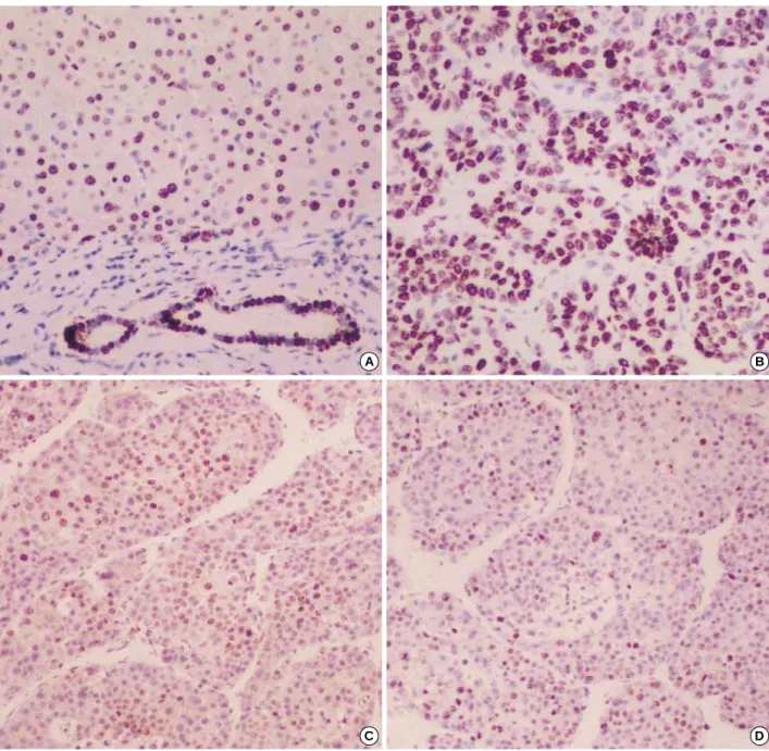
In the nontumorous areas, hepatocytes showed weak to mod- erate membranous staining with little cytoplasmic expression, and the bile ductules showed more prominent membranous expression (Fig. 1A). In the HCCs, altered expression, includ- ing two distinct patterns of nuclear expression and nonnucle- ar overexpression, was observed in 45 cases (58.4%). In 16 cases (20.8%) showing nuclear expression, nuclear immuno- staining was present in 20-60% of neoplastic cells and con- siderable intracytoplasmic expression was accompanied (Fig.
1B). In 29 cases (27.7%) showing nonnuclear overexpression, -catenin expression was localized in the cytoplasm, especial- ly close to the cell membrane, without nuclear expression and the intensity was obviously stronger than those of nonneoplas- tic hepatocytes (Fig. 1C, D). No overexpression was observed in 32 of the 77 HCCs (41.6%), and none of the tumors was completely negative for -catenin throughout the tumor.
According to associated viral status, altered expression was noted in 44 of 73 HBV-associated tumors (60.3%) and one of 4 HCV-associated tumors (25%).
Tumors with altered expression showed significant corre- lations with large tumor size (p=0.027), poor histologic grade (p=0.032), and high tumor stage (p=0.028). Tumors with nonnuclear overexpression showed a positive correlation with portal vein invasion (p=0.006) and tumor size (p=0.044), while nuclear expression showed no correlation with any of patho- logic parameters examined (Table 1).
Mitotic Index and Cyclin D1 Labeling Index (Table 2) The average MI in HCCs was 2.4±2.3 (range 0.1-11). The MI showed a strong correlation with poor histologic grade (p=0.004), large tumor size (p=0.002), presence of portal vein invasion (p<0.001), and high tumor stage (p<0.001).
The average cyclin D1 LIs in the nontumorous areas and HCCs were 13.8 (±13.2; range 1-48) and 26.2 (±18.2; range 1-80), respectively (Fig. 2A, B). There was a positive corre-
lation between cyclin D1 LIs and tumor size (p=0.02). How- ever, the cyclin D1 LIs were strikingly variable according to the histologic differentiation, and the average cyclin D1 LIs in HCCs with well-differentiated histologic grade (28.3±
17.6) were higher than those in cases with poor histologic grade (24.3±18.7), although statistically not significant.
Interestingly, we observed fifteen cases of HCC with high cyclin D1 expression localized in the central portions of the thick trabeculae or pseudoglandular pattern (Fig. 2C). When we applied Ki-67 immunostaining in these cases, we found
a noticeable difference between the topologic distribution of cyclin D1 and Ki-67 stainings (Fig. 2C, D). Furthermore, the cyclin D1 LIs showed no correlation with the MI (p=0.298, Pearson’s correlation test).
Relationships of Mitotic Index and Cyclin D1 Labeling Index with -Catenin Expression
According to -catenin expression patterns (Table 3), the average MIs (±SD) in cases with nuclear expression, nonnu-
Fig. 1. -catenin immunohistochemical staining. (A) Nonneoplastic hepatocytes showing homogeneous weak membranous staining. The bile ducts show moderate membranous staining, compared with hepatocytes, (B) HCCs showing strong nuclear and considerable cyto- plasmic stainings, and (C, D) Low and high magnification photographs of HCCs showing nonnuclear overexpression when compared to the adjacent nontumorous area. Strong membranous staining with occasional cytoplasmic staining is uniformly distributed throughout the tumor.
A B
C D
214 M. Joo, H.K. Lee, Y.K. Kang
clear overexpression, and no overexpression were 3.2 (±3.0), 2.7 (±2.5), and 1.7 (±1.4), respectively, and there was a significant difference between them (p=0.049). HCCs with nuclear expression and nonnuclear overexpression of -catenin showed a significantly higher mitotic index than cases with no overexpression (p=0.018 and 0.038, respectively). The average MIs of cases showing nuclear expression were slight- ly higher than those of cases showing nonnuclear overex- pression, but there was no statistical significance. No signif- icant difference was found in the average cyclin D1 LIs accord- ing to the -catenin expression patterns.
DISCUSSION
-Catenin plays a fundamental role in the regulation of the E-cadherin-catenin cell adhesion complex as well as in the Wnt signaling pathway (1-5). The expression of -catenin in cells is controlled by a multiprotein complex, and mutations in the glycogen synthase kinase 3 (GSK-3 ) phosphorylation sites of the -catenin gene (CTNNB1) result in nuclear and/or cyto- plasmic accumulation of -catenin and constitutive transac- tivation of TCF/LEF target genes, a mechanism occurring in many cancers (3-8). The significances of subcellular localiza-
Fig. 2.Expression of Cyclin D1 protein (A, B, and C) and Ki-67 immunostaining (D). (A) Nonneoplastic hepatocytes showing scattered cyclin D1-positive nuclei. (B) Well-differentiated HCCs showing high cyclin D1 labeling index, and (C) Localization of cyclin D1-positive cells to central portion of trabecular or pseudoglandular pattern, discordant to distribution of Ki-67-positive cells (D).
A B
C D
tion of -catenin using immunohistochemistry have been re- ported to be variable according to the tumor types. In colorec- tal cancer, increased cytoplasmic and nuclear staining seems to be an independent predictor of poor survival (28), whereas decrease or loss of expression correlated with poor prognosis in gastric or pancreatic adenocarcinoma (29, 30). To date, the
reported mutations of -catenin in HCC involve predominant- ly GSK-3 phosphorylation sites in exon 3, suggesting inap- propriate activation of the Wnt signaling pathway, and a high association between the nuclear -catenin expression and gene mutation was previously shown (9-16).
Most of the previous studies had focused on the nuclear ex-
n=77
Altered expression present absent
n=45 n=32 p-value
Nonnuclear overexpression present absent
n=29 n=48 p-value
Nuclear expression present absent
n=16 n=61 p-value Etiology of underlying liver disease
HBV (+) 73 44 29 28 45 16 57
HCV (+) 4 1 3 NS 1 3 NS 0 4 NS
Tumor size
Small (<3 cm) 32 14 18 8 24 6 26
Large (≥3 cm) 45 31 14 0.027 21 24 0.044 10 35 NS
Portal vein invasion
Absent 31 14 17 6 25 8 23
Present 46 31 15 NS 23 23 0.006 8 38 NS
Histologic grade
Well 37 17 20 10 27 7 30
Poor 40 28 12 0.032 19 21 NS 9 31 NS
Stage
T1 7 1 6 1 6 0 7
T2 27 16 11 8 19 8 19
T3 24 13 11 9 15 4 20
T4 19 15 4 0.028 11 8 NS 4 15 NS
Table 1.Relationship between -catenin expression patterns and pathologic parameters
HBV: HBsAg positive; HCV: anti-HCV positive; Well: Edmondson-Steiner grade I+II; Poor: Edmondson-Steiner grade III+IV; NS: not significant.
Variables Average mitotic index p-value Average cyclin D1 LI p-value
Tumor size
Small (<3 cm) 1.43±1.82 20.59±15.35
Large (≥3 cm) 3.07±2.37 0.002 30.24±19.07 0.020
Portal vein invasion
Absent 1.06±1.22 27.64±21.67
Present 3.28±2.43 <0.001 25.28±15.53 NS
Histologic grade
Well 1.61±2.16 28.32±17.57
Poor 3.11±2.21 0.004 24.3±18.69 NS
Stage
T1 0.33±0.43 32.86±15.77
T2 1.2±1.19 26.37±22.12
T3 2.43±1.14 26.33±13.80
T4 4.77±2.98 <0.001 23.47±18.25 NS
Table 2.Relationships of mitotic index and cyclin D1 labeling index with pathologic parameters
Well: Edmondson-Steiner grade I+II; Poor: Edmondson-Steiner grade III+IV; NS: not significant.
-catenin expression patterns Average MI p-value Average cyclin D1 LI p-value
No overexpression 1.66±1.37 26.72±14.4
Nuclear expression 3.21±3.03 22.44±22.4
Nonnuclear overexpression 2.74±2.5 27.79±19.6
Table 3.Relationships of mitotic index and cyclin D1 labeling index with -catenin expression
NS: not significant.
0.018
0.038 NS
NS NS
NS
216 M. Joo, H.K. Lee, Y.K. Kang
pression pattern of -catenin (11, 12, 14, 15). There are sev- eral conflicting reports concerning the association of -catenin with tumor progression and patient’s survival: the results pub- lished by Hsu et al. (12) and Mao et al. (13) demonstrated that expression of mutant nuclear -catenin correlated with low stage of HCC and favorable prognosis. However, accord- ing to the results of some recent studies (15, 16), nuclear ex- pression seems to correlate with tumor progression and poor prognosis. In our study, altered expressions of -catenin includ- ing nonnuclear overexpression and nuclear expression were detected in 58.4% of HCC and showed significant correla- tions with large tumor size, poor histologic grade, and high tumor stage. However, we are not able to find any relation- ship between nuclear expression of -catenin and pathologic features of HCCs. In contrast, nonnuclear overexpression out of two altered expression patterns was more frequent (37.7%
versus 20.8%) as well as pathologically more significant than nuclear expression. With regard to proliferative activity, not only nuclear expression but also nonnuclear overexpression of -catenin correlated significantly with high mitotic index.
Nonnuclear overexpression of -catenin had been previous- ly mentioned by Endo et al. (31), Nhieu et al. (15), Wong et al. (11), and Wei et al. (32). Especially, Wong et al. (11) stressed that the nonnuclear type -catenin overexpression correlat- ed significantly with poor prognostic factors in predominant- ly HBV-associated HCCs as noted in our study, however, they did not suggest any possible explanation. According to our results, nonnuclear overexpression of -catenin appeared to be a frequent finding in predominantly HBV-associated HCC and can contribute tumor progression by stimulating tumor cell proliferation.
Previous studies have provided evidence that nuclear expres- sion of -catenin stimulates tumor cell proliferation or tumor progression (15), however, little is known about the effect of cytoplasmic -catenin. Although molecular mechanism of nonnuclear overexpression pattern remains elusive, it is pos- sibly different from that of the nuclear expression pattern due to the following reasons. First, it is generally accepted that subcellular distribution of -catenin regulates its function;
membrane-bound -catenin predominantly mediates cell- cell adhesion, whereas elevation of the cytoplasmic and nucle- ar pool of the protein is associated with an oncogenic func- tion. Nonnuclear overexpression pattern noted in our study was mainly membranous staining. Second, in our study, HCCs with nonnuclear overexpression showed a high association with portal vein invasion (p=0.006), suggesting that this pheno- menon might be an effect related to alteration of cell-cell adhe- sion facilitating vascular invasion. Wei et al. (32) also report- ed that vascular invasion was frequently noted in HCCs show- ing enforced expression of the membranous E-cadherin/ - catenin complex, and they proposed that dynamic up-and down-regulations of these cell adhesion molecules might be required for the malignant progression of HCC. Third, most of the previous studies had focused on the nuclear expression
pattern and showed a high association between the nuclear expression of -catenin and HCV-related HCCs (11, 12, 14- 16), whereas HCCs examined in the present study and by Wong et al. (11) were predominantly HBV-associated, in which nonnuclear overexpression pattern was predominant and showed significant correlations with pathologic para- meters. Although these facts are not sufficient to suggest a close relationship between nonnuclear overexpression and HBV-related HCCs, nonnuclear overexpression might be fre- quently associated with HBV-related HCCs. Further investi- gation is necessary to resolve this issue.
Although the exact target genes of -catenin/TCF4 are still unknown, cyclin D1, one of the major cell cycle regulators, has been reported as a possible target (19, 20). In our study, we fail- ed to find an association between the expressions of -catenin and the cyclin D1. Considering the reports that overexpression of an oncogenic form of -catenin in the liver did not induce an up-regulation of cyclin D1 in transgenic mice (33), there might be another pathway or target genes at work in hepato- carcinogenesis.
Cyclin D1 function is generally presumed to be associated with cell proliferation. However, contrary to our expectations, the results in our study showed no correlation between cyclin D1 expression and the mitotic index as well as different topo- logic distribution between cyclin D1 and Ki-67 in some cases.
Meanwhile, cyclin D1 LI showed a significant correlation to large tumor size, which was partially in line with the previous reports showing that cyclin D1 overexpression exhibits an advanced clinicopathologic appearance (22, 23). Based on our results, we could reasonably infer that cyclin D1 expression may be unrelated to active cell proliferation, and may confer additional growth advantages to the tumor progression.
In conclusion, our results indicate that the altered expres- sion of -catenin in HCC may play an important role in tumor progression by stimulating tumor cell proliferation, and non- nuclear overexpression may have pathologic significance.
REFERENCES
1. Aberle H, Schwartz H, Kelmer R. Cadherin-catenin complex: pro- tein interactions and their implications for cadherin function. J Cell Biochem 1996; 61: 514-23.
2. Provost E, Rimm DL. Controversies at the cytoplasmic face of the cadherin-based adhesion complex. Curr Opin Cell Biol 1999; 11:
567-72.
3. Behrens J, von Kries JP, Kuhl M, Bruhn L, Wedlich D, Grosschedl R, Birchmeier W. Functional interaction of beta-catenin with the trans- cription factor LEF-1, Nature 1996; 382: 638-42.
4. Clevers H, van de Wetering M. TCF/LEF factor earn their wings.
Trends Genet 1997; 13: 485-9.
5. Omer CA, Miller PJ, Diehl RE, Kral AM. Identification of Tcf4 re- sidues involved in high-affinity beta-catenin binding. Biochem Bio- phys Res Commun 1999; 256: 584-90.
6. Morin PJ, Sparks AB, Korinek V, Barker N, Clevers H, Vogelstein B, Kinzler KW. Activation of -catenin-Tcf signaling in colon can- cer by mutations in -catenin or APC. Science 1997; 275: 1787-90.
7. Iwao K, Nakamori S, Kameyama M, Imaoka S, Kinoshita M, Fukui T, Ishiguro S, Nakamura Y, Miyoshi Y. Activation of the -catenin gene by interstitial deletions involving exon 3 in primary colorectal carcinomas without adenomatous polyposis coli mutations. Cancer Res 1998; 58: 1021-6.
8. Polakis P. The oncogenic activation of -catenin. Curr Opin Genet Dev 1999; 9: 15-21.
9. de La Coste A, Romagnolo B, Billuart P, Renard CA, Buendia MA, Soubrane O, Fabre M, Chelly J, Beldjord C, Kahn A, Perret C. Somat- ic mutations of the -catenin gene are frequent in mouse and human hepatocellular carcinomas. Proc Natl Acad Sci USA 1998; 95: 8847- 51.
10. Miyoshi Y, Iwao K, Nagasawa Y, Aihara T, Sasaki Y, Imaoka S, Mu- rata M, Shimano T, Nakamura Y. Activation of the -catenin gene in primary hepatocellular carcinomas by somatic alterations involving exon 3. Cancer Res 1998; 58: 2524-7.
11. Wong CM, Fan ST, Ng IO. -Catenin mutation and overexpression in hepatocellular carcinoma. Cancer 2001; 92: 136-45.
12. Hsu HC, Jeng YM, Mao TL, Chu JS, Lai PL, Peng SY. -Catenin mutations are associated with a subset of low-stage hepatocellular carcinoma negative for hepatitis B virus and with favorable prognosis.
Am J Pathol 2000; 157: 763-70.
13. Mao TL, Chu JS, Jeng YM, Lai PL, Hsu HC. Expression of mutant -catenin correlates with non-invasive hepatocellular carcinoma, absence of portal vein spread, and good prognosis. J Pathol 2001; 193:
95-101.
14. Laurent-Puig P, Legoix P, Bluteau O, Belghiti J, Franco D, Binot F, Monges G, Thomas G, Bioulac-Sage P, Zucman-Rossi J. Genetic al- terations associated with hepatocellular carcinomas define distinct pathways of hepatocarcinogenesis. Gastroenterology 2001; 120: 1763- 73.
15. Nhieu JT, Renard CA, Wei Y, Cherqui D, Zafrani ES, Buendia MA.
Nuclear accumulation of mutated -catenin in hepatocellular carci- noma is associated with increased cell proliferation. Am J Pathol 1999; 155: 703-10.
16. Inagawa S, Itabashi M, Adachi S, Kawamoto T, Hori M, Shimazaki J, Yoshimi F, Fukao K. Expression and prognostic roles of -catenin in hepatocellular carcinoma: correlation with tumor progression and postoperative survival. Clin Cancer Res 2002; 8: 450-6.
17. Weinberg RA. The retinoblastoma protein and cell cycle control. Cell 1995; 81: 323-30.
18. Sherr CJ. Cancer cell cycles. Science 1996; 274: 1672-7.
19. Tetsu O, McCormick F. -Catenin regulates expression of cyclin D1 in colon carcinoma cells. Nature 1999; 398: 422-6.
20. Shtutman M, Zhurinsky J, Simcha I, Albanese C, D’Amico M, Pestell R, Ben-Ze’ev A. The cyclin D1 gene is a target of the -catenin/LEF- 1 pathway. Proc Natl Acad Sci USA 1999; 96: 5522-7.
21. Lin SY, Xia W, Wang JC, Kwong KY, Spohn B, Wen Y, Pestell RG, Hung MC. -Catenin, a novel prognostic marker for breast cancer:
Its roles in cyclin D1 expression and cancer progression. Proc Natl Acad Sci USA 2000; 97: 4262-6.
22. Zhang YJ, Jiang W, Chen CJ, Lee CS, Kahn SM, Santella RM, Wein- steinIB. Amplification and overexpression of cyclin D1 in human he- patocellular carcinoma. Biochem Biophys Res Commun 1993; 196:
1010-6.
23. Nishida N, Fukuda Y, Komeda T, Kita R, Sando T, Furukawa M, Am- enomori M, Shibagaki I, Nakao K, Ikenaga M. Amplification and overexpression of the cyclin D1 gene in aggressive human hepato- cellular carcinoma. Cancer Res 1994; 54: 3107-10.
24. Joo M, Kang YK, Kim MR, Lee HK, Jang JJ. Cyclin D1 overexpres- sion in hepatocellular carcinoma. Liver 2001; 21: 89-95.
25. Edmondson HA, Steiner PE. Primary carcinoma of the liver: A study of 100 cases among 48,900 necropsies. Cancer 1954; 7: 462-503.
26. Yumoto Y, Hanafusa T, Hada H, Morita T, Ooguchi S, Shinji N, Mi- tani T, Hamaya K, Koide N, Tsuji T. Loss of heterozygosity and anal- ysis of mutation of p53 in hepatocellular carcinoma. J Gastroenterol Hepatol 1995; 10: 179-85.
27. Sobin LH, Wittekind C, eds. UICC: TNM classification of malignant tumors. 5th edn. New York: Wiley-Liss, 1997; 74-7.
28. Hugh TJ, Dilon SA, Taylor BA, Pignatelli M, Poston GJ, Kinsella AR. Cadherin-catenin expression in primary colorectal cancer. Br J Cancer 1999; 80: 1046-51.
29. Qiao Q, Ramadani M, Gansauge S, Gansauge F, Leder G, Beger HG.
Reduced membranous and ectopic cytoplasmic expression of -catenin correlate with cyclin D1 overexpression and poor prognosis in pan- creastic cancer. Int J Cancer 2001; 95: 194-7.
30. Woo DK, Kim HS, Lee HS, Kang YH, Yang HK, Kim WH. Altered expression and mutation of beta-catenin gene in gastric carcinomas and cell lines. Int J Cancer 2001; 95: 108-13.
31. Endo K, Ueda T, Ueyama J, Ohta T, Terada T. Immunoreactive E- cadherin, alpha-catenin, beta-catenin, and gamma-catenin proteins in hepatocellular carcinoma: relationships with tumor grade, clini- copathologic parameters, and patients’ survival. Hum Pathol 2000;
31: 558-65.
32. Wei Y, Nhieu TV, Pringent S, Srivatanakul P, Tiollais P, Buendia MA. Altered expression of E-cadherin in hepatocellular carcinoma:
correlations with genetic alterations, -catenin expression, and clin- ical features. Hepatology 2002; 36: 692-701.
33. Cadoret A, Ovejero C, Saadi-Kheddouci S, Souil E, Fabre M, Romag- nolo B, Kahn A, Perret C. Hepatomegaly in transgenic mice express- ing an oncogenic form of beta-catenin. Cancer Res 2001; 61: 3245-9.