INTRODUCTION
Tubular aggregate myopathy is a rare inherited primary myopathy with an autosomal dominant or recessive pattern (1). It usually presents with slowly progressive proximal mus- cular weakness or stiffness. Diagnostic hallmark is ultrastruc- tural presence of tubular aggregates. To date, 24 Caucacian patients from 7 families have been reported (1-8). Here, we describe the first Korean case of presumably dominantly inher- ited primary tubular aggregate myopathy occurring in a 19- yr-old man.
CASE REPORT
A 19-yr-old Korean man presented with progressive weak- ness and muscle stiffness without atrophy since the age of 13 yr. He was born as a full-term baby with 2.2 kg of birth weig- ht. No perinatal events or developmental delay was document- ed. He had no history of alcohol intake or medication. The stiffness began in the lower extremities, spreading gradually to the upper parts. It was symmetrical or asymmetrical and the proximal musculature was more affected than the distal.
Gower sign was positive, but pseudohypertrophy of the calf muscle was absent. Exertional myalgia, cramps or myotonia were not found. Sensation, coordination and deep tendon re- flexes were unaffected. Ranges of movement were normal. No
triggering factors were found prior to the onset of the symp- tom. His condition continued to deteriorate slowly for 6 yr.
Electromyogram of the biceps brachii showed brief small action potentials, indicating a slightly myopathic pattern. Nerve conduction velocity and Jolly test revealed no abnormal find- ings. Laboratory findings including serum creatine kinase, aldolase, lactate, lactate dehydrogenase, electrolytes and thyroid function test were normal. Forearm ischemic exercise test showed a normal rise of the lactate level. His mother has a his- tory of periodic stiffness once or twice a year for the past sever- al years. However, she declined muscle biopsy and further studies. His father and younger brother had no complaints of neuromuscular symptoms at the time of biopsy from the patient. The initial clinical impression was limb-girdle mus- cular dystrophy. Muscle biopsy was obtained from the vastus medialis. Muscle samples were treated as follows: (i) snap-fro- zen in isopentane pre-cooled in liquid nitrogen, and stored at -80℃until use; (ii) placed in formaldehyde fixative and paraffin embedded for routine histology; (iii) for conventional transmission electron microcopy (TEM), specimens were fixed in 2.5% glutaraldehyde in 0.1 M phosphate-buffered saline, post-fixed with 1% osmic acid and embedded in Epikote. Ultrathin sections were double stained with uranyl acetate and lead citrate and examined with a TEM (Hitachi model 7100) at an accelerating voltage of 75 kv. Six- m serial cryostat sections of submitted biopsied muscle were processed for a routine battery of usual histologic stains and enzyme
135
Tubular Aggregate Myopathy
: A Case Report
We report a first Korean case of presumably dominantly inherited primary tubu- lar aggregate myopathy in a 19-yr-old man, who presented with slowly progres- sive proximal muscle stiffness and weakness. In hematoxylin and eosin stain, it showed subsarcolemmal, or central pale basophilic granular vacuoles, which stained red with modified Gomori’s trichrome and intensive blue with nicotinamide ade- nonine dinucleotide-tetrazolium reductase, respectively. Ultrastructurally, aggre- gates of 60 nm-sized hexagonal tubules were found in both type 1 and type 2 fibers.
We briefly review the pathologic findings of the previously reported cases of tubu- lar aggregate myopathy and discuss the possible pathogenesis of this disease.
We briefly discuss the possible pathogenesis of sarcoplasmic reticulum and review the ultrastructural characteristics.
Key Words : Tubular Aggregates; Muscular Diseases; Sarcoplasmic Reticulum; Genes, Dominant
Na Rae Kim, Yeon-Lim Suh
Department of Pathology, Samsung Medical Center, Sungkyunkwan University School of Medicine, Seoul, Korea
Address for correspondence Yeon-Lim Suh, M.D.
Department of Pathology, Samsung Medical Center, 50 Ilwon-dong, Kangnam-gu, Seoul 135-710, Korea
Tel : +82-2-3410-2761, Fax : +82-2-3410-0025 E-mail: [email protected]
*Current address: Department of Pathology, Kang- nam General Hospital Public Corporation, 171-1 Samsung-dong, Kangnam-gu, Seoul 135-090, Korea Received : 7 February 2002
Accepted : 8 April 2002
136 N.R. Kim, Y.-L. Suh
histochemistry. Immunohistochemistry was performed on the paraffin-embedded sections (5 m-thick) using the monoclonal antibody against dystrophin (MS/486/P1, Neomarkers, Pre- mont, Calif, U.S.A., 1:20 dilution). Peripheral blood sampling was done for polymerase chain reaction (PCR) screening of the dystrophin gene at Xp21 (exon 3, 6, 13, 43, 47, 49, 50, 52, and 60). PCR for the dystrophin gene at Xp21 showed amplification of exon 3, 6, 13, 43, 47, 49, 50, 52, and 60 of the dystrophin gene without abnormalities.
PATHOLOGIC FINDINGS
On frozen cryostat sections stained by hematoxylin and
eosin, pale granular sarcoplasmic changes were observed in ap- proximately 30% of myofibers. The pale zones were round or moth-eaten, and varied in size, measuring up to 90 m in diameter (Fig. 1A). They were single or multiple, and situ- ated in the center and periphery of the myofibers. They showed an intense staining with nicotinamide adenine dinucleotide- tetrazolium reductase [NADH-TR] (Fig. 1B) and periodic acidic Schiff [PAS], stained bright red with Gomori’s trich- rome (Fig. 1C), but not stained with succinyl dehydrogenase [SDH] and myofibrillar adenosine triphosphatase [ATPase]
at pH 9.4, 4.6, and 4.3. They occurred in both type 1 and 2 fibers. In paraffin sections the pale zones appeared to harbor irregular spaces or vacuoles with or without granular mate- rials. There were nonspecific changes, including variation of
Fig. 1.(A) Myofibers of variable size reveal multiple intracytoplas- mic granularities and vacuolation (H&E, ×200). (B, C) NADH-TR and modified Gomori’s trichrome reveal intense subsarcolemmal and interfibrillar reaction, corresponding to tubular aggregates (arrows) (B: NADH-TR, ×400, C: modified Gomori’s trichrome stain, ×400).
A
C
B
myofiber size ranging from 15 to 90 m, atrophic fibers, hy- pertrophic and split fibers, frequent internal nuclei, and a few inflammatory cells. Neither necrotic nor regenerating changes were found. Type 1 fiber predominance and type 2 fiber atro- phy were also observed in ATPase reactions. Immunohisto- chemistry for dystrophin revealed a normal linear staining along the sarcoplasmic membrane. Ultrathin sections of skele- tal muscle fibers showed focal disorganization of myofilaments with a disarray of Z-bands and papillary appearance of sarco- lemmal membrane. Tubular aggregates were found in sub- sarcolemmal and intermyofibrillar areas of almost all myofibers (Fig. 2A). On longitudinal section, they consisted of collec- tions of long, straight tubules, 60 nm in diameter. On trans- verse section, the tubules were in hexagonal array and contain-
ed a smaller 25 nm-sized central tubule-like structure, i.e., tubule in tubule (Fig. 2B). The central tubule was separated by a clear space from the outer wall of the tubules. There were many dysmorphic mitochondria such as megamitochondria and intramitochondrial crystal inclusions (Fig. 2C). The his- tologic and electron microscopic findings of the muscle were consistent with primary tubular aggregate myopathy.
DISCUSSION
Tubular aggregates have been observed in many diseased skeletal muscle fibers though minor in proportion. The dis- eases include periodic paralysis (9), hyperaldosteronism (10),
Fig. 2.(A) Transverse section of a muscle fiber shows compactly packed collection of tubules along the subsarcolemmal spaces (×20,000; bar represents 400 nm), (B) Numerous single- or dou- ble-membrane bound tubules contain inner small microtubules (×30,000; bar represents 267 nm), (C) Rectangular mitochondria contain altered intramitochondrial crystals (×20,000; bar repre- sents 500 nm).
A
C
B
138 N.R. Kim, Y.-L. Suh
facioscapulohumeral dystrophy with aminoaciduria (11), myas- thenia gravis (12), exercise-induced cramps (13), alcohol, caf- feine, or drugs such as zidovudine (14, 15). However, patients with these tubular aggregates may present as a specific type of primary myopathy (1-8). Primary tubular aggregate myopa- thy of familial occurrence was first reported by De Groot and Arts (1) in 1982. To date, 24 patients from 7 families have been reported (1-8). Clinically, the age of onset ranged from 6 to 45 yr (mean: 25.4 yr). All were Caucacians. Progressive proximal weakness (40%) and periodic paralysis or exertional muscle cramps (40%) were the most common presenting sym- ptom. Entirely asymptomatic family members having prima- ry tubular aggregate myopathy accounted for 8% (6). Al- though facial muscular weakness (4%) and limb girdle con- tracture (4%) have been rarely described (1, 3), facial sparing is regarded as an important clue to distinguishing primary tubular aggregate myopathy from facioscapulohumeral dys- trophy. Muscle pain was accompanied in 24% of inherited tubular aggregate myopathy. Autosomal dominant inheritance is the most common (2-6, 8), but less commonly autosomal recessive inheritance or rarely sporadic occurrences have been reported (1, 7, 16). Although the histologic confirmative diag- nosis of his mother could not be made, the present case sug- gested mother-to-son transmission (Fig. 3), implying a pre- sumably autosomal dominant inheritance or remote possibili- ty of autosomal recessive inheritance. Molecular genetic stud- ies have not yet been performed.
Light microscopic examination or clinical history alone can- not exclude the presence of the affected family members be- cause ultrastructural study can show tubular aggregates in pa- tients with normal histology and enzyme histochemistry of
the muscle. Therefore, diagnostic hallmark of this dissease is the ultrastructural demonstration of tubular aggregates of variable morphology. The tubular aggregates usually consist of closely packed double-membrane tubules often containing a single inner tubular structure or amorphous material (8). Tubu- lar aggregates have been divided into three types (17); type I with intraluminal coaxial inner tubules (70 nm): type II with a granular core (70-400 nm), and type III tubules with sub- sarcolemmal filaments of approximately 10 nm (130-400 nm).
In dominantly inherited tubular aggregate myopathy, all three types of tubules have been observed. Recently, Muller and his colleagues (8) described additional findings of tubulofilamen- tous structures, proliferated terminal cisternae or tubulovesic- ular structures, in cases of familial tubular aggregates. Inter- estingly, tubulofilamentous structures were found in the his- tologically normal looking myofibers of asymptomatic fam- ily members. The present case showed many dysmorphic mito- chondria such as megamitochondria and intramitochondrial crystal inclusions, which have been described in other reports (10.5%) (8). Other minor ultrastructural changes described in the reported cases included lipid vacuoles (26.3%), prolif- erated terminal cisternae (15.8 %), increased size and number of mitochondria (10.5%), filamentous bodies (5.3%), lipofus- cin pigments (5.3%), dense bodies (5.3%), and large lysoso- mes (5.3%). It is noteworthy that these minor ultrastructural changes can be found even in asymptomatic family members, whereas tubular aggregates are exclusively observed in the symptomatic patients.
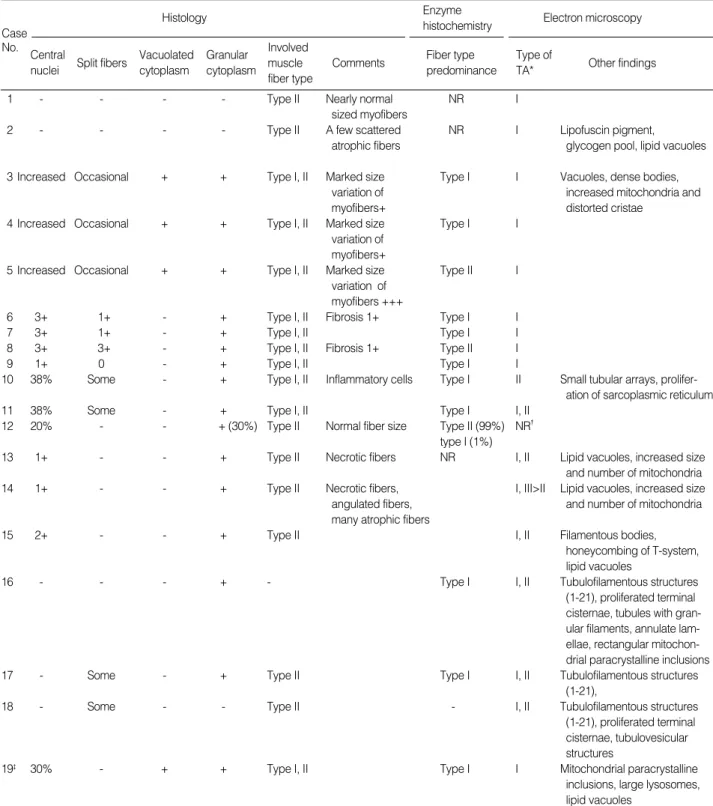
Light microscopically, vacuolated granular to flocculent cyto- plasm is the most salient finding of primary tubular aggregate myopathy, and is observed in 64% of previously reported cases including the present patient. Vacuolated pale unstained areas were intensely stained with NADH-TR and modified Go- mori’s trichrome, but not stained with mitochondrial enzymes such as SDH, phosphorylase or cytochrome c oxidase (2, 6, 8, 18). Furthermore, the lanthanum staining of T-system also failed to show stainability (3, 15). Pathologic findings of 19 cases of tubular aggregate myopathy previously reported are summarized in Table 1. Increased internal nuclei and split fibers were commonly found in 73.7% and 52.6% of 19 cases of tubular aggregate myopathy. Inflammatory cell infiltration, fibrosis, and necrotic/regenerative fibers were rarely observed (3, 5, 6). Type I fiber predominance was found in 52.6% of tubular aggregate myopathy. Both type I and II fibers were commonly involved by tubular aggegates (52.6%).
As the origin of the tubules, modified degenerated mito- chondria were initially suggested (19, 20). Altered sarcoplas- mic reticulum membrane is now widely accepted as the origin of tubular aggregates. Sarsoplasmic reticulum is related to calcium uptake channel during muscle contraction and relax- ation. Tubular aggregates are the sites of calcium accumulation for ATP-dependent calcium uptake and increasing the cal- cium and oxalate loading capacity of the affected fibers. This finding was demonstrated by anti-calcium pump protein IgG
Fig. 3.Pedigree of the family. A shaded circle indicates a suspi- cious patient and an arrow indicates the present patient.
indirect immunofluorescence technique as well as calsequestrin antibody (4). Tubular aggregates may represent an adaptive mechanism aiming at regulating an increased intracellular
level of calcium in order to prevent the muscle fibers from hypercontraction and fiber necrosis (4). The fact that cases sparing type I fibers are not rare (42.1%) might be explained
1 - - - - Type II Nearly normal NR I
sized myofibers
2 - - - - Type II A few scattered NR I Lipofuscin pigment,
atrophic fibers glycogen pool, lipid vacuoles
3 Increased Occasional + + Type I, II Marked size Type I I Vacuoles, dense bodies,
variation of increased mitochondria and
myofibers+ distorted cristae
4 Increased Occasional + + Type I, II Marked size Type I I
variation of myofibers+
5 Increased Occasional + + Type I, II Marked size Type II I
variation of myofibers +++
6 3+ 1+ - + Type I, II Fibrosis 1+ Type I I
7 3+ 1+ - + Type I, II Type I I
8 3+ 3+ - + Type I, II Fibrosis 1+ Type II I
9 1+ 0 - + Type I, II Type I I
10 38% Some - + Type I, II Inflammatory cells Type I II Small tubular arrays, prolifer-
ation of sarcoplasmic reticulum
11 38% Some - + Type I, II Type I I, II
12 20% - - + (30%) Type II Normal fiber size Type II (99%) NR�
type I (1%)
13 1+ - - + Type II Necrotic fibers NR I, II Lipid vacuoles, increased size
and number of mitochondria
14 1+ - - + Type II Necrotic fibers, I, III>II Lipid vacuoles, increased size
angulated fibers, and number of mitochondria
many atrophic fibers
15 2+ - - + Type II I, II Filamentous bodies,
honeycombing of T-system, lipid vacuoles
16 - - - + - Type I I, II Tubulofilamentous structures
(1-21), proliferated terminal cisternae, tubules with gran- ular filaments, annulate lam- ellae, rectangular mitochon- drial paracrystalline inclusions
17 - Some - + Type II Type I I, II Tubulofilamentous structures
(1-21),
18 - Some - - Type II - I, II Tubulofilamentous structures
(1-21), proliferated terminal cisternae, tubulovesicular structures
19� 30% - + + Type I, II Type I I Mitochondrial paracrystalline
inclusions, large lysosomes, lipid vacuoles
NR, not recorded; TA, tubular aggregates; *, type according to Cameron et al. (5); �, Electron microscopic examination was done, but the type of tubules was not recorded or illustrated; �, The present case.
Histology Enzyme
Electron microscopy
Case histochemistry
No. Central Vacuolated Granular Involved
Fiber type Type of nuclei Split fibers
cytoplasm cytoplasm muscle Comments
predominance TA* Other findings fiber type
Table 1.Summary of pathologic findings of 19 cases of dominantly inherited tubular aggregate myopathy on which biopsies findings were described
140 N.R. Kim, Y.-L. Suh
by relative abundance of sarcoplasmic reticulum in type II, compared to type I fibers (21). Bendahan et al. (7) demonstrat- ed by magnetic resonance spectroscopy the association bet- ween tubular aggregates and hyperacidosis, indicating altered proton handling as a mechanism of pathogenesis. Martin et al.
(22) also suggested that the heat shock protein contributes to the formation of tubular aggregates. Although tubular aggre- gates can be found as a nonspecific change representing an ad- aptive response, it remains unclear whether the same mecha- nism is ascribed to the primary inherited tubular aggregate myopathy.
In summary, the diagnosis of this unique tubular aggregate myopathy can be made on the basis of clinical and pathologic examination, using skeletal muscle enzyme histochemistry and electron microscopy including asymptomatic family mem- bers. The present case is the first Korean case of presumably dominantly inherited tubular aggregate myopathy.
REFERENCES
1. de Groot JG, Arts WF. Familial myopathy with tubular aggregates.
J Neurol 1982; 227: 35-41.
2. Rohkamm R, Boxler K, Ricker K, Jerusalem F. A dominantly inher- ited myopathy with excessive tubular aggregates. Neurology 1983;
33: 331-6.
3. Pierobon-Bormiooli S, Arani M, Ringel SP, Angelini C, Vergani L, Betto R, Salviati G. Familial neuromuscular disease with tubular aggregates. Muscle Nerve 1985; 8: 291-8.
4. Salviati G, Pierobon-Bormioli S, Betto R, Damiani E, Angelini C, Ringel SP, Salvatori S, Margreth A. Tubular aggregates: sarcolem- mal reticulum origin, calcium storage ability, and functional impli- cations. Muscle Nerve 1985; 8: 299-306.
5. Cameron CH, Allen IV, Patterson V, Avaria MA. Dominantly inherit- ed tubular aggregate myopathy. J Pathol 1992; 168: 397-403.
6. Martin JJ, Ceuterick C, Van Goethem G. On a dominantly inherited myopathy with tubular aggregates. Neuromuscul Disord 1997; 7: 512- 20.
7. Bendahan D, Pouget J, Pellissier JF, Figarella-Branger D, Cozzone PJ. Magnetic resonance spectroscopy and histological study of tubular aggregates in a familial myopathy. J Neurol Sci 1996; 139: 149-55.
8. Muller HD, Vielhaber S, Brunn A, Schroder JM. Dominantly inher-
ited myopathy with novel tubular aggregates containing 1-21 tubulofil- amentous structures. Acta Neuropathol 2001; 102: 27-35.
9. Takizawa S, Ishihara T, Shinohara Y. A case of hypokalemic peri- odic paralysis with tubular aggregates in type 2A fibers and type 2B fibers Rinsho Shinkeigaku 1986; 26: 81-6 (Jpn).
10. Meyers KR, Gilden DH, Rinaldi CF, Hansen JL. Periodic muscle weakness, normokalemia, and tubular aggregates. Neurology 1972;
22: 269-79.
11 Sipila I, Simell O, Rapola J, Sainio K, Tuuteri L. Gyrate atrophy of the choroid and retina with hyperornithinemia: tubular aggregates and type 2 fiber atrophy in muscle. Neurology 1979; 29: 996-1005.
12. Dobkin BH, Verity MA. Familial neuromuscular disease with type I fiber hypoplasia, tubular aggregates, cardiomyopathy, and myas- thenic features. Neurology 1978; 28: 1135-40.
13. Lazaro RP, Fenichel GM, Kilroy AW, Saito A, Fleischer S. Cramps, muscle pain, and tubular aggregates. Arch Neurol 1980; 37: 715-7.
14. Negro AV, Angulo M, Pomar JMR, Errasti CA. Tubular aggregates in skeletal muscle of chronic alcoholic patients. Acta Neuropathol 1982; 56: 250-4.
15. Doriguzzi C, Mongini T, Jeantet A, Monga G. Tubular aggregates in a case of osteomalacic myopathy due to anticonvulsant drugs. Clin Neuropathol 1984; 3: 42-5.
16. Alonso-Losada G, Cimas I, Pego R, La Torre P, Teijeira S, Navarro C. Isolated progressive muscle weakness with tubular aggregates. Clin Neuropathol 1998; 17: 50-4.
17. Cullen MF, Johnson MA, Mastaglia FL. Pathological reactions of skeletal muscle. In: Mastaglia FL, Walton JN, editors, Skeletal mus- cle pathology, 2nd edn, London: Churchill Livingstone 1992: 155.
18. Meijer AE. Histochemical features of tubular aggregates in dis- eased human skeletal muscle fibers. J Neurol Sci 1988; 86: 73-82.
19. Lewis PD, Pallis C, Pearse AG. “Myopathy”with tubular aggregates. J Neurol Sci 1971; 13: 381-8.
20. Thomas PK, Cooper JM, King RH, Workman JM, Schapira AH, Goss- Sampson MA, Muller DP. Myopathy in vitamin E deficient rats; mus- cle fiber necrosis associated with disturbances of mitochondrial func- tion. J Anat 1993; 183: 451-61.
21. Niakan E, Harati Y, Danon MJ. Tubular aggregates: their association with myalgia. J Neurol Neurosurg Psychiatry 1985; 48: 882-6.
22. Martin JE, Mather K, Swash M, Gray AB. Expression of heat shock protein epitopes in tubular aggregates. Muscle Nerve 1991; 14: 219- 25.