DOI : 10.3341/jkos.2008.49.8.1303
HRT2와 HRT3의 녹내장의 진단에의 유용성 비교
이미연․이나영․박찬기 가톨릭대학교 의과대학 안과 및 시과학교실
목적: Heidelberg Retina Tomograph (HRT) 2와 HRT3의 녹내장 진단에의 유용성을 비교하고자 하였다.
대상과 방법: 정상안 52안, 녹내장 의증 62안, 녹내장 172안 총 286안을 대상으로 하여 후향적 조사를 실시하였다.
Areas Under the Receiver Operating Characteristics (AROCs) curves를 이용하여 각 지표의 유용성을 알 아보았고, κ분석을 이용하여 분류의 일치도를 분석하였다.
결과: 정상안과 녹내장 의증안을 녹내장안과 비교시 HRT2에서는 유두 함몰 형태치(Cup shape measure)가 Areas Under the ROC curve (AUC) 0.784로 가장 높았고, HRT3에서는 Global probability score (GPS) 가 AUC 0.718로 가장 높았다. 녹내장 의증안과 녹내장안을 정상안과 비교하였을 때는 HRT2에서는 유두 함몰 형태 치가 AUC 0.785로 가장 높았고, HRT3에서는 GPS가 AUC 0.696로 가장 높았다. 최종적 분류를 비교한 결과는 민감도는 HRT3가 95%로, HRT2의 91%보다 높았으며, 특이도는 HRT2가 61%로 HRT3의 59%보다 높았다.
결론: HRT3는 HRT2와 유사하였다. 하지만 HRT3는 주관적인 입력이 배제된 결과가 보장되었다.
<한안지 49(8):1303-1308, 2008>
<접수일 : 2007년 11월 20일, 심사통과일 : 2008년 5월 2일>
통신저자 : 박 찬 기
서울시 서초구 반포동 505 가톨릭대학교 강남성모병원 안과 Tel: 02-590-1523, Fax: 02-590-2044 E-mail: [email protected]
전세계 실명 원인 중 2번째를 차지하는 녹내장은 시 야 손상 이전에 시신경의 형태학적 손상이 증상 없이 선행되며 이는 비가역적이므로 시신경유두 형태 분석 은 매우 중요하다.1 시신경유두 모양을 분석하는 Heidelberg Retina Tomograph (HRT) (Heidel berg Engineering GmbH. Heidelberg. Germa ny)는 검사 결과가 빠르고, 산동이 불필요하며, 재현성 이 높고, 3차원적 계측치를 알 수 있어 시신경유두 모 양 분석에 최근 유용하게 많이 사용되고 있다. 주로 사 용되고 있는 Moorfield regression analysis (MRA) 를 이용한 HRT2는 재현성이 높은 특징을 가지고 있 다. 2-4 또한 HRT2의 여러 가지 계측치들은 기존 연구 들에 의해 정상안과 녹내장안을 구별하는데 의미가 있 다고 밝혀져 있다.5-7 하지만 HRT2 소프트웨어는 검사 자가 수동적, 주관적으로 시신경유두 경계를 따라 유두 경계선(contour line)을 그리며, 이에 의해 결정되는 기준면(reference plane)에 의해 시신경유두가 결정 된다. 즉, 많은 HRT2의 지표들은 유두 경계선에 영향
을 받아 주관적인 요소가 결과에 포함되게 된다. 유두 경계선을 그리는 것은 시간적 소모 뿐 아니라 검사자에 따른 변수를 결과에 제공하게 된다.8-10 HRT3소프트웨 어에서는 검사자가 유두 경계선을 그리는 과정이 제외 되었다. HRT3에서는 Swindale et al이 제시한 삼차 원적 시신경유두 모양 모델11과 대상안을 비교하여 인 공 지능 기계에 의한 분석을 통해 Glaucoma probability score (GPS)라는 결과를 도출하게 된 다. 또한 HRT3는 HRT2보다 더 큰 정상군의 데이터 가 입력되어 있는 특징이 있다. 하지만 HRT3에서도 동양인의 데이터는 입력되어 있지 않은 문제를 가지고 있다. HRT2에 대해서는 한국인에게 적용할 판별식이 제시된 적 있다.12 하지만 HRT3에 대해서는 아직 한국 인에의 적용에 더 많은 연구가 필요할 것이다. 저자들 은 HRT2와 HRT3의 임상에서의 녹내장 진단에의 유 용성을 비교하고 HRT2와 HRT3의 여러 지표 중 이에 가장 유용한 지표를 알아보고자 하였다.
대상과 방법
2006년 10월부터 2006년 12월까지 본원 안과 외래 에 통원중인 한국인 환자 중 총 286안을 대상으로 하였 으며 후향적으로 관찰하였다. 모든 환자에서 시력 검 사, 굴절 검사, 세극등 검사, 안압 검사, 전방각 검사, 안저 검사를 시행하였고, Humphrey standard
Normal (n=52)
Glaucoma suspect (n=62)
Glaucoma (n=172) Age (yrs)
Mean deviation (dB) Pattern deviation (dB)
44.7±12.8 -1.8±1.1
1.7±0.3
46.5±14.0 -1.7±1.2
1.7±0.4
48.7±15.4 -9.9±6.4
4.8±3.7 Table 1. Characteristics of the study population
automated perimetry (Allergan Humphrey Ins. U.S.A.) central 24-2를 이용하여 시야 검사를 시행하였다. 또한 HRT2와 HRT3 모두를 이용하여 시 신경유두 분석을 시행하였다. 20세 이상, 교정시력 20/40이상, 굴절력(구면 렌즈 대응치) +7.0~-7.0D 이며 난시 3D 이내의 환자를 대상으로 하였으며, 검사 에 영향을 미칠 수 있는 다른 안질환 및 뇌질환이 없는 환자를 대상으로 하였다. 시행한 검사에 따르면 정상안 은 52안, 녹내장의증 안은 62안, 녹내장 안은 172안이 었다. 정상안은 녹내장성 시신경유두의 변화를 보이지 않으며 시야 검사상 녹내장성 결손이 없고 안압을 측정 한 중에 한번도 21 mmHg를 넘지 않은 경우로 정의하 였다. 녹내장의증안은 시야 손상이 없으면서 안압이 22 mmHg 이상이거나 시신경유두의 녹내장성 변화 즉, 비대칭적 시신경유두함몰(양안의 유두함몰비 차이가 0.2 이상) 혹은 증가된 유두함몰(수직 유두함몰비가 0.6 이상)이 있는 경우를 포함하였다. 녹내장안은 안압 과 관계없이 녹내장성 시야 손상과 시신경유두 변화 혹 은 시신경 섬유층의 손상이 있는 경우를 대상으로 하였 다. 녹내장성 시야 손상은 Anderson criteria를 만족 시키거나, Corrected pattern stan dard deviation (CPSD)이 5% 미만에서 나타나거나 Glaucoma hemifield test (GHT)상 이상 소견 (Outside normal limits)이 나타나는 경우로 정의 하였다. 30% 이하의 주시 상실(fixation loss), 가양 성(false-positive response), 가음성(false nega tive response)일 경우 의미 있는 것으로 판단하였 다. 검사자는 시신경의 유두 경계를 공막륜의 안쪽 가 장자리로 하여 유두 경계선을 그렸고, 한 검사자에 의 해서 이루어졌다. HRT2에 서는 측정한 12가지 유두 측정치들인 시신경유두 면적(disc area), 유두함몰 면 적(cup area), 시신경테 면적(rim area), 유두함몰 용적(cup volume), 신경테 용적(rim volume), 유 두함몰 면적비(cup/disc area ratio), horizon tal cup/disc ratio, vertical cup/disc ratio, 평균 유 두 함몰 깊이(mean cup depth), 최대 유두 함몰 깊 이(maximum cup depth), 유두 함몰 형태 측정 (cup shape measurement), 유두 경계 변이 (height variation contour), 평균 망막 신경섬유층 단면적(mean RNFL thickness), 망막 신경 섬유층 단면 면적(RNFL cross sectional area) 결과를 정 상 데이터와 비교하여 정상 소견(Within normal lmits), 경계 소견(Borderline), 이상 소견(Outside normal limits)으로 최종적으로 분류하였다. HRT3 에 의해서는 자동으로 시신경 이미지를 분석하였으며, 이에 의한 유두 측정치들은 수평 시신경 섬유 굽이
(Horizontal RNFL curvature), 수직 시신경 섬유 굽이(Vertical RNFL curvature), 유두 함몰 크기 (cup size), rim steepness, 유두 함몰 깊이(cup depth) 총 5가지이다. 또한 HRT3에 의한 최종적 진 단은 Glaucoma probability score (GPS)로 0%
에서 100%까지 분포하며, 0%에서 27%까지는 정상 소견(Within normal limits)으로, 28%에서 64%
까지는 경계 소견(Borderline)으로, 그리고 64%에서 100%까지는 이상 소견(Outside normal limits)으 로 판단하였다.
통계적 분석은 SPSS/PC 프로그램을 이용하였으 며, HRT2와 HRT3의 각 지표가 정상안, 녹내장의증 안, 녹내장안의 판별에 이용되는 능력은 Areas Under the Receiver operating characteristic (AROC) curve를 그린 후, Areas under the ROCs curves (AUC)를 비교하여 판단하였으며, HRT2와 HRT3의 최종적 분류의 임상적 분류와의 일치성에 대 해서는 k analysis로 분석하였다. P-value<0.05를 통계적으로 유의하다고 판정하였다.
결 과
환자의 평균 나이는 정상군은 44.7±12.8, 녹내장의 증군은 46.5±14.0, 녹내장 군은 48.7±15.4였으며, Mean deviation (MD) 값은 정상군은 -1.8±1.1, 녹 내장의증군은 -1.7±1.2, 녹내장 군은 -9.9±6.4였으 며, Pattern standard deviation (PSD) 값은 정 상군은 1.7±0.3, 녹내장의증군은 1.7±0.4, 녹내장군 은 4.8±3.7였다(Table 1).
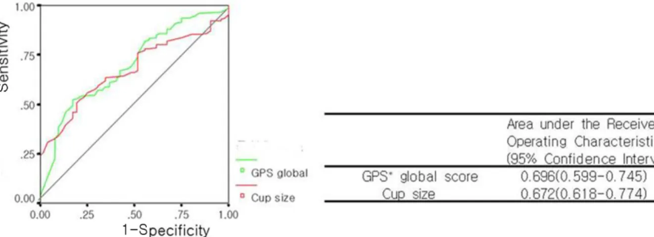
정상안과 녹내장의증안을 한군으로 하여 녹내장안 과 비교했을 때 HRT2에서는 유두함몰 형태치가, AUC 0.784 (95% CI,0.720-0.851)로 가장 높았고, 다음으로는 신경테 용적이 AUC 0.775 (95% CI, 0.713-0.838)로 높았다(Fig. 1). HRT3에서는 유두 함몰 크기가, AUC 0.672 (95% CI, 0.618-0.774) 로 계측치 중에는 가장 높았으나, GPS가 AUC 0.696 (95% CI, 0.599-0.745)로 실제 가장 높은 결과를 보 였다(Fig. 2).
Figure 2. Comparision of glaucoma and non-glaucoma eyes with HRT (Heidelberg Retina Tomograph) 3.
* GPS=Global probability score.
Figure 1. Comparision of glaucoma and non-glaucoma eyes with HRT (Heidelberg Retina tomography) 2.
Figure 3. Comparision of normal and non-normal eyes with HRT (Heidelberg Retina Tomograph) 2.
HRT3
HRT2 WNL BL ONL TOTA L
WNL* BL† ONL‡
20 10 4
19 17 29
16 27 144
55 54 177
TOTAL 34 65 187 286
HRT Heidelberg Retina Tomograph; * WNL=within normal limits; †BL=borderline; ‡ONL=outside normal limits.
Table 2. Subjects' Classification by Overall Heidelberg Retina Tomograph (HRT) 2 and HRT (Heidelberg Retina Tomograph) 3
Sensitivity Specificity HRT2
HRT3
91%
95%
61%
59%
Table 3. Sensitivity, Specificiy for discrimination between Normal vs Glaucoma with HRT (Heidelberg Retina Tomograph) 2, HRT (Heidelberg Retina Tomograph) 3
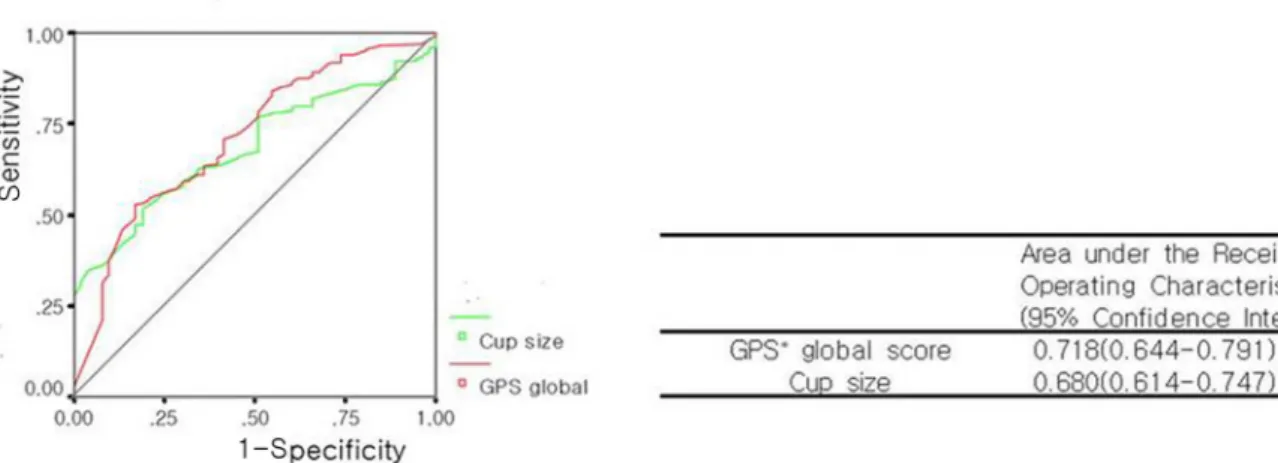
Figure 4. Comparision of normal and non-normal eyes with HRT (Heidelberg Retina Tomograph) 3.
* GPS=Global probability score
녹내장의증안과 녹내장안을 합친 군을 정상안과 비 교하였을 때는 HRT2에서는 유두함몰 형태 치가 AUC 0.785 (95% CI, 0.727-0.844)로 가장 높았고, 다음 으로는 유두함몰 면적비가 AUC 0.782 (95% CI, 0.720-0.845)로 높았다(Fig. 3). 정상안과 녹내장의 증안을 한군으로 하여 녹내장안과 비교했을 때 두번째 로 높은 AUC를 보였던 지표인 신경테 용적은 AUC 0.767 (95% CI, 0.703-0.831)로, 세번째로 높은 결 과를 보였다. HRT3에서는 유두 함몰 크기가 AUC 0.680 (95% CI, 0.614-0.747)로 가장 높고, GPS 는 AUC 0.718 (95% CI, 0.644-0.791)였다(Fig.
4). 최종적 분류는 HRT2와 HRT3의 최종적 분류를 분석하였을때는 통계학적으로 유의하게 일치하였다(κ
=0.307, 95% CI : 0.261-0.335, p=0.046)(Table 2). 정상, 녹내장의증, 녹내장의 임상적 진단과 HRT2, HRT3의 최종적 분류를 비교하였을 때, 민감도는 녹내 장의증안과 녹내장안을 합친 군을 정상안과 비교한 결 과에서 도출하였으며, HRT3가 95%로 HRT2의 91%
보다 높은 결과를 보였다. 그리고 특이도는 정상안과
녹내장의증안을 한군으로 하여 녹내장안과 비교한 결과 에서 도출하였으며, HRT2가 61%로 HRT3의 59%보 다 높은 결과를 보였다(Table 3).
고 찰
녹내장의 진단을 위해 사용되는 안압 측정, 시야 검 사, 시신경유두 검사 등은 그 진단적 가치 및 조기 진단 적 유용성에서 한계점을 가지고 있다. 시신경유두의 녹 내장성 변화, 특히 시신경 축삭의 손실은 시야 결손에 선행한다고 알려져 있으므로 녹내장 진단과 진행을 알 기 위해서는 시신경유두의 미세 변화를 알아내는 것이 중요하다.12-16 HRT 검사는 시신경유두의 미세한 녹내 장성 초기 변화를 알아낼 수 있다는 장점이 있어서 시 신경유두의 미세 변화를 조기에 발견하는데 많은 도움 을 주고 있다. 새로운 소프트웨어인 HRT3는 시신경유 두 경계를 수동적으로 정의하는 과정 없이 자동으로 시 신경유두 모양 분석을 할 수 있도록 고안된 프로그램이 다. 본 연구의 결과에 의하면 기존의 HRT2에 의한 최 종적 분류와 HRT3에 의한 최종적 분류는 통계학적으 로 유의하게 일치 하고 있었으며, 각 프로그램에서 진 단 능력이 가장 우수하다고 판단된 시신경유두 계측치 는 정상안과 녹내장의증안을 한군으로 하여 녹내장안과 비교했을 때는 HRT2에서는 유두 함몰 형태치였으며,
HRT3에서는 GPS였다. 녹내장의증안과 녹내장안을 합친 군을 정상안과 비교하였을 때도 마찬가지로 HRT2에서는 유두 함몰 형태치였으며, HRT3에서는 GPS였다. HRT2, HRT3의 최종적 분류와 임상적 분 류를 비교하여 민감도, 특이도를 비교하여 보았을 때 민감도는 HRT3가 95%로 HRT2의 91%보다 높은 결과를 보였고, 특이도는 HRT2가 61%로 HRT3의 59%보다 높은 결과를 보였다. 즉 HRT2의 최종적 분 류는 녹내장 확진에 HRT3의 최종적 분류는 녹내장 선 별 검사로 유용하다고 판단된다. HRT3는 HRT2 프로 그램보다 더 많은 수의 정상인 데이터가 입력되어 있는 프로그램이다. 많은 수의 정상 데이터와 더 많은 인종 이 포함되어 있지만, 아직 한국인을 포함한 동양인의 데이터는 포함되어 있지 않다. 시신경유두의 국소 형태 는 인종간에 차이가 있는 것으로 알려져 있다. 이런 점 을 참조할 때 한국인에게 적용할 경우의 문제점에 대해 서 연구가 더 필요할 것으로 사료된다. HRT3에 의한 최종적 분류가 HRT2에 의한 분류보다 이상 소견으로 판단한 경우가 더 많았지만, HRT2와 HRT3의 최종 분류는 통계학적으로 유의하게 서로 일치하고 있었으 며, 본 연구의 결과를 분석하면 HRT2와 HRT3가 녹 내장안과 정상안의 감별 능력에 차이가 있다고 보기 어 렵다. 즉 HRT3가 HRT2보다 녹내장 진단에 더 우수 하다고 말할 수 없다. HRT3는 형태학적인 검사에 주 관적인 요소가 배제되었다는 점이 주목할만한 장점이 다. 본 연구에서는 HRT의 결과에 영향을 줄 수 있는 근시안 등은 연구 데이터에서 배제하였으므로, 이런 근 시안 등에서의 HRT의 검사 능력에 대해서는 확정적으 로 논하기 어렵다. 요약적으로 새로운 HRT3 프로그램 은 수동적으로 유두 경계선을 그리지 않고 자동적으로 시신경유두 형태 검사가 가능하며, 이에 의해 기존의 HRT에 변수를 주던 요소를 배제시킬 수 있게 되었다.
즉, HRT3는 HRT2와 유사한 녹내장 진단 능력을 유 지한 채 자동적인 방법에 의해 주관적 요소를 배제한 새로운 소프트웨어이다. 또한 두 검사를 임상에 응용할 경우 최종적 분류를 HRT2는 녹내장 확진 검사로, HRT3는 선별 검사로 이용하는 것이 적합하다고 판단 된다.
참고문헌
1) Quigley HA. Number of people with glaucoma worldwide. Br J Ophthalmol 1996;80:389-93.
2) Kruse FE, Burk RO, Volcker HE, et al. Reproducibility of topographic measurements of the optic nerve head with laser tomographic scanning. Ophthalmology 1989;96:1320-4.
3) Dreher AW, Tse PC, Weinreb RN. Reproducibility of topographic measurements of the normal and glaucomatous optic nerve head with the laser tomographic scanner. Am J Ophthalmol 1991;111:221-9.
4) Rohrschneider K, Burk RO, Volcker HE. Reproducibility of topometric data acquisition in normal and glaucomatous optic nerve heads with the laser tomographic scanner. Graefes Arch Clin Exp Ophthalmol 1993;231:457-64.
5) Miqlior S, Casula M, Guareschi M, et al. Clinical ability of Heidelberg retinal tomograph examination to detect glauco matous visual field changes. Ophthalmology 2001;108:1621-7.
6) Uchida H, Brigatti L, Caprioli J. Detection of structural damage from glaucoma with confocal laser image analysis.
Invest Ophthalmol Vis Sci 1996;37:2393-401.
7) Hatch WV, Flanagan JG, Etchells EE, et al. Laser scanning tomogrphy of the optic nerve head in ocular hypertension and glaucoma. Br J Ophthalmol 1997;104:545-8.
8) Miglior S, Albe E, Guareschi M, et al. Intraobserver and interobserver reproducibility in the evaulation of optic disc steremetric parameters by Heidelberg Retina Tomograph.
Ophthalmology 2002;109:1072-7.
9) Garway-Heath DF, Poinoosawmy D, Wollstein G, et al.
Interobserver and intraobserver variation in the analysis of optic disc images : comparision of the Heidelberg retina tomograph and computer assisted planimetry. Br J Ophthalmol 1999;83:664-9.
10) Iester M, Mikelberg FS, Courtright P, et al. Interobserver variability of optic disc variables measured by confocal scanning laser tomography. Am J Ophthalmol 2001;132:57-62.
11) Swindale NV, Stjepanovic G, Chin A, Mikelberg FS.
Automated analysis of normal and glaucomatous optic nerve head topography images. Invest Ophthalmol Vis Sci 2000;41:
1730-42.
12) Park YJ, Park CK, Moon JI, Baek NH. The Diagnostic Precision of Glaucoma Classification with New HRT Discriminant Formula in Koreans. J Korean Ophthalmol Soc 1999;40:175-81.
13) Sommer A, Pollack I, Maumence AE. Optic disc parameters and onset of glaucomatous field loss. Arch Ophthalmol 1979;97:1444-8.
14) Quigley HA, Addicks EM, Gree WR. Optic nerve damage in human glaucoma. Quantitative correlation of nerve fiber loss and visual field defect in glaucoma, ischemic neuropathy, papilledema, and toxic neuropathy. Arch Ophthalmol 1982;
100:135-46.
15) Pederson JE, Anderson DR. The mode of progressive disc cupping in ocular hypertension and glaucoma. Arch Ophthalmol 1980;98:490-5.
16) Caprioli J, Miller JM, Sears M. Quantitative evaluation of the optic nerve head in patients with unilateral visual field loss from primary open angle glaucoma. Ophthalmology 1987;
94:1484-7.
=ABSTRACT=
Comparison between Heidelberg Retina Tomograph 2 and Heidelberg Retina Tomograph 3 for Glaucoma Detection
Mee Yon Lee, M.D., Na Young Lee, M.D., Chan Kee Park, M.D.
Department of Ophthalmology and Visual Science, College of Medicine, The Catholic University of Korea, Seoul, Korea
Purpose: To compare the ability of Heidelberg Retina Tomograph (HRT) 2 and the new version, HRT3, to discriminate between glaucoma and non-glaucoma eyes and to evaluate the most useful parameters for glaucoma detection.
Methods: Fifty-two healthy eyes, 62 glaucoma-suspect eyes, and 172 glaucoma eyes (a total of 286 eyes) were studied retrospectively. The discrimination capabilities of healthy and glaucomatous eyes were compared using areas under the receiver operating characteristics (AROCs) curves. Agreement between classifications was defined by κ analysis.
Results: The stereometric parameters that best discriminated between glaucoma and non-glaucoma eyes were cup shape measure at HRT2 (areas under the ROCs curves (AUC) 0.784) and global probability score (GPS) at HRT3 (AUC 0.718). The stereometric parameters that best discriminated between normal and non-normal eyes were cup shape measure at HRT2 (AUC 0.785) and GPS at HRT3 (AUC 0.696). The sensitivity was 95% at HRT3 and 91% at HRT2, and specificity was 61% at HRT2 and 59% at HRT3.
Conclusions: The glaucoma-discriminating ability of the HRT3 software is similar to that of HRT2; however, HRT3 provided results without the need for subjective operator input.
J Korean Ophthalmol Soc 49(8):1303-1308, 2008 Key Words: HRT, Glaucoma
Address reprint requests to Chan Kee Park, M.D.
Department of Ophthalmology, The Catholic University, Gangnam St. Mary’s Hospital
#505 Banpo-dong Seocho-gu, Seoul 137-701, Korea
Tel: 82-2-590-1523, Fax: 82-2-590-2044, E-mail: [email protected]