ISSN 0378-6471 (Print)⋅ISSN 2092-9374 (Online)
http://dx.doi.org/10.3341/jkos.2015.56.10.1544
Original Article
백내장 수술 후 난시교정용 인공수정체를 이용한 난시 교정 효과
Effect of Toric Intraocular Lens Implantation on Astigmatism in Cataract Surgery
차용재1⋅김미금1,2⋅위원량1,2
Yong Jae Cha, MD1, Mee Kum Kim, MD, PhD1,2, Won Ryang Wee, MD, PhD1,2
서울대학교 의과대학 서울대학교병원 안과학교실1, 서울대학교 의과대학 서울대학교병원 의생명연구원2 Department of Ophthalmology, Seoul National University Hospital, Seoul National University College of Medicine1, Seoul, Korea Biomedical Research Institute, Seoul National University Hospital, Seoul National University College of Medicine2, Seoul, Korea
Purpose: To evaluate the efficacy of Tecnis® toric intraocular lens (IOL) implantation for the correction of astigmatism and rota- tional stability during cataract surgery in patients with cataract and astigmatism.
Methods: We prospectively analyzed 17 eyes of 14 patients with 1 to 4 diopters (D) of corneal astigmatism who underwent pha- coemulsification and Tecnis® toric IOL implantation at Seoul National University Hospital from June 2013 to May 2014. Informed consent was obtained from all participants before the clinical trial. We evaluated the changes in visual acuity, refraction, astigma- tism, IOL axis and higher order aberration for 3 months postoperatively. Power vector analysis was used to analyze astigmatism.
Results: The mean uncorrected visual acuity (log MAR) significantly improved from 0.58 ± 0.34 to 0.26 ± 0.43 at 3 months postoperatively. The mean refractive astigmatism was significantly decreased by 77.9% from a mean value of -2.67 ± 0.89 D to -0.59 ± 0.48 D at 3 months postoperatively. According to power vector analysis, M, B, J0, and J45 were significantly reduced after the surgery. The mean difference between achieved and intended IOL axis was 3.26 degrees clockwise at postoperative 3 months, which was statistically insignificant. Most of the rotational changes were observed within a month after the surgery.
Conclusions: Phacoemulsification and Tecnis® toric IOL implantation in patients with cataracts and astigmatism showed efficacy for the correction of astigmatism and rotational stability.
J Korean Ophthalmol Soc 2015;56(10):1544-1551
Key Words: Astigmatism, Cataract, Tecnis® toric IOL, Vector analysis
■Received: 2015. 3. 20. ■ Revised: 2015. 6. 24.
■Accepted: 2015. 9. 22.
■Address reprint requests to Mee Kum Kim, MD, PhD Department of Ophthalmology, Seoul National University Hospital, #101 Daehak-ro, Jongno-gu, Seoul 03080, Korea Tel: 82-2-2072-2665, Fax: 82-2-741-3187
E-mail: [email protected]
* This study was presented as a narration at the 111th Annual Meeting of the Korean Ophthalmological Society 2014.
* This study was supported for grant by Abbott Medical Optics (AMO, Santa Ana, CA, USA) (grant number 06-2013-3210).
ⓒ2015 The Korean Ophthalmological Society
This is an Open Access article distributed under the terms of the Creative Commons Attribution Non-Commercial License (http://creativecommons.org/licenses/by-nc/3.0/) which permits unrestricted non-commercial use, distribution, and reproduction in any medium, provided the original work is properly cited.
최근에는 백내장 수술 후 시력 개선에 대한 환자의 요구 도가 높아지고, 또한 기술의 발달에 힘입어 다양한 인공수 정체가 개발되면서, 난시 및 노안의 병합 교정에 대한 관심 이 증가하고 있다.1,2 특히 백내장 환자에서 각막 난시가 1.25D 이상인 인구가 22-27%임을 고려하면,3,4 각막 난시 가 1.25D 이상의 환자에서 단순히 백내장 수술과 일반 인 공수정체를 삽입하는 경우 수술 후 안경 교정이 필요할 가 능성이 높고, 이로 인한 환자 만족도가 저하될 우려가 백내 장 수술이 필요한 인구의 약 4분의 1에서 발생할 수 있음을 예측할 수 있다.
백내장 수술과 병합한 난시 교정으로는 각막윤부이완절 개술의 병합이 가능하며, 난시교정용 인공수정체 삽입 또
는 각막에 굴절교정각막절제술을 시행할 수 있다. 이 중 굴 절교정각막절제술은 난시교정 효과는 우수하나 백내장 수 술과 동시에 하기가 어렵고 추후 시행해야 하는 불편함이 있고,5 백내장 수술과 동시에 시행이 가능한 각막윤부이완 절개술 병합 또는 난시교정용 인공수정체 삽입술이 많이 시 행되고 있다.6-11 각막윤부이완절개술보다는 난시교정용 인 공수정체 삽입술이 교정 효과가 더 우수한 것으로 보고되고 있으며,9 비교적 초기에 널리 공급된 회절형의 AcrySof® toric IOL (Alcon Laboratories Inc., Fort Worth, TX, USA) 의 교정 효과가 국내외에서 많이 보고되었다.11,12
난시교정용 인공수정체의 난시 교정 효과는 여러 가지 요인에 의해서 영향을 받는데,13 술전 난시 검사의 오차,14,15 인공수정체 축의 회전,16,17 각막 후면난시18 등이 영향을 줄 수 있고, 도수 결정 시 반영되는 전방 깊이 및 각막 굴절력, 안구축장19 등이 영향을 줄 수 있다. AcrySof® toric IOL은 난시 인공수정체 도수 결정 프로그램에 안구축장, 전방 깊 이가 반영되어 있지 않아, 교정 효과에 영향을 줄 수 있음이 밝혀지면서,10 최근 개발되어 난시 도수결정 프로그램에 안 구축장이 반영된 Tecnis® ZCT toric IOL (Abbott Medical Optics, Santa Ana, CA, USA)의 난시교정 효과가 국외에 보고되었다.20 그러나 전체적인 보고가 아직은 많지 않아 컨센서스가 확립되어 있지 않고 또 국내에는 전혀 보고가 없어 Tecnis® ZCT toric IOL의 난시교정 효과에 대한 증거 를 축적하고자 전향적 연구를 진행하였다.
대상과 방법
본 연구는 전향적 연구로 헬싱키선언을 준수하여 시행되 었고, 서울대학교병원 임상시험 심사위원회의 심의 및 승 인을 받았다(IRB number: 1305-011-485). 서울대학교병원 에서 2013년 6월부터 2014년 5월까지 임상시험 설명 후 자 발적 동의서를 받았으며, 백내장 수술이 필요한 환자를 대 상으로 하였다. 연구 포함 기준은 21세 이상의 노년성 백내 장 환자 중 자동굴절검사계(Automated refractor, KR890, TOPCON, Tokyo, Japan) 및 IOL-Master (Carl Zeiss, Meditec AG, Jena, Germany)의 각막굴절계로 측정한 각막 난시가 1-4디옵터(diopter, D) 이내의 환자를 대상으로 하 였다. 임부, 수유부, 수정체 낭의 안정성에 영향을 줄 수 있 는 기타 다른 질환이 있는 경우(거짓비늘증후군, 녹내장, 외상성 백내장, Marfan 증후군 등), 피험자 중 스스로 동의 서를 읽을 수 없는 자와 연구기간 동안 예정된 경과관찰 및 검사를 받지 못한 경우는 대상에서 제외하였다. 환자군은 총 14명(17안)이었으며, 한 명의 술자(K.M.K)에 의하여 초 음파유화술 및 Tecnis® ZCT toric IOL (a single-piece toric
hydrophobic acrylic IOL, Abbott Medical Optics, Santa Ana, CA, USA) 후낭삽입술을 시행 받았고, 포함된 모든 환자에서 수술 시 합병증은 발생하지 않았다.
대상 수 산출에 필요한 수치를 확인하기 위해 “난시교정 인공수정체 삽입 후 임상 결과”12를 근거로 표준편차를 2.92로 가정하였으며, 허용오차는 1.5로 설정하고 연구대상 수를 산출하여, 산출된 연구대상 수는 15안으로 탈락률을 10%로 고려하여 17안이었다.
대상 환자는 수술 전에 나안시력 및 최대교정시력(logMAR), 자동굴절검사(Automated refractor, KR890, TOPCON), 세 극등현미경검사, 안저검사, IOL master 및 A-scan (Quantel medical, Clemont-Ferrand, France)을 이용한 인공수정체 도 수 측정 검사, 각막지형도 및 고위수차(Orbscan II, Bausch and Lomb Inc., Rochester, NY, USA)를 시행하였다.
인공수정체
Tecnis® toric IOL은 아크릴 재질의 접힘형 연성 인공수 정체인데, 앞면이 난시를 교정할 수 있는 toric 비구면이다.
인공수정체의 도수를 결정하기 위하여 숙련된 검사자를 통 하여 A-scan (Quantel medical, Clemont-Ferrand, France)과 IOL master (Carl Zeiss, Meditec AG, Jena, Germany)를 이 용하여 안구축장을 3회 측정하였고, 인공수정체 도수를 구 하는 공식에서 A값은 A scan은 118.8, IOL master는 119.3 을 이용하였다. 각막 곡률과 각막 난시의 측정은 자동굴절검 사계(Automated refractor, KR890, TOPCON), IOL Master (Carl Zeiss, Meditec AG, Jena, Germany) 및 각막 지형도 (Orbscan II, Bausch and Lomb Inc., Rochester, NY, USA) 를 이용하여 각각 3회 측정하였으며, 각 검사의 굴절력, 난 시 축 및 난시 양의 3회 평균값을 이용하였다.
Tecnis® toric 인공수정체의 종류 및 삽입 축은 toric IOL calculator 프로그램(https://www.amoeasy.com/calc)에 각막곡 률, 절개 방향, 수술로 인하여 유발되는 각막 난시(surgically- induced astigmatism, SIA), 인공수정체의 spherical power, 안축장 등을 포함한 환자의 정보를 입력하여 산출되는 결 과에 따라 결정하였다. 위에서 언급한 세 가지 검사의 평균 굴절력을 각각 대입하여 얻은 결과 중 검사 간 일치도가 높 으면서(세 가지 중 2개 이상 일치하는 값) 가장 난시가 0에 근접하고, 축은 방향이 변하지 않는 값으로 인공수정체 난 시 도수를 선정하여 과교정을 피하고자 하였다. 각막 절개 방향은 steep-on axis로 시행하였고(직난시: 상측, 도난시:
이측), 상측 각막 절개의 경우(직난시) 과교정을 피하기 위 해 SIA를 0.5로 설정하였고, 이측 각막 절개의 경우(도난 시) 부족교정을 줄이기 위해 SIA를 0으로 설정하였다.
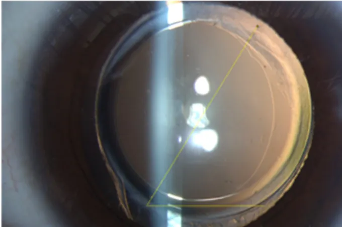
Figure 1. Image analysis for the rotation of the cylindrical
axis on the toric intraocular lens.Table 1. Demographics and preoperative status of the enrolled
patientsDemographics Data
No. of eyes (patients) 17 (14)
Mean age (years, range) 64.5 (32-84)
Sex (male:female) 4:13
Laterality (right:left) 9:8
UCVA (log MAR) 0.58 ± 0.34
Spherical equivalent (diopters) -0.65 ± 3.52
Axial length (mm) 24.08 ± 1.26
WTR versus ATR 10:7
Values are presented as mean ± SD unless otherwise indicated.
UCVA = uncorrected visual acuity; WTR = with the rule; ATR = against the rule.
수술 방법
기준 표지자는 수술 직전 환자를 세극등현미경 앞에 앉 혀 정면을 보게 한 상태에서 얼굴의 위치가 기울어지지 않았 는지 확인한 후, 각막 윤부의 3시, 9시 방향에 기준점을 표시 했다. 수술 중 환자가 누운 후에 Mendez ring (K3-7900, Katena)과 toric axis marker (K3-7910, Katena)를 이용하여 목표 난시축을 표시하였다. 모든 수술은 숙련된 한 명의 술 자(K.M.K)에 의해 이루어졌으며, 각막에 2.8 mm의 각막절개 (clear corneal incision)를 시행하고, Infinity Vision System® (Alcon Laboratories Inc., Fort Worth, TX, USA)과 OZilTM Torsional Handpeice (Alcon Laboratories Inc., Fort Worth, TX, USA)를 이용하여 수정체유화술을 시행하였다. Monarch injector (Alcon Laboratories Inc., Fort Worth, TX, USA)로 인공수정체를 후낭에 삽입하였다. 점탄 물질을 제거 후 tor- ic axis marker (K3-7910, Katena)로 축 방향을 다시 확인 하였으며, 인공수정체를 회전시켜 미리 표시해 둔 표지자 (marking)를 기준으로 인공수정체의 축을 원하는 방향으로 교정하였다. 수술을 마칠 때 각막 봉합술은 시행하지 않았 고, 수술 중 후낭 파열 등의 합병증은 발생하지 않았다.
수술 후 검사
수술 후 1일, 1주, 1달, 3달에 나안 시력과 최대교정시력 (logMAR), 굴절 이상의 변화, 인공수정체 축 위치 변화, 그 리고 고위 수차의 변화를 분석하였다. 난시의 벡터 분석은 Cartesian astigmatism (J0)과 oblique astigmatism (J45)을 이 용한 power vector 분석법을 통해 M=S+C/2, J0=(-C/2)×
cos2θ, J45=(-C/2)×sin2θ, B
M J J와 같은 변수 들을 이용하여 분석하였다.21 인공수정체의 회전안정성(축 의 변화)에 대한 분석은 다음과 같이 시행하였다. 수술 후 1주, 1달과 3달째에 수술한 눈을 산동시킨 후, 환자를 세극 등현미경 앞에 앉혀 가능한 최대한 똑바로 정면을 보게 하 여 역조명을 비추어 전안부 사진을 촬영하였다. Image J 소 프트웨어를 이용하여 모든 사진에서 인공수정체 면에 표시 되어 있는 기준 표지자(reference marks)를 참고하여 두 표 식을 서로 잇는 선을 그려 원래 의도하였던 삽입 축과 인공 수정체가 실제로 삽입된 축의 차이를 분석하였다(Fig. 1).모든 사진은 무작위로 선택하여 검사자의 편견이 개입되지 않도록 하였다.
통계 분석 방법
통계 분석은 SPSS 21.0 (IBM Inc., Armonk, NY, USA) 통계프로그램을 이용하여 수술 전과 후의 차이에 대하여 Wilcoxon’s signed ranks test, Independent student t-test 등 을 사용하였다. 모든 통계 분석에서 유의도(p-value)는 0.05
미만인 경우만을 통계적으로 유의한 것으로 설정하였다.
결 과
총 17안 14명의 환자들의 평균 나이는 64.5(32-84)세였 으며, 총 17안 중 남자가 4안, 여자가 13안이었고, 우안이 9안, 좌안이 8안이었다. 술전 시력 및 굴절력, 각막 난시, 안축장은 Table 1에 제시하였다.
시력
나안시력(logMAR)은 수술 전 평균 0.58 ± 0.34에서 수 술 후 1주에 0.22 ± 033, 수술 후 1개월에는 0.21 ± 0.33, 그리고 수술 후 3개월에 0.26 ± 0.43으로 각각의 경과 관찰 시기에 모두 수술 전에 비해 유의하게 호전되었다(p=0.023, p=0.013, p=0.033, Independent student t-test; Fig. 2).
굴절이상의 변화 및 고위 수차
평균 굴절난시는 수술 전 -2.67 ± 0.89D였으며, 수술 후
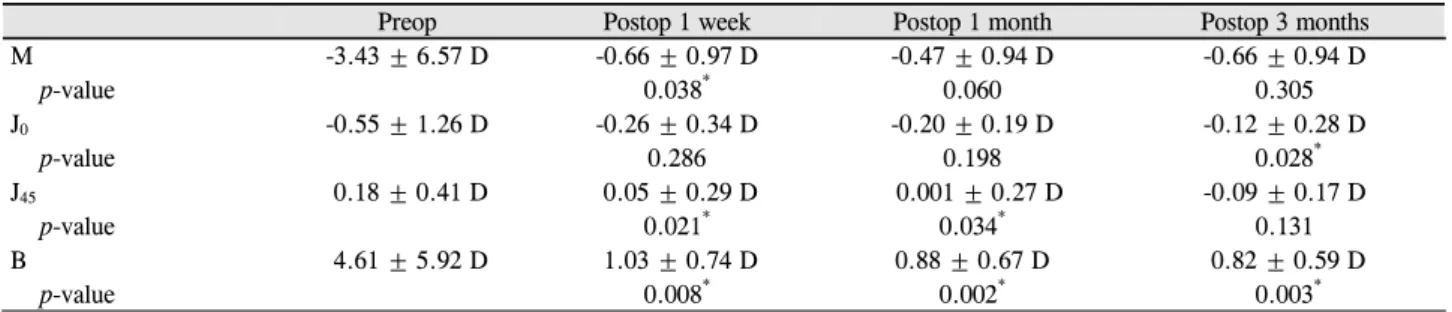
Table 2. Vector analysis of astigmatism
Preop Postop 1 week Postop 1 month Postop 3 months
M -3.43 ± 6.57 D -0.66 ± 0.97 D -0.47 ± 0.94 D -0.66 ± 0.94 D
p-value 0.038* 0.060 0.305
J0 -0.55 ± 1.26 D -0.26 ± 0.34 D -0.20 ± 0.19 D -0.12 ± 0.28 D
p-value 0.286 0.198 0.028*
J45 0.18 ± 0.41 D 0.05 ± 0.29 D 0.001 ± 0.27 D -0.09 ± 0.17 D
p-value 0.021* 0.034* 0.131
B 4.61 ± 5.92 D 1.03 ± 0.74 D 0.88 ± 0.67 D 0.82 ± 0.59 D
p-value 0.008* 0.002* 0.003*
Values are presented as mean ± SD unless otherwise indicated. p-value by Wilcoxon’s signed-ranks test.
Preop = preoperation; Postop = postoperation; J0=x-coordinate of the astigmatism vector; J45=y-coordinate of the astigmatism; M = spher- ical equivalent refraction; B = total dioptric power.
*Statistically significant values.
Figure 2. Changes in visual acuity. There were statistically
significant improvements in visual acuity at postoperative 1 week, 1 month, and 3 months compared with pre-operative values. log MAR = logarithm of the minimum angle of reso- lution; Preop = preoperative.Figure 3. Change in refractive astigmatism. There were stat-
istically significant decreases in refractive astigmatism at post- operative 1 week, 1 month, and 3 months compared with pre- operative values. Preop = preoperative.Figure 4. Changes in the corneal astigmatism between pre-
and post- operation. There is no significant changes between the follow-ups. Preop = preoperation.Figure 5. Changes in spherical equivalents between pre- and
post- operation. Preop = preoperation.1주에 -0.85 ± 0.54D, 수술 후 1개월에 -0.66 ± 0.39D, 그리 고 수술 후 3개월에는 -0.59 ± 0.48D로 수술 전에 비해 유 의하게 줄어들었다(p=0.008, p=0.002, p=0.003, Wilcoxon’s signed-ranks test; Fig. 3). 굴절 난시의 변화는 수술 후 1개 월부터 안정적으로 유지됨이 관찰되었다. 평균 각막 난시 는 수술 전 -1.85 ± 0.56D였으며, -1.64 ± 0.53D, 수술 후
1개월에 -1.53 ± 0.44D, 수술 후 3개월에 -1.54 ± 0.62D로 술 전과 비교하여 유의한 변화는 없었다(Fig. 4). 평균 구면 대응치(spherical equivalent, SE)는 수술 전 -0.65 ± 3.52D 에서, 수술 후 1주에 -0.23 ± 1.06D, 수술 후 1개월에 -0.14
± 1.07D, 그리고 수술 후 3개월에는 -0.37 ± 0.98D로 관찰 되었다(Fig. 5). 난시를 Power vector analysis로 분석하여 각 변수들의 변화를 살펴보면(Table 2), M값은 수술 전 -3.43 ± 0.57D와 비교하여 수술 후 1주에 -0.66 ± 0.97D, 수술 후 1개월에는 -0.47 ± 0.94D로 유의한 변화가 있었다
Figure 7. Absolute amount of rotation of a single-piece toric
acrylic intraocular lens (IOL) compared with preoperative IOL axis. There was significant rotation between 1 week and 1 month after surgery. However, axis was stable after 1 month.Table 3. Post-operative high order aberration of the enrolled patients
High order aberration Mean ± SD
Mean postoperative 3 months vertical coma (μm) -0.17 ± 0.12
Mean postoperative 3 months horizontal coma (μm) -0.29 ± 0.20
Mean postoperative 3 months SA (μm) 0.16 ± 0.12
Mean postoperative 3 months HOA RMS (μm) 0.36 ± 0.16
Values are presented as mean ± SD.
SA = spherical aberrations; HOA = high-ordered aberration; RMS = root mean square.
Figure 6. Diagram showing refractive astigmatism in diopters
measured before surgery and 1 week, 1 month, and 3 months after implanting a single-piece toric acrylic intraocular lens.Astigmatism is shown as vectors at J0 and J45. Each ring repre- sents 1 diopter.
(p=0.038, p=0.040, Wilcoxon’s signed-ranks test). J0값은 수술 전 -0.55 ± 1.26D와 비교하여 수술 후 3개월에 -0.12
± 0.28D로(p=0.028, Wilcoxon’s signed-ranks test), J45값은 수술 전 0.18 ± 0.41D와 비교하여 수술 후 1주에 0.05 ± 0.29D, 수술 후 1개월에는 0.001 ± 0.27D로 유의한 변화가 있었다(p=0.021, p=0.034, Wilcoxon’s signed-ranks test). B 값은 수술 전 4.61 ± 5.92와 비교하여 수술 후 1주에 1.03
± 0.74, 수술 후 1개월에는 0.88 ± 0.67, 수술 후 3개월에는 0.82 ± 0.59로 유의한 변화가 있었다(p=0.008, p=0.002, p=0.003, Wilcoxon’s signed-ranks test; Fig. 6). 고위수차는 수술 전에는 백내장으로 측정이 안 되었고, 수술 후 3개월 의 수차를 살펴보면 구면 수차가 0.16D, 종축 코마수차가 -0.17D, 횡축 코마수차가 -0.29D의 값을 보였다(Table 3).
인공수정체의 회전 안정성
수술 전 의도하였던 축과 비교하여 수술 후 인공수정체 의 회전 움직임을 분석해 보면, 방향성을 포함하여 분석 시 수술 전 의도한 축과 수술 후 3개월째 실제 인공수정체의
축 사이의 평균 차이는 3.26 ± 8.96도만큼 시계 방향 회전 을 보였으며, 이는 통계적으로 유의하지는 않았다(p=0.221, independent t-test). 회전 값의 절대값rotated degree
을 관찰 시 수술 후 1주에는 5.17 ± 5.27도, 수술 후 1개월 과 3개월에는 각각 8.24 ± 6.76도, 8.45 ± 6.74도만큼의 회 전 움직임이 관찰되었다. 수술 후 1주와 1개월 사이에서 유 의한 인공수정체 축의 변화를 관찰할 수 있었으나(3.07도, p=0.017, independent t-test), 수술 후 1개월과 3개월 사이 의 축의 변화는 유의하지 않았다(Fig. 7). 수술 후 3달까지 의 전체 회전 양의 62.7%는 수술 후 1달까지의 기간에 발 생함을 알 수 있었다. 수술 후 3개월에 인공수정체 축의 움 직임의 방향은 55.5%에서 시계 방향이었다.
고 찰
본 연구는 난시가 1D에서 4D 이내인 백내장 환자에서 백내장 수술 시 Tecnis® toric IOL 삽입이 시력 교정 효과 가 우수하고, 난시 감소 효과는 78%를 보여, 최근 국외에 서 난시 감소 효과 44.9%를 보고한 연구16보다 우수하였고, 75%를 보고한 Tecnis® toric IOL의 결과20에 교정 효과가 필적함을 확인하였다. Tecnis® toric IOL의 난시 교정 효과 는 가장 널리 사용되고 있는 AcrySof® toric IOL (Alcon Laboratories Inc., Fort Worth, TX, USA) 삽입의 난시 감소 효
[rotated degree]2
과 63.3%에서 80.1% 정도에도 필적함을 알 수 있었다.12,22,23 고위수차에 미치는 영향도 AcrySof® toric IOL 삽입의 보 고와 필적하였다.24
실제로 난시교정용 인공수정체의 난시교정 효과는 술 전 검사 간 오차, 난시축 표기 시의 오차, 환자의 술 전 각막 전면 난시 상태(직난시 또는 도난시), 환자의 각막 후면 난 시, 술 중 또는 술 후 인공수정체 축의 회전, 각 렌즈별 도 수 결정 프로그램 등 매우 다양한 요인에 의해서 복합적으 로 영향을 받기 때문에,13-19 다양하게 소개되고 있는 난시 교정용 인공수정체의 교정 효과를 직접적으로 비교하기는 매우 어렵다. 저자들은 비교적 국내에 초기에 공급된 AcrySof® toric IOL을 백내장 환자의 난시교정 목적으로 많이 삽입하였는데, 교정 효과가 기대했던 것보다는 부족 교정이 되는 경향을 보여(unpublished data), 여러 가지 요 인에 관심을 갖고 분석 중에 난시 도수 결정 프로그램의 요 인이 영향을 미치는 것에 주목하였다. 각막의 난시에서 인 공수정체의 난시를 환산하는 과정에 ratio는 Effective lens position (ELP)에 따라 영향을 받게 된다.19,25,26 ELP는 전방 깊이에 의해 결정되고 전방 깊이는 각막 굴절력과 안구축 장의 영향을 받으므로, 이 두 가지 요소가 난시 도수를 결 정하는 데 고려되어야 하는데, Alcon사의 난시 환산 프로 그램은 안구축장 및 전방 깊이의 요인이 배제되어 있어, 실 제로 각막 굴절력이 매우 가파르거나 안구축장이 길어 전 방 깊이가 깊은 환자에서는 부족 교정이 될 수밖에 없다는 것이 알려졌다. 비교적 후발 주자로 시장에 출시된 Tenis toric IOL의 난시 환산 프로그램은 이 점을 보완하여 안구 축장을 고려하게 되어 있어, 저자들은 이러한 차이가 부족 교정의 개선에 도움이 될 것이라 판단하여, Tenis toric IOL 을 이용한 난시교정 효과 임상 시험을 진행하게 되었다. 저 자들이 초기에 삽입했던 AcrySof® toric IOL의 난시 감소 효과가 56%였음에 비해 Tecnis® toric IOL은 난시 감소효 과가 75%로 통계적으로 유의하지는 않았으나, 교정 효과가 증가하는 경향을 확인할 수 있었다(Supplementary Table 1).
Tecnis® toric IOL의 난시 환산 프로그램의 개선에도 불 구하고 난시 교정 효과가 90% 내지 100%에 미치지 못하 는 것은 그 외에도 여러 가지 요인이 여전히 작용하고 있기 때문이라고 의심된다. 난시 환산 프로그램이 직접적으로 전방깊이를 측정하여 적용하는 것이 아니라, 각막굴절력과 안구축장으로 환산하는 과정 중에도 오차가 발생할 가능성 이 있다. 최근에는 난시 환산 프로그램에 직접 전방 깊이를 측정하여 적용하는 난시교정용 인공수정체가 개발되어 연 구가 되고 있어, 관심을 모으고 있다.27 그 외에도 술전 검 사가 영향을 미칠 수 있으며, 많은 논란이 있으나 기종에 따른 검사 간 오차에 대해서는 학계에서 인정하고 있는 부
분이다.15,28,29 최근 각막 후면난시의 중요도가 많이 논의되
고 있으며, 후면 난시를 측정할 수 있는 기종이 아닌 경우 후면 난시가 고려되지 않을 때에 도난시에서는 부족 교정 이 많이 되고 있다는 것은 널리 인정되고 있다.18,30,31 본 연 구진의 기종은 후면 난시를 측정할 수 없는 Orbscan의 난 시값을 참조하였기에, 부족 교정을 줄이고자 도난시의 경 우는 SIA를 0으로 설정하였지만, 그럼에도 각막 전면난시 의 방향과 후면난시의 방향이 100% 일치하는 것은 아니므 로,32 임의 설정으로 인한 오차 발생의 가능성이 있다고 판 단한다. 그리고 술 전 검사의 난시값 및 축의 다양성은 술 자가 선택을 함에 있어 많은 혼란을 야기한다. 본 연구진의 분 석에 의하면 난시가 1.5D 이상 되면 automated keratometry, IOL master 및 Orbscan의 난시축이 많이 일치하기는 하지 만, 여전히 난시값에는 차이가 있고 IOL master의 측정값 이 유의하게 가장 높게 측정되었다.14 본 연구진은 기존 난 시렌즈의 교정 효과가 부족 교정의 경향이 있음을 주지하 고 있음에도 불구하고, 과교정을 항상 우려하여 세 검사값 중 가장 높은 값을 선택하지는 못하였고, 일치하는 최소 두 검사값을 선택하여 난시 양을 정하였기 때문에 이 부분에 서도 오차가 발생할 수 있었을 것으로 판단한다. 이러한 부 분은 각 술자가 본인의 환자 데이터 다량 수집 및 분석을 통해(맞춤식 최적화, customization) 어떤 값을 취할지 결정 하는 것이 궁극적으로는 필요할 것으로 사료된다. 또 한 가 지 수술 전 오차는 난시의 기준축을 표시하는 과정에서 발 생이 가능하다. 본 연구팀은 가장 간단한 방법인 세극등하 에서 횡축세극을 이용하여 각막에 술자 또는 보조의가 기 준점을 표시하는 방법을 선택하였는데, 검사자 간 오차가 발생 가능하다고 판단한다. 이러한 오차를 줄이기 위해 혈 관을 이용한 세점 기준축 방법, 세극등 스트립을 이용하거 나 software program을 이용하는 등 다양한 방법으로 개선 된 검사 방법이 소개되고 있다.33-36
마지막으로 수술 후에 발생하는 렌즈의 회전이 오차의 요인 중 하나이다. 본 연구는 실제 인공수정체 축 사이의 평균 차이는 3.26도 시계 방향 회전이 관찰되었고, 이는 통 계적으로 유의하지는 않았으며, 5.0도 및 3.15도를 보고한 기존의 국외 연구와 필적하는 결과였다.22,23 그러나 회전 절대값 관찰 시 수술 후 1주에는 5.17도, 수술 후 1개월에 는 8.24도로, 국외에 보고된 테크니스 토릭 인공수정체 최 근 연구의 1.8-2.7도보다 높은 값을 보였다.20 수술 후 1주 이내에는 수술 전 검사의 오차 및 렌즈의 낭 내 자유 회전 에 의한 오차 가능성이 의심되고, 재질의 특성상 AcrySof® toric IOL보다 덜 끈적거림(sticky)이 원인일 가능성이 있다 고 판단된다. 수술 후 1일에 렌즈 회전이 가장 크게 발생한 기존의 국외 연구와 달리, 본 연구의 렌즈 회전은 주로 술
후 1주에서 한 달 이내에 유의하게 변화하였는데, 국외 보 고에서 수술 후 1일까지의 변화가 컸던 이유는 수술 중 인 공수정체 축의 위치를 맞추는 과정에서의 오차 또는 수술 마지막에 점탄 물질 등의 제거와 연관이 있다고 추측되 며,20 이와는 달리 본 연구에서 수술 후 1주에서 한 달 이내 의 변화는 낭 내 수축에 의할 가능성이 의심되고, 본 연구 진의 전낭원형 절개술의 크기가 낭 내 수축의 유발과 연관 이 있을 가능성이 있다. 이를 분석함으로써, 본 연구진은 다시 한 번 난시교정 효과에 전낭원형절개술의 크기가 중 요할 가능성을 확인하였으며, 앞으로의 전낭원형절개술 시 행 시 낭 내 수축을 최소화할 수 있는 크기로 최적화할 예 정이다.
결론적으로, 난시를 동반한 백내장 환자에서 Tecnis® toric IOL 삽입은 시력 및 난시교정 효과가 우수함을 확인 하였으며, 이는 국제적으로 최근 발표된 국외 보고 결과를 지지하는 증거를 축적하여 학문적 컨센서스를 이루는 데 도 움이 되었고, 또한 국내에서는 최초의 보고라는 점에서 그 의의가 있다고 하겠다. 가능한 오차 요인을 줄이기 위해 술 자의 환자 결과 분석을 통한 맞춤식 최적화(customization) 가 난시 교정렌즈 삽입술에 필요하다고 사료된다.
참고문헌
1) Rubenstein JB, Raciti M. Approaches to corneal astigmatism in cataract surgery. Curr Opin Ophthalmol 2013;24:30-4.
2) Kretz FT, Bastelica A, Carreras H, et al. Clinical outcomes and sur- geon assessment after implantation of a new diffractive multifocal toric intraocular lens. Br J Ophthalmol 2015;99:405-11.
3) Chen W, Zuo C, Chen C, et al. Prevalence of corneal astigmatism before cataract surgery in Chinese patients. J Cataract Refract Surg 2013;39:188-92.
4) Ferrer-Blasco T, Montés-Micó R, Peixoto-de-Matos SC, et al.
Prevalence of corneal astigmatism before cataract surgery. J Cataract Refract Surg 2009;35:70-5.
5) Fouda S, Kamiya K, Aizawa D, Shimizu K. Limbal relaxing in- cision during cataract extraction versus photoastigmatic keratec- tomy after cataract extraction in controlling pre-existing corneal astigmatism. Graefes Arch Clin Exp Ophthalmol 2010;248:1029- 35.
6) Kim DH, Wee WR, Lee JH, Kim MK. The short term effects of a single limbal relaxing incision combined with clear corneal incision. Korean J Ophthalmol 2010;24:78-82.
7) Kaufmann C, Peter J, Ooi K, et al. Limbal relaxing incisions versus on-axis incisions to reduce corneal astigmatism at the time of cata- ract surgery. J Cataract Refract Surg 2005;31:2261-5.
8) Lim R, Borasio E, Ilari L. Long-term stability of keratometric as- tigmatism after limbal relaxing incisions. J Cataract Refract Surg 2014;40:1676-81.
9) Hirnschall N, Gangwani V, Crnej A, et al. Correction of moderate corneal astigmatism during cataract surgery: toric intraocular lens versus peripheral corneal relaxing incisions. J Cataract Refract
Surg 2014;40:354-61.
10) Jeon JH, Hyung Taek Tyler R, Seo KY, et al. Comparison of re- fractive stability after non-toric versus toric intraocular lens im- plantation during cataract surgery. Am J Ophthalmol 2014;157:
658-65.e1.
11) Visser N, Beckers HJ, Bauer NJ, et al. Toric vs aspherical control intraocular lenses in patients with cataract and corneal astigma- tism: a randomized clinical trial. JAMA Ophthalmol 2014;132:
1462-8.
12) Na JH, Lee HS, Joo CK. The clinical result of acrySof toric intra- ocular lens implantation. J Korean Ophthalmol Soc 2009;50:831-8.
13) Jeon HM, Lee KH. Analysis of miscorrection after implantation of the toric intraocular lens. J Korean Ophthalmol Soc 2014;55:1636- 41.
14) Han JM, Choi HJ, Kim MK, et al. Comparative analysis of corneal refraction and astigmatism measured with autokeratometer, IOL Master, and topography. J Korean Ophthalmol Soc 2011;52:1427- 33.
15) Lee H, Kim TI, Kim EK. Corneal astigmatism analysis for toric in- traocular lens implantation: precise measurements for perfect correction. Curr Opin Ophthalmol 2015;26:34-8.
16) Hirnschall N, Maedel S, Weber M, Findl O. Rotational stability of a single-piece toric acrylic intraocular lens: a pilot study. Am J Ophthalmol 2014;157:405-11.e1.
17) Mencucci R, Favuzza E, Guerra F, et al. Clinical outcomes and ro- tational stability of a 4-haptic toric intraocular lens in myopic eyes.
J Cataract Refract Surg 2014;40:1479-87.
18) Preussner PR, Hoffmann P, Wahl J. Impact of posterior corneal sur- face on toric intraocular lens (IOL) calculation. Curr Eye Res 2015;
40:809-14.
19) Savini G, Hoffer KJ, Carbonelli M, et al. Influence of axial length and corneal power on the astigmatic power of toric intraocular lenses. J Cataract Refract Surg 2013;39:1900-3.
20) Waltz KL, Featherstone K, Tsai L, Trentacost D. Clinical outcomes of TECNIS toric intraocular lens implantation after cataract re- moval in patients with corneal astigmatism. Ophthalmology 2015;
122:39-47.
21) Kaye SB, Campbell SH, Davey K, Patterson A. A method for as- sessing the accuracy of surgical technique in the correction of astigmatism. Br J Ophthalmol 1992;76:738-40.
22) Miyake T, Kamiya K, Amano R, et al. Long-term clinical outcomes of toric intraocular lens implantation in cataract cases with preex- isting astigmatism. J Cataract Refract Surg 2014;40:1654-60.
23) Ferreira TB, Almeida A. Comparison of the visual outcomes and OPD-scan results of AMO Tecnis toric and Alcon Acrysof IQ toric intraocular lenses. J Refract Surg 2012;28:551-5.
24) Hayashi K, Kondo H, Yoshida M, et al. Higher-order aberrations and visual function in pseudophakic eyes with a toric intraocular lens. J Cataract Refract Surg 2012;38:1156-65.
25) Eom Y, Kang SY, Song JS, et al. Effect of effective lens position on cylinder power of toric intraocular lenses. Can J Ophthalmol 2015;
50:26-32.
26) Goggin M, Moore S, Esterman A. Outcome of toric intraocular lens implantation after adjusting for anterior chamber depth and in- traocular lens sphere equivalent power effects. Arch Ophthalmol 2011;129:998-1003.
27) Bascaran L, Mendicute J, Macias-Murelaga B, et al. Efficacy and stability of AT TORBI 709 M toric IOL. J Refract Surg 2013;
= 국문초록 =
백내장 수술 후 난시교정용 인공수정체를 이용한 난시 교정 효과
목적: 난시를 동반한 백내장 환자에서 백내장 수술 시 테크니스 토릭 인공수정체 삽입술의 난시교정 효과 및 안정성을 분석하고자 하였다.
대상과 방법: 1D에서 4D의 각막 난시를 동반한 백내장 환자 중 2013년 6월부터 2014년 5월까지 서울대학교병원에서 임상시험 동의하 에 백내장 제거술 및 테크니스 토릭 인공수정체 삽입술을 받은 14명 17안을 전향적으로 관찰하여 분석하였다. 수술 전과 후의 시력, 굴절력, 난시, 인공수정체 축, 고위수차의 변화를 술후 3개월까지 관찰하였다. 난시 분석에는 power vector analysis가 사용되었다.
결과: 나안시력(logMAR)은 술 전 평균 0.58 ± 0.34에서 3개월에 0.26 ± 0.43으로 유의하게 향상되었다. 평균 굴절난시는 술 전 -2.67 ± 0.89D였고, 술후 3개월에 -0.59 ± 0.48D로 최종 77.9%의 유의한 감소 효과를 보였다. 난시벡터 분석 시 M, B, J0, J45
모두 술 전과 비교하여 유의하게 감소하였다. 수술 전 의도한 축과 술 후 3개월째 실제 인공수정체 축 간의 평균 차이는 3.26도만큼 시계 방향으로 회전하였지만 통계적으로 유의하지 않았고, 회전은 대부분 술 후 한 달 이내에 발생하였다.
결론: 난시를 동반한 백내장 환자에서 초음파 유화술 및 테크니스 토릭 인공수정체 삽입술은 우수한 난시교정 효과 및 회전 안정성을 보였다.
<대한안과학회지 2015;56(10):1544-1551>
Supplementary Table 1. Comparison of changes in astigmatism between Tecnis® toric IOL implantation and Acrysof® IQ toric IOL
implantation in cataract surgeryTecnis® toric IOL (n = 17)
Acrysof® IQ toric IOL
(n = 14) p-value
Mean preoperative refractive astigmatism (diopters) -2.67 ± 0.89 -1.96 ± 0.85 0.072
Mean preoperative corneal astigmatism (diopters) -1.85 ± 0.56 -1.68 ± 0.25 0.468
Mean postoperative 3 months refractive astigmatism (diopters) -0.65 ± 0.54 -0.86 ± 0.55 0.278 Values are presented as mean ± SD unless otherwise indicated. p-value by Mann-Whitney U-test.
IOL = intraocular lens.
29:194-9.
28) Ale Magar JB, Cunningham F, Brian G. Comparison of automated and partial coherence keratometry and resulting choice of toric IOL. Optom Vis Sci 2013;90:385-91.
29) Chang M, Kang SY, Kim HM. Which keratometer is most reliable for correcting astigmatism with toric intraocular lenses? Korean J Ophthalmol 2012;26:10-4.
30) Savini G, Næser K. An analysis of the factors influencing the re- sidual refractive astigmatism after cataract surgery with toric intra- ocular lenses. Invest Ophthalmol Vis Sci 2015;56:827-35.
31) Hasegawa Y, Okamoto F, Nakano S, et al. Effect of preoperative corneal astigmatism orientation on results with a toric intraocular lens. J Cataract Refract Surg 2013;39:1846-51.
32) Miyake T, Shimizu K, Kamiya K. Distribution of posterior corneal
astigmatism according to axis orientation of anterior corneal astigmatism. PloS One 2015;10:e0117194.
33) Teichman JC, Baig K, Ahmed II. Simple technique to measure tor- ic intraocular lens alignment and stability using a smartphone. J Cataract Refract Surg 2014;40:1949-52.
34) Kasthurirangan S, Feuchter L, Smith P, Nixon D. Software-based evaluation of toric IOL orientation in a multicenter clinical study. J Refract Surg 2014;30:820-6.
35) George VE, George DS. Axis measurement strip for Haag-Streit BM900 series slitlamp. J Cataract Refract Surg 2014;40:1584-7.
36) Cha D, Kang SY, Kim SH, et al. New axis-marking method for a toric intraocular lens: mapping method. J Refract Surg 2011;27:
375-9.