pISSN: 0378-6471 eISSN: 2092-9374
http://dx.doi.org/10.3341/jkos.2013.54.11.1688
= 증례보고 =
AL-Scan Ⓡ 을 이용한 술 전 안구생체계측과 백내장 수술 후 굴절력 예측의 정확성
김성인⋅강석재⋅오태훈⋅최진석⋅백남호 새빛안과병원
목적: AL-ScanⓇ으로 측정한 안축장을 IOL masterⓇ 및 A-scan과 비교하였고, 전방깊이, 각막굴절력의 값을 IOL masterⓇ와 비교하 여, 백내장 수술 후 굴절력 예측의 정확성을 검토하고자 하였다.
대상과 방법: 본원에서 백내장 수술이 계획된 40안을 대상으로 AL-ScanⓇ, IOL masterⓇ와 A-scan을 이용하여 안축장, 전방깊이, 각막굴절력을 측정하였다. 인공수정체의 도수결정은 SRK-T 공식을 이용했으며, 백내장 수술 4주 후 굴절검사를 시행하였고, 예측오 차는 최종굴절력에서 예상굴절력을 뺀 차이로 구하였다.
결과: AL-scanⓇ, IOL masterⓇ, A-scan에서 안축장은 각각 23.08 ± 0.62 mm, 23.09 ± 0.62 mm, 22.99 ± 0.62 mm였고, A-scan 의 값이 통계적으로 유의하게 짧게 측정되었다(p<0.001, p<0.001). 전방 깊이와 각막굴절력은 AL-scanⓇ에서 각각 3.11 ± 0.06 mm, 44.82 ± 1.34 D였고, IOL masterⓇ에서 3.13 ± 0.06 mm, 44.85 ± 1.26 D이며, 유의한 차이는 없었다(p=0.226, 0.331). 절대값 평균 예측오차는 AL-scanⓇ에서 0.44 ± 0.35 D, IOL masterⓇ에서 0.40 ± 0.34 D였으며, A-scan에서 0.39 ± 0.30 D로 세 군간에 차이 가 없었다(p=0.843, 0.847, 1.000).
결론: AL-ScanⓇ을 이용한 안축장, 전방깊이는 IOL masterⓇ를 이용한 측정치와 높은 일치도를 보였고, 술 후 예측오차에서도 차이가 없었다.
<대한안과학회지 2013;54(11):1688-1693>
■Received: 2013. 3. 29. ■ Revised: 2013. 6. 15.
■Accepted: 2013. 9. 8.
■Address reprint requests to Tae Hoon Oh, MD Department of Ophthalmology, Saevit Eye Hospital,
#1065 Jungang-ro, Ilsandong-gu, Goyang 410-817, Korea Tel: 82-31-900-7700, Fax: 82-31-900-7777
E-mail: [email protected]
* This study was presented as a e-poster at the 109th Annual Meeting of the Korean Ophthalmology Society 2013.
백내장 수술 시 삽입되는 인공수정체의 도수 값 결정에 있어서 과거에는 주로 초음파를 이용해 오다가 1999년에 부분결합간섭(partial coherence interferometry) 원리를 이용한 측정방법이 소개된 후, 현재는 임상에서 널리 쓰이 고 있다. 부분결합간섭계 원리를 이용한 측정법들은 국소마 취제의 점안 없이 비접촉 측정이 가능하여 감염의 위험이 없고, 검사가 쉬운 장점이 있다.1현재 널리 이용되고 있는 부분결합간섭의 원리를 이용한 장비인 IOL master®(Carl Zeiss, Jena, Germany)는 760 μm 파장의 적외선 광선을 이용하여 전방깊이, 각막굴절력, 안축장 등을 측정할 수 있 으며, 이를 통해 인공수정체의 정확한 굴절예측치를 얻을 수 있다.2,3이미 발표된 논문들에서도 백내장 수술 후 굴절
예측치나 안축장, 전방 깊이 같은 안구 생체계측 측정에 있 어 IOL master®의 정확성과 재현성이 보고되었고, A-scan 으로 측정된 값과도 높은 일치도를 나타냈다고 보고하였 다.4,5IOL master®가 개발된 이후 광학을 이용한 안구 생체 계측 장비인 Lenstar®(Haag Streit AG, Koeniz, Switzerland) 및 AL scan® (Nidek, Japan) 등이 임상에 도입되었는데, AL scanⓇ은 830 μm 파장의 적외선 광선을 이용한 부분결 합간섭원리와 샤임플러그(scheimpflug) 이미지를 이용하 여 안축장, 전방 깊이, 각막 두께, 각막굴절력, 동공 크기, 각막크기(white to white diameter) 등을 20초 동안 짧은 시간 안에 측정하는 장점이 있다. 아직 국내외 보고에서 AL scan®과 IOL master®를 비교 분석한 연구는 없었다. 이에 본 연구에서는 백내장안에서 AL scan®과 IOL master®로 측정한 안축장, 전방깊이, 각막굴절력을 비교하고, AL scan®을 사용하여 결정한 인공수정체 도수의 정확성을 IOL master®, A-scan과 비교하여 알아보고자 하였다.
대상과 방법
2012년 12월부터 2013년 1월까지 백내장 수술을 위해 본원에 내원한 환자 중 안과 질환 및 수술, 외상의 기왕력
A
0.06B
0.04
0.02
-0.00
-0.02
-0.04
-0.06
-0.08
-0.10
-0.12
21.5 22.0 22.5 23.0 23.5 24.0 24.5 25.0 25.5
AVERAGE of AL-scan and IOL master® ® +1.96 SD
0.036
Mean -0.006
-1.96 SD -0.049
AL-scanand IOL master®®
0.16
0.14
0.12
0.10
0.08
0.06
0.04
0.02
0.00
-0.02
21.5 22.0 22.5 23.0 23.5 24.0 24.5 25.0 25.5
AVERAGE of AL-scan and A-scan® ®
+1.96 SD 0.141
Mean 0.094
-1.96 SD 0.048
AL-scan and A-scan®®
Figure 1. Bland-Altman plot of axial length between AL scan® and IOL master® (A), and A-scan (B) (95% limits of agreement for
axial length difference: AL scan® -IOL master® [-0.049, 0.036]; AL scan® - A-scan [0.048, 0.141]).이 없는 30명 40안을 대상으로 의무기록을 후향적으로 분 석하였다. 후낭 혼탁, 심한 백내장, 환자가 주시를 못하는 등의 이유로 IOL master®혹은 AL scan®으로 계측이 불가 능하였던 경우는 제외하였고, 수술 중 후낭 파열이 있거나, 수정체를 후낭에 넣지 않은 경우, 각막 절개 부위에 봉합을 시행했던 경우는 제외하였다.
수술 전 검사로 먼저 AL scan®을 이용하여 안구 생체 계 측을 시행한 후 IOL master®를 나중에 검사하였으며 각각 의 기기에서 안축장, 전방깊이, 각막굴절력을 측정하였다.
그리고 모든 경우에서 가장 마지막으로 A-scan (AB- 5500, Sonomed Inc., Ohio, USA)을 시행하였다. 인공수정 체의 도수 결정은 SRK/T공식을 이용했으며,6IOL master® 및 AL scan®의 경우 내장된 소프트웨어를 이용해 계산하였 고, A-scan은 자동굴절검사(KR-8800®, Topcon, Japan) 를 통한 각막굴절력 계측치를 이용해 계산하였다. 목표 도 수는 AL-scan 예측치를 기준으로 SRK/T 공식으로 정시 에 가장 가까운 근시값으로 정하였다. 백내장 40안은 한 명 의 술자에 의해 2.2 mm로 이측 투명 각막절개를 이용한 수 정체유화술 및 인공수정체 낭내 삽입술을 시행하였다. 또한 인공수정체에 따른 오차를 최소화하기 위해 동일한 인공수 정체 AcrysofⓇ SN60AT (Alcon Laboratories Inc. Fort Worth, TX, USA)를 사용하였다. 인공수정체의 A-con- stant는 A-scan은 118.4, IOL master®와 AL scan®에서는 119.0을 사용하였다. 최종 굴절력(final spherical equiv- alent)은 백내장 수술 후 4주째 현성굴절검사를 이용해 측 정하였고, 예측오차(prediction error)는 최종 굴절력에서 예상 굴절력을 뺀 차이로 구하였다. 양의 예측오차는 최종 굴절력이 예상한 것보다 원시임을 의미하며, 음의 예측오차 는 근시임을 의미하지만, 각 군간의 예측오차의 비교시에는
양의 오차와 음의오차가 서로 상쇄되는 문제가 있어서 예 측오차 절대값을 비교하였다. 통계학적인 분석은 SPSS 18.0 version을 이용하였고, 각 기기 간 안축장, 전방깊이, 각막 굴절력의 일치도를 알아보기 위하여 Bland와 Altman의 방 법을 이용하였으며, Bland-Altman plot에서 독립변수는 두 검사의 평균으로 종속변수는 두 검사의 차이로 하였다.
각 군간에서 세기기 간의 예측오차 및 평균의 차이를 비교 하기 위해 ANOVA 및 paired t-test를 이용하였다. p값의 유의수준은 0.05 미만으로 하였다.
결 과
총 30명 40안(남자 18안, 여자 22안)을 대상으로 하였으 며, 평균 연령은 68.7 ±9.1세(51-95세)였다. AL scan®, IOL master®, A-scan에서 측정된 안축장은 각각 23.08 ± 0.62 mm, 23.09 ±0.62 mm, 22.99 ±0.62 mm였고, AL scan®과 IOL master®는 통계적으로 유의한 차이가 없었으 나(ANOVA, p=0.999), AL scan®과 A-scan, IOL mas- ter®와 A-scan의 안축장 측정치 간에는 A-scan의 안축장 값이 통계적으로 유의하게 짧게 측정되었다(ANOVA, p<0.001, p<0.001). 또한, 각 군간의 안축장 평균은 높은 상관 관계 를 갖는 것으로 나타났다(correlation coefficiency=0.999, 0.998, 0.995). 측정 기기 간 안축장의 일치도를 비교한 Bland-Altman plot에서 AL scan®과 IOL master®, AL scan®과 A-scan 사이의 95% limit of agreement는 -0.049~+0.036, 0.048~0.141이며, 각각에서 1안을 제외 한 모든 측정치는 이 범위 내에 존재했다(Fig. 1).전방깊이 와 반경 2.4 mm에서의 각막굴절력은 AL scan®에서 각각 3.11 ±0.06 mm, 44.82 ±1.34 D이고, IOL master®에서
Table 1. Demographics and Biometry measurements by AL scan®, IOL master®, A-scan
AL scan® IOL master® A-scan
Age (years) 68.7 ± 9.1 (51-95) - -
Sex (M/F) 40 (18/22) - -
AL* (mm) 23.08 ± 0.62 23.09 ± 0.62 22.99 ± 0.62
ACD† (mm) 3.11 ± 0.06 3.13 ± 0.06 -
K 2.4‡ mm (diopter) 44.82 ± 1.34 44.85 ± 1.26 -
K 3.0‡ mm (diopter) 44.75 ± 1.34 - -
Values are presented as mean ± SD.
*Axial length; †Anterior chamber depth; ‡Keratometry was calculated at corneal radius 2.4 mm and 3.0 mm.
0.6
0.5
0.4
0.3
0.2
0.1
0.0
-0.1
-0.2
-0.3
-0.4
2.0 2.5 3.0 3.5 4.0
AVERAGE of AL-scan and IOL master (anterior chamber depth)
®
®
AL-scan-IOL m (anteior chamber depth)
+1.96 SD 0.28
Mean -0.03
-1.96 SD -0.34
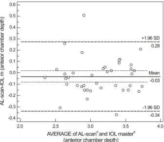
Figure 2. Bland-Altman plot of anterior chamber depth be-
tween AL scan® and IOL master® (95% limits of agreement for axial length difference: AL scan® - IOL master® [-0.34, 0.28]).3.13 ±0.06 mm, 44.85 ±1.26 D이며, 두 측정치 사이에 통계적으로 유의한 차이는 없었다(paired t-test, p=0.226, 0.331). 또한 반경 3 mm에서 측정한 각막굴절력은 AL scan® 에서 44.75 ±1.34 D였고, 자동굴절검사에서는 44.78 ±1.37 D 였으며 두 군간의 통계적 차이는 없었다(paired t-test, p=0.322)(Table 1). 전방 깊이에 대한 AL scan®과 IOL master®의 측정치 비교(Fig. 2)와 각막굴절력에 대한 AL scan®과 IOL master®, AL scan®과 자동굴절검사의 측정치 비교(Fig. 3)에서도 Bland-Altman plot 상 대부분이 95%
limit of agreement 구간 내에 존재해 높은 일치율을 보였다 (각각 -0.34~0.28, -0.49~0.41, -0.44~ 0.38). 인공수정 체 삽입술 후 AL scan®을 이용해 계산된 평균 예측오차는 +0.01 ± 0.57 D이며, 절대값 평균 예측오차는 0.44 ± 0.35 D였다. IOL master®의 경우 평균 예측오차는 +0.03
±0.53 D이며, 절대값 평균 예측오차는 0.40 ±0.34 D이 며, A-scan을 이용했을 경우는 평균 예측오차 +0.21 ± 0.46 D이며, 절대값 평균예측오차는 0.39 ±0.30 D였다.
예측오차가 0.5 D 이내의 비율은 A scan에서 72.5%로 가
장 높게 나타났으며, 1.0 D 이내는 IOL master®에서 97.5%로 가장 높게 나타났다(Table 2). ALscan®과IOL master®, AL scan®과 A-scan, IOL master®와 A-scan 사 이에서 비교한 절대값 예측오차의 평균은 통계적으로 유의 한 차이가 없었다(각각 p=0.843, 0.847, 1.000 ANOVA).
고 찰
백내장 수술 후 굴절상태는 환자의 삶의 질에 큰 영향을 미친다. 더욱이 최근에는 다초점 인공수정체 및 난시교정 인공수정체의 개발과 더불어 수술자와 환자들의 결과에 대한 기대가 커져 정확한 굴절력 예측이 필요하게 되었다.
수술 후 굴절상태는 여러 요인들에 의해 영향을 받는데, Olsen7은 백내장 수술 후 요구되는 굴절력에 이르지 못하 는 이유로 안축장의 측정오차(54%), 수술 후 전방깊이의 예측오차(38%), 각막굴절력의 측정오차(8%) 등이 영향을 준다고 하였고, 안축장계측 시 0.1 mm의 오차가 생기면 수 술 후 굴절력의 오차는 평균 0.28 D가 생긴다고 하였다. 따 라서 백내장 환자에서 정확한 안축장 측정이 필요하며, 보 다 정확한 안축장 측정을 위해 여러 가지 방법이 이용되어 왔다. 본 연구에서 AL scan®을 이용해 측정한 안축장은 기 존의 부분결합간섭을 이용한 IOL master®와 높은 일치도 를 보였으나, 초음파를 이용한 A-scan의 측정값보다 0.09 mm 길게 측정되었고, 이는 통계적으로 유의하게 차이가 있 었다. AL scan®측정치와 나머지 두 기기 간의 상관계수는 각각 0.999, 0.998로 강한 상관 관계를 보였으며, Bland Altman plot에서 매우 좁은 범위의 95% limit of agree- ment 구간을 확인할 수 있었다. A-scan의 경우 초음파 탐 침자를 각막에 대고 검사하는 과정상에서 생기는 각막의 함입으로 인해 안축장이 짧게 측정될 수 있고,8 초음파를 이용한 안축장이 각막에서 망막의 내경계막까지의 거리를 측정하는 반면, 광원을 이용한 기기는 각막에서 망막색소상 피층까지의 거리를 측정하게 되기 때문에 안축장이 서로 다르게 측정될 수 있다.9,10본 연구에서도 비접촉 부분결합 간섭계인 AL scan®, IOL master®보다 A-scan에서 측정
0.6
0.4
0.2
0.0
-0.2
-0.4
-0.6
-0.8
42 43 44 45 46 47 48
AVERAGE of AL scan 2.4 and IOL m (keratometry)
®
®
49 AL scan 2.4 - IOL master (keratometry)
+1.96 SD 0.41
Mean -0.04
-1.96 SD -0.49
0.6
0.4
0.2
0.0
-0.2
-0.4
-0.6
-0.8
-1.0
41 42 43 44 45 46 47 48 49
AVERAGE of AL scan 3.0 mm and ARK (keratometry)
®
AL scan 3.0 mm - ARK (keratometry)
®
+1.96 SD 0.38
Mean -0.03
-1.96 SD -0.44
A B
Figure 3. Bland-Altman plot of keratometry between AL scan® and IOL master® (A), and Auto refractive keratometry (ARK) (B)
(95% limits of agreement for axial length difference: AL scan® -IOL master® [-0.49, 0.41]; AL scan® - ARK [-0.44, 0.38]).Table 2. Comparison of Absolute prediction error among AL scan®, IOL master®, A-scan
Prediction error (diopter) Number of eyes (%)
Absolute PE* Range 0.5D 1.0D 1.5D 2.0D
AL scan® 0.44 ± 0.35 0.04-1.59 22†,‡(55%) 38 (95%) 39 (97.5%) 40 (100%)
IOL master® 0.40 ± 0.34 0.03-1.58 27 (67.5%) 39 (97.5%) 39 (97.5%) 40 (100%)
A-scan 0.40 ± 0.30 0.01-1.17 29 (72.5%) 38 (95%) 40 (100%) 40 (100%)
Values are presented as mean ± SD.
*Prediction error; †Denotes significant difference compared to A-scan group (ANOVA, p < 0.05); ‡Denotes significant difference compared to IOL master® group (ANOVA, p < 0.05).
된 안축장이 평균 0.1 mm 정도 짧게 측정되었는데, 이 결 과는 과거에 발표된 보고들과 큰 차이가 없었다.1,11-14하지 만 A-scan과 부분결합간섭계 사이의 안축장 차이가 없었 다는 보고도 있어,5,15 초음파 및 IOL master®를 측정하는 검사자의 숙련도 및 여러가지 요인들이 결과에 영향을 미 치는 것으로 생각한다.
AL scan®은 샤임플러그(Scheimpflug) 원리를 이용하여 얻은 안구의 전방영상을 분석하여 전방깊이 및 각막 중심 두께를 측정하는 반면, IOL master®는 전방깊이 측정을 위 해 0.7 mm의 비스듬히 비추는 슬릿 광원을 이용해 각막의 바깥 표면에서 수정체 사이의 영상을 분석하여 거리를 측 정한다.16 샤임플러그를 이용한 전방깊이가 IOL master® 보다 0.11 mm 높게 측정되었다는 보고도 있으나, 대부분 의 결과에서 통계적으로 전방깊이의 큰 차이가 없었다 하 였고, 전방깊이가 0.2 mm 차이가 날 때 인공수정체 도수가 0.1 D 차이가 나기 때문에 임상적으로 큰 의미는 없다고 하 였다.17-20본 연구에서는 AL scan®과 IOL master®사이에 전방 깊이는 각각 3.11 ±0.06 mm, 3.13 ±0.06 mm로 측 정되어 두군 사이 차이는 없었고(p=0.226), Bland Altman plot 상 좁은 범위의 95% limit of agreement를 보여 높은 일치도를 보이는 것을 확인하였다.
술 후 굴절력의 예측오차의 절대값의 평균은 A-scan, IOL master®, AL scan® 순으로 나타났으나(0.39D, 0.40D, 0.44D), 통계적으로 의미 있는 차이를 보이지 않는 것으로 조사되었다. 또한 0.5D 미만 예측오차에서는 AL scan®, IOL master®, A-scan 순으로 나타났고, AL scan®만이 두 군보다 통계적으로 유의하게 차이가 있었다(Table 2). IOL master®에서 생체계측의 원리는 760 μm의 적외선 광선이 같은 축을 갖는 두 개의 coaxial beam으로 나누어져 안구로 들어가서 각막표면과 망막색소상피층에 각각 반사되며, 이 두 개의 광선 사이의 시간적 연장을 간섭계(interferometer) 에 의해 포착하여 안구 계측 정보로 전환하는 것이다.15AL scan®은 파장의 중첩을 통한 시간의 차이를 간섭계로 포착 하는 원리는 IOL master®와 유사하지만 830 μm의 super luminescent diode (SLD) laser를 사용하여 생체 계측을 하기 때문에 더 높은 침투력을 가진다. 본 연구에는 포함되 지 않았지만, 핵경화 및 후낭하 혼탁이 심한 백내장에서 IOL master®로 계측되지 않았지만, AL scan®에서 계측된 경우가 일부 존재하였다. 이는 AL scan® 광원의 파장이 760 μm인 IOL master®보다 길어 침투력이 더 좋기 때문 이다. AL scan®은 안구 생체 계측에 있어 IOL master®가 제공하는 각막굴절력, 안축장, 전방 깊이, 각막크기(white
to white diameter) 외에도 중심각막두께, 동공크기 등의 정보도 측정할 수 있는 장점이 있다.21-24 또한 환자 정보 입력 시작부터 측정 후 결과물을 출력하는 데까지 총 소요 시간이 AL scan®의 경우에는 20초로, 다른 광학을 이용한 안구 생체 계측 장비인 Lenstar®의 203.2초, IOL master® 의 84.2초보다 빠른 시간 안에 결과를 얻어내는 장점이 있
다.21,22 백내장 수술 시 굴절력 예측을 위한 각막굴절력의
측정은 AL scan®의 경우 반경 2.4 mm의 K값과 3.3 mm 두 곳의 평균치 K값을 구하는 반면, IOL master®는 반경 2.4 mm 의 평균치 K값을 구한다.25-27평균치 값을 위한 측정데이 터 갯수에서도 AL scan®은 반경 2.4 mm와 3 mm에서 각각 360개의 점을 분석하고 IOL master®는 반경 2.4 mm에서 6개의 점을 분석하는 차이가 있다. 본 연구에서는 AL scan®과 IOL master® 사이에 각막굴절력은 각각 44.82 ± 1.34 D, 44.85 ± 1.26 D이고(p=0.331), Bland Altman plot 상 좁은 범위의 95% limit of agreement를 보여 높은 일치도를 보여 두 군간의 차이를 보이지는 않았다. 하지만 굴절교정수술 후 백내장 수술을 하는 것과 같이 비정상적 인 각막굴절력일 경우에 차이가 나타날 수 있을 것으로 예 상되어, 이를 임상적으로 비교하는 것도 추후 필요할 것으 로 생각한다.
본 연구는 새로운 부분결합간섭기계인 AL scan®과 기존 의 IOL master®, A-scan에서의 안축장, 전방깊이, 각막굴 절력 등의 안구 생체 계측치를 비교하였고, 백내장 수술 후 세 기기 간의 인공수정체 도수 예측의 정확성을 비교하였 다. 각 기기 간의 측정치는 높은 일치도를 보였고, 비교적 정확하게 인공 수정체도수예측이 가능하여 AL scan®이 백 내장 수술에 있어서 유용한 술 전 계측방법이 될 수 있을 것으로 생각한다.
REFERENCES
1) Drexler W, Findl O, Menapace R, et al. Partial coherence inter- ferometry: a novel approach to biometry in cataract surgery. Am J Ophthalmol 1998;126:524-34.
2) Gimbel HV, Sun R. Accuracy and predictability of intraocular lens power calculation after laser in situ keratomileusis. J Cataract Refract Surg 2001;27:571-6.
3) Holzer MP, Mamusa M, Auffarth GU. Accuracy of a new partial coherence interferometry analyser for biometric measurements. Br J Ophthalmol 2009;93:807-10.
4) Drexler W, Hitzenberger CK, Baumgartner A, et al. Investigation of dispersion effects in ocular media by multiple wavelength parti- al coherence interferometry. Exp Eye Res 1998;66:25-33.
5) Lam AK, Chan R, Pang PC. The repeatability and accuracy of axial length and anterior chamber depth measurements from the IOLMaster. Ophthalmic Physiol Opt 2001;21:477-83.
6) Retzlaff JA, Sanders DR, Kraff MC. Development of the SRK/T
intraocular lens implant power calculation formula. J Cataract Refract Surg 1990;16:333-40.
7) Olsen T. Sources of error in intraocular lens power calculation. J Cataract Refract Surg 1992;18:125-9.
8) Giers U, Epple C. Comparison of A-scan device accuracy. J Cataract Refract Surg 1990;16:235-42.
9) Haigis W, Lege B, Miller N, Schneider B. Comparison of im- mersion ultrasound biometry and partial coherence interferometry for intraocular lens calculation according to Haigis. Graefes Arch Clin Exp Ophthalmol 2000;238:765-73.
10) Hoffer KJ, Shammas HJ, Savini G. Comparison of 2 laser instru- ments for measuring axial length. J Cataract Refract Surg 2010;36:644-8.
11) Findl O, Drexler W, Menapace R, et al. Improved prediction of in- traocular lens power using partial coherence interferometry. J Cataract Refract Surg 2001;27:861-7.
12) Rajan MS, Keilhorn I, Bell JA. Partial coherence laser inter- ferometry vs conventional ultrasound biometry in intraocular lens power calculations. Eye (Lond) 2002;16:552-6.
13) Rose LT, Moshegov CN. Comparison of the Zeiss IOLMaster and applanation A-scan ultrasound: biometry for intraocular lens calculation. Clin Experiment Ophthalmol 2003;31:121-4.
14) Eleftheriadis H. IOLMaster biometry: refractive results of 100 consecutive cases. Br J Ophthalmol 2003;87:960-3.
15) Santodomingo-Rubido J, Mallen EA, Gilmartin B, Wolffsohn JS.
A new non-contact optical device for ocular biometry. Br J Ophthalmol 2002;86:458-62.
16) Buckhurst PJ, Wolffsohn JS, Shah S, et al. A new optical low co- herence reflectometry device for ocular biometry in cataract patients. Br J Ophthalmol 2009;93:949-53.
17) Utine CA, Altin F, Cakir H, Perente I. Comparison of anterior chamber depth measurements taken with the Pentacam, Orbscan IIz and IOLMaster in myopic and emmetropic eyes. Acta Ophthalmol 2009;87:386-91.
18) Lackner B, Schmidinger G, Skorpik C. Validity and repeatability of anterior chamber depth measurements with Pentacam and Orbscan. Optom Vis Sci 2005;82:858-61.
19) Rabsilber TM, Becker KA, Frisch IB, Auffarth GU. Anterior chamber depth in relation to refractive status measured with the Orbscan II Topography System. J Cataract Refract Surgy 2003;
29:2115-21.
20) Reddy AR, Pande MV, Finn P, El-Gogary H. Comparative estima- tion of anterior chamber depth by ultrasonography, Orbscan II, and IOLMaster. J Cataract Refract Surg 2004;30:1268-71.
21) Shin JW, Seong MC, Kang MH, et al. Comparison of ocular bio- metry and postoperative refraction in cataract patients between Lenstar® and IOL master®. J Korean Ophthalmol Soc 2012;53:833-8.
22) Kwag JY, Choi SH. Comparison of ocular biometry measured by ultrasound and two kinds of partial coherence interferometers. J Korean Ophthalmol Soc 2011;52:169-74.
23) Kim DY, Kim MJ, Kim JY, Tchah HW. Comparison of formulas for intraocular lens power calculation installed in a parital coherence interferometer. J Korean Ophthalmol Soc 2009;50:523-8.
24) Jun RM, Kang SY, Kim BY. Accuracy of biometry and intraocular lens power calculation with partial coherence interferometry in high myopia. J Korean Ophthalmol Soc 2008;49:1746-51.
25) Hwang JS, Lee JH. Comparison of the IOL master® and A-scan ul- trasound: refractive results of 96 consecutive cases. J Korean
=ABSTRACT=
Accuracy of Ocular Biometry and Postoperative Refraction in Cataract Patients with AL-Scan
ⓇSung In Kim, MD, Sug Jae Kang, MD, Tae Hoon Oh, MD, Jin Seok Choi, MD, PhD, Nam Ho Baek, MD, PhD
Department of Ophthalmology, Saevit Eye Hospital, Goyang, Korea
Purpose: To compare the axial lengths, anterior chamber depths, and keratometric measurements and to predict post- operative refractions of AL-Scan®, IOL master®, and ultrasound.
Methods: A total of 40 eyes in 30 patients who received cataract surgery were included in the present study. The axial length, anterior chamber depth, and keratometry were measured by 2 types of partial coherence interferometry (AL-Scan® and IOL master®) and ultrasound. The SRK/T formula was used to calculate IOL power, and the predictive error which sub- tracts predictive refraction from postoperative refraction was compared among the ocular biometry devices.
Results: Axial lengths were 23.08 ± 0.62 mm, 23.09 ± 0.62 mm, and 22.99 ± 0.62 mm measured by AL scan®, IOL master®, and ultrasound, respectively. Axial length measured by ultrasound was statistically significantly shorter than AL scan® and IOL master® (p < 0.001, p < 0.001, respectively). The anterior chamber depth and keratometry were 3.11 ± 0.06 mm and 44.82 ± 1.34 D measured by AL scan®, and 3.13 ± 0.06 mm and 44.85 ± 1.26 D measured by IOL master®, respectively.
The differences of anterior chamber depth and keratometry between the 2 devices were not statistically significant (p = 0.226, p = 0.331, respectively). The mean absolute prediction errors were 0.44 ± 0.35 D, 0.40 ± 0.34 D, and 0.39 ± 0.30 D in AL-Scan®, IOL master® and ultrasound, respectively, and were not statistically significantly different (p = 0.843, p = 0.847, p = 1.000, respectively).
Conclusions: The ocular biometric measurements and prediction of postoperative refraction using AL-Scan® were as ac- curate as IOL master® and ultrasound.
J Korean Ophthalmol Soc 2013;54(11):1688-1693
Key Words: AL-Scan®, Axial length, IOL master®, Partial coherence interferometry
Address reprint requests to Tae Hoon Oh, MD Department of Ophthalmology, Saevit Eye Hospital
#1065 Jungang-ro, Ilsandong-gu, Goyang 410-817, Korea
Tel: 82-31-900-7700, Fax: 82-31-900-7777, E-mail: [email protected] Ophthalmol Soc 2007;48:27-32.
26) Chung JK, Choe CM, You YS, Lee SJ. Biometry with partial co- herence interferometry and ultrasonography in high myopes. J Korean Ophthalmol Soc 2006;47:355-61.
27) Song BY, Yang KJ, Yoon KC. Accuracy of partial coherence inter- ferometry in intraocular lens power calculation. J Korean Ophthalmol Soc 2005;46:775-80.
![Figure 1. Bland-Altman plot of axial length between AL scan® and IOL master® (A), and A-scan (B) (95% limits of agreement for axial length difference: AL scan® -IOL master® [-0.049, 0.036]; AL scan® - A-scan [0.048, 0.141]).](https://thumb-ap.123doks.com/thumbv2/123dokinfo/5341327.180126/2.892.88.805.780.1066/figure-bland-altman-length-master-limits-agreement-difference.webp)
![Figure 3. Bland-Altman plot of keratometry between AL scan® and IOL master® (A), and Auto refractive keratometry (ARK) (B) (95% limits of agreement for axial length difference: AL scan® -IOL master® [-0.49, 0.41]; AL scan® - ARK [-0.44, 0.38]).](https://thumb-ap.123doks.com/thumbv2/123dokinfo/5341327.180126/4.892.89.804.105.378/figure-bland-altman-keratometry-refractive-keratometry-agreement-difference.webp)
