시신경유두오목 황반병증에 대한 유리체절제술의 장기 결과
전체 글
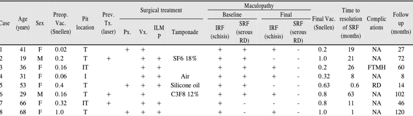
수치
관련 문서
Continued to the entropy cycle and energy balance with entropy changes. Reversible cycle consisting of two isothermal branches, AB and CD, and two isentropic branches,
Students needing additional information about grading policies and procedures should meet with their faculty advisor, Executive Director/Chairperson or a
For this study—our third on global workforce trends, follow- ing studies in 2014 and 2018—Boston Consulting Group and The Network surveyed some 209,000 people in 190 countries
Basic aspects of AUTOSAR architecture and methodology Safety mechanisms supported by AUTOSAR.. Technical safety concepts supported by AUTOSAR Relationship to ISO
GDP impact of COVID-19 spread, public health response, and economic policies. Virus spread and public
Phase profile of conventional DOE Obtained diffraction image. Optical vortices appear in the diffraction image generated by
Micro- and nano-sized pores were formed on the surface of the alloy using PEO and anodization methods, and the pore shape change according to the Zr
The Incomplete diffuse type PVD found primarily in patients with diabetic retinopathy and retinal detachment and complete diifuse type PVD found primarily in patients
