Introduction
The posterior root of the medial meniscus (MM) can serve as an anchor to regulate the meniscal shift during the knee motion and load bearing. Injuries to the MM posterior root, including complete radial and/or oblique tears adjacent to the ligamentous insertion and posterior horn, lead to accelerated degeneration of the knee joint articular cartilage by disrupting meniscal func
tions1). In addition, the medial meniscus posterior root tear (MMPRT) leads to abnormal biomechanics of the tibiofemoral joint and the inability to convert axial loads into hoop stresses by inducing radial displacement of the MM, also called medial meniscus extrusion (MME)2,3). Extrusion (subluxation, bulging, and radial/extraarticular displacement) of the MM is associated with a loss of medial compartment cartilage volume4), medial joint space narrowing5), severity of osteoarthritis as reflected by radiographic KellgrenLawrence grade6,7), degenerative knee ab
normalities8), and knee joint pain9) in patients with osteoarthritic knees. In addition, meniscal extrusion predicts an increase in subchondral bone lesions and tibial plateau expansion in osteoar
thritic knees10). MME is also a characteristic finding on magnetic resonance imaging (MRI) of patients having the MMPRT11). However, the timedependent extent of MME in patients suffer
ing from the MMPRT remains unclear. Moreover, the relation
ship among the MME severity, treatment choice, and optimal timing for surgical intervention has not been elucidated.
Meniscal Extrusion Progresses Shortly after the Medial Meniscus Posterior Root Tear
Takayuki Furumatsu, MD, Yuya Kodama, MD, Yusuke Kamatsuki, MD, Tomohito Hino, MD, Yoshiki Okazaki, MD, and Toshifumi Ozaki, MD
Department of Orthopaedic Surgery, Okayama University Graduate School, Okayama, Japan
Purpose: Medial meniscus posterior root tears (MMPRT) induce medial meniscus extrusion (MME). However, the timedependent extent of MME in patients suffering from the MMPRT remains unclear. This study evaluated the extent of MME after painful popping events that occurred at the onset of the MMPRT.
Materials and Methods: Thirtyfive patients who had an episode of posteromedial painful popping were investigated. All the patients were diagnosed as having an MMPRT by magnetic resonance imaging (MRI) within 12 months after painful popping. Medial meniscus body width (MMBW), absolute MME, and relative MME (100×absolute MME/MMBW) were assessed among three groups divided according to the time after painful popping events: early period (<1 month), subacute period (1–3 months), and chronic period (4–12 months).
Results: In the early period, absolute and relative MMEs were 3.0 mm and 32.7%, respectively. Absolute MME increased up to 4.2 mm and 5.8 mm during the subacute and chronic periods, respectively. Relative MME also progressed to 49.2% and 60.3% in the subacute and chronic periods, respectively.
Conclusions: This study demonstrated that absolute and relative MMEs increased progressively within the short period after the onset of symptomatic MMPRT. Our results suggest that early diagnosis of an MMPRT may be important to prevent progression of MME following the MMPRT.
Keywords: Meniscus, Tear, Root, Extrusion pISSN 2234-0726 · eISSN 2234-2451
Knee Surgery & Related Research
Received May 4, 2017; Revised June 6, 2017;
Accepted July 3, 2017
Correspondence to: Takayuki Furumatsu, MD
Department of Orthopaedic Surgery, Okayama University Graduate School, 251 Shikatacho, Kitaku, Okayama 7008558, Japan Tel: +81862357273, Fax: +81862239727
Email: [email protected]u.ac.jp
295
This is an Open Access article distributed under the terms of the Creative Commons Attribution NonCommercial License (http://creativecommons.org/licenses/bync/4.0/) which permits unrestricted noncommercial use, distribution, and reproduction in any medium, provided the original work is properly cited.
Copyright © 2017 KOREAN KNEE SOCIETY www.jksrr.org
The MMPRT can occur especially in middleaged or older patients who have a single event of posteromedial painful pop
ping sensation during light activities such as using stairs and squatting12,13). The positive predictive value of painful popping in identifying the MMPRT is 96.5% and the specificity is 99.5%13). In the treatment of the MMPRT, early diagnosis and appropriate surgical intervention are important to obtain a successful clini
cal outcome and prevent rapid progression of degenerative knee diseases1,14). MRIbased characteristic findings such as a radial tear, cleft/truncation, ghost, and giraffe neck signs provide high diagnostic accuracy and specificity11,15). However, we cannot ob
tain information about the exact onset of the MMPRT from these MRI findings. Although the sensitivity of a painful popping for the detection of an MMPRT is low (35%)13), the memory of hear
ing a painful popping sound seems to be necessary to determine the timing of the MMPRT onset and MRI examination and treat
ment strategy for the symptomatic MMPRT.
Repair of the MMPRT can reduce the mean tibiofemoral con
tact pressure by increasing the tibiofemoral contact area as dem
onstrated in a human cadaveric knee study16). Several repair tech
niques such as transtibial pullout repair, suture anchordependent repair, direct allinside repair, and posterior reattachment of the MM posterior root have been developed for arthroscopic treat
ment of the MMPRT1,14). MMPRT repairs produce more favor
able clinical outcomes compared with conservative treatments17). Despite the favorable clinical results of transtibial pullout repair of the MMPRT, several metaanalyses have demonstrated that many patients show no significant reduction in MME follow
ing the MMPRT repair18,19). On the other hand, there are few reports that demonstrate the MMPRT repair can reduce MME.
For example, MMPRT pullout repairs decreased MME from 3.13 mm to 2.94 mm20). Patients with decreased MME following the MMPRT pullout repair had more favorable clinical scores than those with increased MME at 1 year postoperatively21). Based on these findings, a timely surgical intervention before causing a severe MME may be a critical factor for the treatment of the MMPRT and preventing progression of degenerative knee joint diseases. In this study, we evaluated the timedependent changes in the extent of MME after painful popping events involved in the MMPRT onset.
Materials and Methods
1. Study Materials
This study received the approval of our Institutional Review Board, and written informed consent was obtained from all pa
tients. Thirtyfive patients (32 females and 3 males) who had an episode of posteromedial painful popping between September 2013 and August 2016 were included (Table 1). All the patients were diagnosed as having the MMPRT with MRI examinations.
The presence of an MMPRT was defined according to character
istic MRI findings (cleft/giraffe neck/radial tear signs of the MM posterior root within 9 mm from the attachment, ghost sign, and MM extrusion11,15)). Patients who had an MMPRT without a memory of painful popping (n=3) and an MMPRT with previous meniscal injury and/or knee surgery (n=2) were excluded. The mean age of the patients was 63.5 years. Types of the MMPRT were determined by careful arthroscopic examinations (24 knees) or arthroplastyassociated direct observations (5 knees) accord
ing to the meniscal root tear classification (Table 1)22). Transtibial pullout repairs (16 knees) were performed in patients who met operative indications for the pullout repair2325). Allinside menis
cal repairs (6 knees) were performed in the other patients. There was no patient who underwent meniscectomy alone. Patients were examined by MRI scans prior to surgical treatments and within 12 months after painful popping events. Six patients were treated conservatively because of their low activities and hesita
tion about surgical treatments. Conservative treatments involved partial weight bearing and a stick, crutch, knee brace, and medi
cal guidance were used appropriately.
2. MRI-Based Measurements
MRI evaluation was performed using an Achieva 1.5 T (Phil
ips, Amsterdam, The Netherlands) with a knee coil. Standard
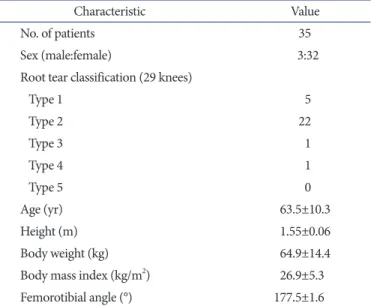
Table 1. Demographics and Clinical Characteristics
Characteristic Value
No. of patients 35
Sex (male:female) 3:32
Root tear classification (29 knees)
Type 1 5
Type 2 22
Type 3 1
Type 4 1
Type 5 0
Age (yr) 63.5±10.3
Height (m) 1.55±0.06
Body weight (kg) 64.9±14.4
Body mass index (kg/m2) 26.9±5.3
Femorotibial angle (°) 177.5±1.6
Values are presented as mean±standard deviation or number.
sequences included sagittal [repetition time (TR)/echo time (TE), 742/18], coronal (TR/TE, 637/18), and axial (TR/TE, 499/18) T2
weighted fastfield echo with a 20° flip angle. The slice thickness was 3 mm with a 0.6mm gap. The field of view was 16 (or 17) cm with an acquisition matrix size of 205×256 (or 200×368)2629). The MRIbased medial meniscus body width (MMBW), absolute MME, and medial meniscus height (MMH) were assessed. The MMBW was measured from the inner border to the outer border of the MM on the coronal image that crossed the midpoint of the anteroposterior length of the MM. The absolute MME was mea
sured from the medial margin of the tibial plateau to the outer border of the MM. The MMH was measured from the inferior margin to the superior margin of the MM on the same coronal image. The relative MME was calculated using the following for
mula: 100×absolute MME/MMBW (%).
3. Statistical Analysis
Data were presented as means±standard deviations. Differences among groups were compared using the oneway analysis of vari
ance (ANOVA) with Tukey post hoc tests. Power and statistical analyses were performed using EZR (Saitama Medical Center, Saitama, Japan), which is a graphical user interface for R (the R Foundation for statistical computing). Significance was set to p<0.05. Two orthopedic surgeons (TH and YK) independently measured MRIbased values of the MM in a blinded manner.
Each observer performed each measurement twice, at least 2 weeks apart. The reliability of the measurements was assessed by examining the interobserver and intraobserver reliabilities with the intraclass correlation coefficient (ICC). An ICC values of
>0.80 was considered to represent a reliable measurement. Linear regression analysis was used to assess the correlation between the absolute MME and the other factors (age, height, body mass
index, and femorotibial angle). A good correlation was defined as R2≥0.60, fair correlation as R2≥0.50, and poor correlation as R2<0.50.
Results
On the MRIbased measurements of the MM, the MMBW showed no significant change among three groups divided ac
cording to the time after painful popping events into the early pe
riod (<1 month), subacute period (1–3 months), and chronic pe
riod (4–12 months) (Table 2 and Fig. 1A). In the early period, the absolute and relative MMEs were 3.0±1.2 mm and 32.7%±12.2%, respectively (Table 2, Fig. 1B and C). However, the absolute MME significantly increased up to 4.2±1.2 mm and 5.8±1.6 mm in the subacute and chronic periods, respectively. The relative MME also progressed to 49.2%±11.9% and 60.3%±14.9% in the subacute and chronic periods, respectively. Significant difference in the MMH was observed between the early and chronic dura
tions (p=0.014) (Table 2 and Fig. 1D). The interobserver and
Table 2. Changes in the MMBW, MME, and MMH after Painful Popping Events
Variable Early (n=15)
<1 month Subacute (n=17)
1−3 months Chronic (n=17) 4−12 months
MMBW (mm) 9.1±1.2 8.5±1.1 9.6±1.3
Absolute MME (mm)a) 3.0±1.2 4.2±1.2 5.8±1.6 Relative MME (%)a) 32.7±12.2 49.2±11.9 60.3±14.9
MMH (mm)a) 6.8±1.7 7.1±1.4 8.4±1.6
Values are presented as mean±standard deviation.
MMBW: medial meniscus body width, MME: medial meniscus extru
sion, MMH: medial meniscus height.
a)Significant differences were observed among groups.
<1 1-3 4-12 12
10 8 6 4 2
MMBW(mm)
Duration (mo) 0
A
<1 1-3 4-12 8
6 4 MMEAbsolute(mm) 2
Duration (mo) 0
B
<1 1-3 4-12 80
60 40 MMERelative(%) 20
Duration (mo) 0
C
<1 1-3 4-12 12
10 8 6 4 2
MMH(mm)
Duration (mo) 0
a) D
a) a)
Fig. 1. Magnetic resonance imagingbased measurements of the medial meniscus after the onset of medial meniscus posterior root tears. (A) Medial meniscus body width (MMBW). (B) Absolute medial meniscus extrusion (MME). (C) Relative MME (100×absolute MME/MMBW). (D) Medial meniscus height (MMH). a)p<0.05.
intraobserver reliabilities for the measurements were considered satisfactory (mean ICC values >0.94).
A poor correlation was observed between the absolute MME and age (R2=0.125). The absolute MME and the other factors (height, body mass index, and tibiofemoral angle) also showed poor correlations (R2=0.009, 0.011, and 0.001, respectively).
Representative sequential MRI scans showed a posttraumatic progression of MME after a painful popping event of the knee (Fig. 2). A 65yearold female had a single event of painful pop
ping of her right knee in the full squatting position. Minor MME (1.3 mm) and specific MRI findings of the MMPRT were ob
served at 1 week after the painful popping event (Fig. 2A). The MME increased to 5.0 mm at 10 weeks after the MMPRT (Fig.
2B). The MME progressed to 5.7 mm at 18 weeks postMMPRT (Fig. 2C). Bone edematous change in the medial femoral con
dyle was detected on MRI at 10 and 18 weeks after the MMPRT onset (Fig. 2B and C). Radiographic findings associated with spontaneous osteonecrosis of the knee progressed to the Koshino classification stage 3 (collapsed stage) at 18 weeks (Fig. 3A)30). She underwent arthroscopic assessment and unicompartmental knee arthroplasty of her right knee at 20 weeks after the painful pop
ping because of severe knee pain (Fig. 3B and C).
A B C
Fig. 3. Spontaneous osteonecrosis of the knee following the medial meniscus posterior root tear (MMPRT) in the 65yearold female shown in Fig. 2 (the right knee). (A) Sclerotic halo (arrowheads) and calcified plate (arrow) were observed in a radiograph at 18 weeks after a painful popping event.
Koshino classification: stage 3. (B) Arthroscopic findings at 20 weeks postMMPRT. Arrowheads: type 2 MMPRT. (C) An unstable osteochondral flap.
MMBW MMBW MMBW
MME MME MME
MMH MMH MMH
A B C
Fig. 2. Medial meniscus extrusion (MME) increased after the medial meniscus posterior root tear (MMPRT) onset in a 65yearold female (the right knee). (A) The coronal view showed minor MME 1week after a painful popping event indicating the MMPRT onset. Doubleheaded arrow: medial meniscus body width (MMBW). Dotted line: absolute MME. Solid line: medial meniscus height (MMH). (B) The MME increased 10 weeks after the MMPRT. Note the bone edema of the medial femoral condyle (arrowheads) and medial shift of the medial collateral ligament (arrow). (C) The MME progressed 18 weeks after the MMPRT. Bars: 1 cm.
Discussion
The most important finding in this study was that the absolute MME increased progressively within 12 months after the onset of symptomatic MMPRT (Table 2 and Fig. 1B). The relative MME also increased during nonoperative treatment period following the posteromedial painful popping event (Table 2 and Fig. 1C).
These results may provide us with an important evaluation cri
terion for the MMPRT to determine the timing of surgical treat
ments. MME of ≥3 mm is more frequent in painful osteoarthritic knees than in contralateral painless knees, and more frequent in osteoarthritic knees that have a higher radiographic grade (KellgrenLawrence grade III)9). A radially displaced MM forms a bulged (or swelled) meniscal shape on the coronal MRI during the progression of osteoarthritic knees31). In addition, the status of MME can affect postoperative clinical outcome of the MMPRT transtibial pullout repair21). Patients with decreased MME (3.5±1.4 mm) at 1 year postMMPRT pullout repairs have more favorable clinical outcomes and radiographic findings at 5year followup than those with increased MME (5.1±1.4 mm) at 1 year postop
eratively21). We consider that pullout repairdependent positional restoration of the MM may be disturbed by a bulged/swelled MM and knee joint space narrowing concomitant with sequen
tial MME increase after the MMPRT onset. Our study demon
strated that the mean absolute MME was 3.0±1.2 mm even in the early period less than 1 month after the MMPRT onset. Based on these findings, it seems that the symptomatic MMPRT should be treated with arthroscopic meniscal repair techniques as soon as possible following the diagnosis of the MMPRT if the patients meet surgical indications for the MMPRT repair.
Nonoperative treatment of the MMPRT was associated with poor clinical outcome, worsening osteoarthritis of the knee, and a relatively high rate (16/52 knees, 31%) of total knee arthroplasty at a mean of 30 months after diagnosis of the MMPRT29). The overall failure rate based on clinical and radiographic criteria was 87% in this literature. In addition, female gender was related to a higher rate of arthroplasty32). In biomechanical studies, the MMPRT led to excessive contact pressure on the articular surface of the knee by reducing the contact area of the knee joint carti
lage2,3). We consider that the MMPRT can suddenly deteriorate the status of articular cartilage and subchondral bone by altering the knee joint kinematics and homeostasis immediately. There
fore, the MMPRTrelated osteonecrotic lesions and spontaneous osteonecrosis of the knee may be associated with female gender having osteoporotic subchondral bone quality.
Many studies have reported that MME is associated with pro
gression of symptomatic knee osteoarthritis4,6,9). Meniscusto
femoral condyle congruity is essential for the development of cir
cumferential hoop stresses and meniscal function. Abnormalities in the position of the MM and its coverage, such as the MMPRT, MME, and meniscectomyrelated meniscal defects, can alter knee joint congruity and are associated with the progression of tibio
femoral osteoarthritis and cartilage degradation4). Sung et al.33) reported that the mean absolute MME and relative MME were 4.1±0.7 mm and 46.1%±9.0% in 36 knees showing the MMPRT at a mean of 5.3months symptom duration, respectively. In ad
dition, MRIbased osteonecrotic lesions were observed in 12/36 knees (33%) of the MMPRT patients33). In our study, the absolute MME significantly increased from 3.0±1.2 mm to 5.8±1.6 mm postMMPRT (Table 2 and Fig. 1). In the subacute period (1–3 months after the painful popping), the absolute and relative MME progressed to 4.2±1.2 mm and 49.2%±11.9%, respectively (Table 2). Based on these findings, the MMPRT should be treated within the subacute period after the MMPRT onset for prevent
ing a substantial MME of more than 4 mm. We consider that surgical intervention at an adequate timing may be important to prevent degenerative knee joint disease following the MMPRT if the patient meets operative indications. Further investigations will be required to determine the precise timing of operative treatment for the MMPRT.
There are several limitations in this study. MRI examinations were performed differently in terms of the number of exami
nations and duration from the onset of MMPRT to MRI as
sessment. Followup MRI scans were not performed in all the patients after the primary MRIbased diagnosis of the MMPRT.
Repeated MRI examinations in the same patients will be needed to precisely assess the timedependent MME progression fol
lowing the MMPRT. In this study, we evaluated the MRIbased MME in a single knee flexion angle (10°) under nonweight bearing condition. Open MRI assessments of meniscal move
ment using thin slices in several knee flexion angles under load
ing condition will be required to enhance the diagnostic value of MME in the MMPRT treatment. In addition, threedimensional reconstruction of the MM using dynamic MRI may be useful to understand the MME increase after the MMPRT onset. Our study was a retrospective comparative study with a small sample size. Additional followup MRI studies involving in a larger sam
ple size will be required to evaluate the real effect of MME on the progression of postMMPRT symptoms.
Conclusions
This study demonstrated that the absolute and relative MME increased progressively within the short period after the onset of symptomatic MMPRT. Our results suggest that the accurate diagnosis of the MMPRT in the early period after the onset may be important to prevent the increase of MME following the MMPRT.
Conflict of Interest
No potential conflict of interest relevant to this article was re
ported.
Acknowledgements
We thank Drs. Shinichi Miyazawa, Takaaki Tanaka, and Hiroto Inoue for their clinical supports.
References
1. Bhatia S, LaPrade CM, Ellman MB, LaPrade RF. Menis
cal root tears: significance, diagnosis, and treatment. Am J Sports Med. 2014;42:301630.
2. Padalecki JR, Jansson KS, Smith SD, Dornan GJ, Pierce CM, Wijdicks CA, Laprade RF. Biomechanical consequences of a complete radial tear adjacent to the medial meniscus pos
terior root attachment site: in situ pullout repair restores derangement of joint mechanics. Am J Sports Med. 2014;42:
699707.
3. Allaire R, Muriuki M, Gilbertson L, Harner CD. Biome
chanical consequences of a tear of the posterior root of the medial meniscus: similar to total meniscectomy. J Bone Joint Surg Am. 2008;90:192231.
4. Berthiaume MJ, Raynauld JP, MartelPelletier J, Labonte F, Beaudoin G, Bloch DA, Choquette D, Haraoui B, Altman RD, Hochberg M, Meyer JM, Cline GA, Pelletier JP. Menis
cal tear and extrusion are strongly associated with progres
sion of symptomatic knee osteoarthritis as assessed by quan
titative magnetic resonance imaging. Ann Rheum Dis. 2005;
64:55663.
5. Bloecker K, Guermazi A, Wirth W, Benichou O, Kwoh CK, Hunter DJ, Englund M, Resch H, Eckstein F; OAI investiga
tors. Tibial coverage, meniscus position, size and damage in knees discordant for joint space narrowing: data from the Osteoarthritis Initiative. Osteoarthritis Cartilage. 2013;21:
41927.
6. Lee DH, Lee BS, Kim JM, Yang KS, Cha EJ, Park JH, Bin SI.
Predictors of degenerative medial meniscus extrusion: radial component and knee osteoarthritis. Knee Surg Sports Trau
matol Arthrosc. 2011;19:2229.
7. Kawaguchi K, Enokida M, Otsuki R, Teshima R. Ultrasono
graphic evaluation of medial radial displacement of the me
dial meniscus in knee osteoarthritis. Arthritis Rheum. 2012;
64:17380.
8. Stehling C, Souza RB, Hellio Le Graverand MP, Wyman BT, Li X, Majumdar S, Link TM. Loading of the knee during 3.0T MRI is associated with significantly increased medial meniscus extrusion in mild and moderate osteoarthritis. Eur J Radiol. 2012;81:183945.
9. Wenger A, Englund M, Wirth W, Hudelmaier M, Kwoh K, Eckstein F; OAI Investigators. Relationship of 3D meniscal morphology and position with knee pain in subjects with knee osteoarthritis: a pilot study. Eur Radiol. 2012;22:21120.
10. Wang Y, Wluka AE, Pelletier JP, MartelPelletier J, Abram F, Ding C, Cicuttini FM. Meniscal extrusion predicts increases in subchondral bone marrow lesions and bone cysts and ex
pansion of subchondral bone in osteoarthritic knees. Rheu
matology (Oxford). 2010;49:9971004.
11. Choi SH, Bae S, Ji SK, Chang MJ. The MRI findings of meniscal root tear of the medial meniscus: emphasis on cor
onal, sagittal and axial images. Knee Surg Sports Traumatol Arthrosc. 2012;20:2098103.
12. Han SB, Shetty GM, Lee DH, Chae DJ, Seo SS, Wang KH, Yoo SH, Nha KW. Unfavorable results of partial meniscec
tomy for complete posterior medial meniscus root tear with early osteoarthritis: a 5 to 8year followup study. Arthros
copy. 2010;26:132632.
13. Bae JH, Paik NH, Park GW, Yoon JR, Chae DJ, Kwon JH, Kim JI, Nha KW. Predictive value of painful popping for a posterior root tear of the medial meniscus in middleaged to older Asian patients. Arthroscopy. 2013;29:5459.
14. Bonasia DE, Pellegrino P, D’Amelio A, Cottino U, Rossi R.
Meniscal root tear repair: why, when and how? Orthop Rev (Pavia). 2015;7:5792.
15. Furumatsu T, Fujii M, Kodama Y, Ozaki T. A giraffe neck sign of the medial meniscus: a characteristic finding of the medial meniscus posterior root tear on magnetic resonance imaging. J Orthop Sci. 2017;22:7316.
16. LaPrade CM, Foad A, Smith SD, Turnbull TL, Dornan GJ, Engebretsen L, Wijdicks CA, LaPrade RF. Biomechanical consequences of a nonanatomic posterior medial meniscal
root repair. Am J Sports Med. 2015;43:91220.
17. LaPrade RF, LaPrade CM, James EW. Recent advances in posterior meniscal root repair techniques. J Am Acad Or
thop Surg. 2015;23:716.
18. Feucht MJ, Kühle J, Bode G, Mehl J, Schmal H, Südkamp NP, Niemeyer P. Arthroscopic transtibial pullout repair for posterior medial meniscus root tears: a systematic review of clinical, radiographic, and secondlook arthroscopic results.
Arthroscopy. 2015;31:180816.
19. Chung KS, Ha JK, Ra HJ, Kim JG. A metaanalysis of clinical and radiographic outcomes of posterior horn medial me
niscus root repairs. Knee Surg Sports Traumatol Arthrosc.
2016;24:145568.
20. Kim SB, Ha JK, Lee SW, Kim DW, Shim JC, Kim JG, Lee MY. Medial meniscus root tear refixation: comparison of clinical, radiologic, and arthroscopic findings with medial meniscectomy. Arthroscopy. 2011;27:34654.
21. Chung KS, Ha JK, Ra HJ, Nam GW, Kim JG. Pullout fixation of posterior medial meniscus root tears: correlation between meniscus extrusion and midterm clinical results. Am J Sports Med. 2017;45:429.
22. LaPrade CM, James EW, Cram TR, Feagin JA, Engebretsen L, LaPrade RF. Meniscal root tears: a classification system based on tear morphology. Am J Sports Med. 2015;43:3639.
23. Kodama Y, Furumatsu T, Fujii M, Tanaka T, Miyazawa S, Ozaki T. Pullout repair of a medial meniscus posterior root tear using a FasTFix® allinside suture technique. Orthop Traumatol Surg Res. 2016;102:9514.
24. Furumatsu T, Kodama Y, Fujii M, Tanaka T, Hino T, Kamat
suki Y, Yamada K, Miyazawa S, Ozaki T. A new aiming guide can create the tibial tunnel at favorable position in transtibial pullout repair for the medial meniscus posterior root tear.
Orthop Traumatol Surg Res. 2017;103:36771.
25. Fujii M, Furumatsu T, Kodama Y, Miyazawa S, Hino T, Ka
matsuki Y, Yamada K, Ozaki T. A novel suture technique using the FasTFix combined with Ultrabraid for pullout re
pair of the medial meniscus posterior root tear. Eur J Orthop Surg Traumatol. 2017;27:55962.
26. Furumatsu T, Miyazawa S, Tanaka T, Okada Y, Fujii M, Ozaki T. Postoperative change in medial meniscal length in concurrent allinside meniscus repair with anterior cruciate ligament reconstruction. Int Orthop. 2014;38:13939.
27. Fujii M, Furumatsu T, Miyazawa S, Okada Y, Tanaka T, Ozaki T, Abe N. Intercondylar notch size influences cyclops formation after anterior cruciate ligament reconstruction.
Knee Surg Sports Traumatol Arthrosc. 2015;23:10929.
28. Narazaki S, Furumatsu T, Tanaka T, Fujii M, Miyazawa S, Inoue H, Shimamura Y, Saiga K, Ozaki T. Postoperative change in the length and extrusion of the medial meniscus after anterior cruciate ligament reconstruction. Int Orthop.
2015;39:24817.
29. Kashihara N, Furumatsu T, Kodama Y, Tanaka T, Ozaki T.
Concurrent lateral meniscal repair with anterior cruciate ligament reconstruction induces the extrusion of the lateral meniscus: assessments of magnetic resonance images. Acta Med Okayama. 2016;70:4418.
30. Koshino T. The treatment of spontaneous osteonecrosis of the knee by high tibial osteotomy with and without bone
grafting or drilling of the lesion. J Bone Joint Surg Am. 1982;
64:4758.
31. Wenger A, Wirth W, Hudelmaier M, NoebauerHuhmann I, Trattnig S, Bloecker K, Frobell RB, Kwoh CK, Eckstein F, En
glund M. Meniscus body position, size, and shape in persons with and persons without radiographic knee osteoarthritis:
quantitative analyses of knee magnetic resonance images from the osteoarthritis initiative. Arthritis Rheum. 2013;65:
180411.
32. Krych AJ, Reardon PJ, Johnson NR, Mohan R, Peter L, Levy BA, Stuart MJ. Nonoperative management of medial me
niscus posterior horn root tears is associated with worsening arthritis and poor clinical outcome at 5year followup. Knee Surg Sports Traumatol Arthrosc. 2017;25:3839.
33. Sung JH, Ha JK, Lee DW, Seo WY, Kim JG. Meniscal extru
sion and spontaneous osteonecrosis with root tear of medial meniscus: comparison with horizontal tear. Arthroscopy.
2013;29:72632.