Copyright © 2016 The Korean Society for Bone and Mineral Research
This is an Open Access article distributed under the terms of the Creative Commons Attribution Non-Commercial Li- cense (http://creativecommons.org/licenses/by-nc/3.0/) which permits unrestricted non-commercial use, distribu- tion, and reproduction in any medium, provided the original work is properly cited.
Multiple Fractures in Patient with Graves’ Disease Accompanied by Isolated Hypogonadotropic
Hypogonadism
Hyon-Seung Yi1,2, Ji Min Kim1,2, Sang Hyeon Ju1, Younghak Lee1, Hyun Jin Kim1,2, Koon Soon Kim1,2
1Department of Internal Medicine, 2Research Center for Endocrine and Metabolic Diseases, Chungnam National University School of Medicine, Daejeon, Korea
Isolated hypogonadotropic hypogonadism (IHH) is known to decrease bone mineral density due to deficiency of sex steroid hormone. Graves’ disease is also an important cause of secondary osteoporosis. However, IHH does not preclude the development of primary hyperthyroidism caused by Graves’ disease, leading to more severe osteoporosis rapidly. Here, we describe the first case of 35-year-old Asian female patient with IHH ac- companied by Graves’ disease and osteoporosis-induced multiple fractures. Endocrine laboratory findings revealed preserved anterior pituitary functions except for secretion of gonadotropins and showed primary hyperthyroidism with positive autoantibodies.
Sella magnetic resonance imaging showed slightly small sized pituitary gland without mass lesion. Dual energy X-ray absorptiometry revealed severe osteoporosis in lumbar spine and femur neck of the patient. Plain film radiography of the pelvis and shoulder revealed a displaced and nondisplaced fracture, respectively. After surgical fixation with screws for the femoral fracture, the patient was treated with antithyroid medication, cal- cium, and vitamin D until now and has been recovering fairly well. We report a patient of IHH with Graves’ disease and multiple fractures that is a first case in Korea.
Key Words: Graves disease, Hypogonadism, Osteoporosis
INTRODUCTION
Isolated hypogonadotropic hypogonadism (IHH) is characterized by impairment of gonadal function secondary to deficient gonadotropin secretion.[1] It can re- sult from a variety of congenital, acquired, and functional defects related to go- nadotropin releasing hormone (GnRH) deficiency. In general, IHH is caused by ge- netic mutation or acquired anatomical abnormalities including infiltrative disor- ders or space-occupying tumors involving the hypothalamic-pituitary axis, subse- quently promoting deficiency of sex hormones.[2,3]
Sex steroid hormones are important factors in bone mineral dynamics and play an essential role in the pathogenesis of osteoporosis. There have been many in- vestigations of the links between sex hormone status and bone mineral density (BMD) for various clinical conditions. In renal transplant recipients, serum levels of estradiol predict BMD in women.[4] Estrogens also play a pivotal role in the regu- lation of bone loss and metabolism in elderly men.[5]
Corresponding author Koon Soon Kim
Department of Internal Medicine and Research Center for Endocrine and Metabolic Diseases Chungnam National University School of Medicine, 282 Munhwa-ro, Jung-gu, Daejeon 35015, Korea Tel: +82-42-280-7134
Fax: +82-42-280-7995 E-mail: [email protected] Received: December 28, 2015 Revised: February 13, 2016 Accepted: February 13, 2016
No potential conflict of interest relevant to this article was reported.
This study was supported by research fund of Chungnam National University in 2014 (2014-2142- 01).
Maintenance of proper BMD requires not only sex ste- roid hormones but also thyroid hormones and vitamin D.
Moreover, abnormal status of thyroid hormone or lower levels of vitamin D can promote pathologic or non-trauma induced fractures.[6-9] Although abnormal thyroid hor- monal status is rare in patients with IHH, IHH accompanied by primary or secondary hypothyroidism including brady- cardia and heart failure was recently reported.[10] Howev- er, to our best knowledge, there has been no report of IHH associated with Graves’ disease. Therefore, we herein report a rare case of IHH accompanied by multiple fractures due
to thyrotoxicosis and sex steroid hor mone deficiency.
CASE
A 35-year-old Asian woman, born from non-consanguin- eous parents, was referred to the department of endocri- nology for evaluation of a patient with multiple fragility fractures, and severe osteoporosis accompanied by diffuse goiter. The patient was an ex-smoker and non-drinker. She was 1.71 m tall and weighed 51.2 kg with a body mass in- dex (BMI) of 17.5 kg/m2. The diagnosis of IHH was estab-
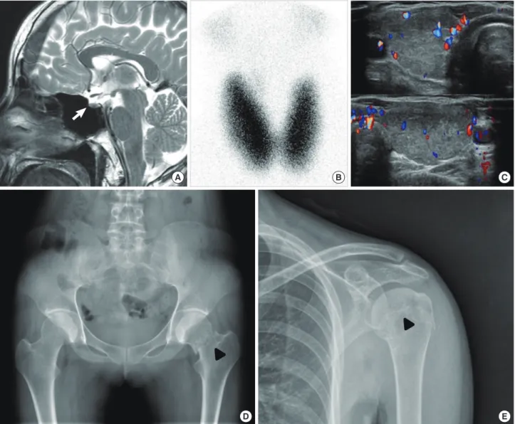
Fig. 1. Image findings of the patient. (A) Mid-sagittal T2-weighted image showing thinning of the lower half of the pituitary stalk (arrow). (B) Tech- netium-99m pertechnetate scintigraphy for thyroid gland. Radioactive thyroid uptake was 8.9% (range, 1.7 to 4.0%). (C) Thyroid ultrasonography with color doppler method (upper panel, right thyroid; lower panel, left thyroid). (D) Antero-posterior X-ray of the pelvis demonstrating a fracture of the left femoral neck (arrow head). (E) Antero-posterior radiograph of the left shoulder showing nondisplaced fracture of proximal humerus (ar- row head).
A B C
D E
lished by suggestive clinical findings with primary amen- orrhea and absence of growth and development of sec- ondary sexual characteristics and laboratory findings at 16 years. The patient had no facial anomaly or olfactory com- plaints. No familial history of anosmia, delayed puberty or hypogonadism was reported by the patient. The karyotype was 46XX and genetic screening for mutations in the hy- pogonadotropic hypogonadism genes was not performed.
At that time, anterior pituitary function was preserved ex- cept for gonadotropin secretion. The patient has been treat- ed with estrogen replacement since she was 16 years old, but she was taken off estrogen by herself several years ago.
Sella magnetic resonance imaging scan revealed a small sized pituitary gland without mass-like lesion and thinning of the lower half of the pituitary stalk (Fig. 1A). In the com- bined pituitary stimulation test at 28 years old, the peak luteinizing hormone (LH) was 1.79 IU/L and peak follicle stimulating hormone (FSH) was 1.50 IU/L, suggesting hy- pogonadotropic hypogonadism (Table 1). In the recent visit, the patient’s blood pressure was 130/82 mmHg and her heart rate was 98 beats/min. On laboratory examina- tion, complete blood count revealed hemoglobin: 12.3 g/
dL; leukocyte count: 4.9×10⁹/L; and platelet: 313×109/L.
Serum levels of total cholesterol (207 mg/dL), triglyceride (85 mg/dL), albumin (5.0 g/dL), aspartate transaminase (AST; 22 IU/L), and alanine aminotransferase (ALT; 20 IU/L) were all within normal range. The levels of basal LH, FSH and estradiol were 0.25 IU/L (range, follicular 0.6-6.2; mid- cycle 12-51; luteal 0.0-6.0), 0.23 IU/L (range, follicular 3.3- 8.8; mid-cycle 5.4-20; luteal 1.6-8.7), and 10 pg/mL (range, follicular 21-251; mid-cycle 38-649; luteal 21-312), respec- tively. Symptoms and sign of thyrotoxicosis including tachy- cardia, smooth skin, and goiter were also developed in the patient. A Technetium-99m (Tc-99m) pertechnetate scin-
tigraphy revealed diffuse enlargement of both lobes of the thyroid gland with markedly increased uptake (Fig. 1B). A thyroid function test showed newly developed primary hyperthyroidism in the patient (Table 2). Moreover, the level of thyrotropin binding inhibiting immunoglobulin was also increased (Table 2). Neck ultrasonography showed an enlarged heterogeneous echo genic thyroid gland with increased vascularity determined by the color doppler method (Fig. 1C). Serum 25-hydroxy-vitamin D level was also decreased in the patient. The antero-posterior pelvic X-ray showed left proximal femoral fracture (Fig. 1D) and shoulder X-ray revealed a non-displaced proximal humeral fracture (Fig. 1E). BMD was measured at the lumbar spine and femoral neck of the patient using dual energy X-ray absorptiometry. The patient had significantly lower BMD
Table 1. Combined pituitary stimulation test
Time (min) 0 30 60 90 120
LH (IU/L) 0.01 1.65 1.79 1.29 0.72
FSH (IU/L) 0.01 0.01 0.77 1.50 1.11
ACTH (pg/mL) 56.7 557.9 127.9 36.7 14.1
TSH (μIU/mL) 5.09 13.42 9.42 7.28 6.20
GH (ng/mL) 1.96 1.71 3.59 1.23 0.38
Cortisol (μg/dL) 1.63 15.19 15.22 12.14 10.82 LH, luteinizing hormone; FSH, follicle stimulating hormone; ACTH, adre- nocorticotropic hormone; TSH, thyroid-stimulating hormone; GH, growth hormone.
Table 2. Laboratory findings related to thyroid and calcium metabolism Values
Reference range treatmentPre- Post-
treatment
TSH (IU/L) 0.01 0.96 0.25-4.0
Free T4 (ng/dL) 3.99 1.01 0.7-1.9
T3 (ng/mL) 5.19 1.27 0.6-1.9
Thyroglobulin antibody (U/mL) 1,317 - 0-30.0 Thyroperoxidase antibody (U/mL) 69,270 - 0-8 Thyrotropin binding inhibiting
immunoglobulin (%) 48 32 -15-15
Calcium (mg/dL) 8.3 9.5 8.5-10.5
Phosphorus (mg/dL) 3.7 3.4 2.5-4.7
25-hydroxy-vitamin D (pg/mL) 8.32 32.74 25-80 Parathyroid hormone (pg/mL) 54.42 43.48 10-65 C-terminal telopeptide of type I
collagen (ng/mL) 0.212 0.119 <0.573
TSH, thyroid-stimulating hormone.
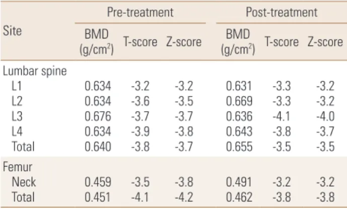
Table 3. Bone mineral density of lumbar spine and proximal femur Site
Pre-treatment Post-treatment (g/cmBMD 2) T-score Z-score BMD
(g/cm2) T-score Z-score Lumbar spine
L1 L2 L3 L4 Total
0.634 0.634 0.676 0.634 0.640
-3.2-3.6 -3.7-3.9 -3.8
-3.2-3.5 -3.7-3.8 -3.7
0.631 0.669 0.636 0.643 0.655
-3.3-3.3 -4.1-3.8 -3.5
-3.2-3.2 -4.0-3.7 -3.5 Femur
Neck
Total 0.459 0.451 -3.5
-4.1 -3.8
-4.2 0.491 0.462 -3.2
-3.8 -3.2 -3.8 BMD, bone mineral density.
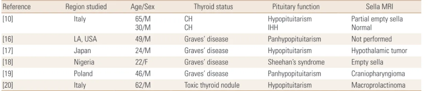
Table 4. Case reports for thyroid dysfunction in hypogonadotropic hypogonadism
Reference Region studied Age/Sex Thyroid status Pituitary function Sella MRI
[10] Italy 65/M
30/M CH
CH Hypopituitarism
IHH Partial empty sella
Normal
[16] LA, USA 49/M Graves’ disease Panhypopituitarism Not performed
[17] Japan 24/M Graves’ disease Hypopituitarism Hypothalamic tumor
[18] Nigeria 22/F Graves’ disease Sheehan’s syndrome Empty sella
[19] Poland 46/M Graves’ disease Panhypopituitarism Craniopharyngioma
[20] Italy 62/M Toxic thyroid nodule Hypopituitarism Macroprolactinoma
CH, central hypothyroidism; IHH, isolated hypogonadotropic hypogonadism; MRI, magnetic resonance imaging.
at both lumbar spine and femur neck (Table 3). She was treated with conservative management for humeral frac- ture and received surgical fixation with screws for the left femoral fracture. The patient was also treated with me- thimazole, estrogen replacement, calcium, and vitamin D for two years, thereby leading to 2.34% and 6.97% increase in BMD of lumbar spine and femur neck, respectively (Table 3). She was maintained in an euthyroid state with 2.5 to 5.0 mg of methimazole per day and has been recovering fairly well with estrogen replacement and treatment of calcium and vitamin D 2,000 IU per day for six months (Table 2).
DISCUSSION
In general, IHH presents as decreased ovarian function leading to menstrual defect, diminished vaginal secretion, infertility, and impaired breast development in premeno- pausal woman. In this case report, we presented IHH ac- companied by Graves’ disease and multiple fractures. To our best knowledge, this case report is the first paper de- scribing severe osteoporosis-induced bone fracture in a patient with IHH accompanied by Graves’ disease.
Pituitary hormone deficiencies causing hypogonadism, hypothyroidism, or hypoadrenalism may induce lower BMD.
[11] People with IHH are also prone to develop osteoporo- sis or fragile bones leading to higher risk of fractures in- duced by otherwise minor injuries.[9] Althou gh the mech- anisms underlying the relationship between central hypo- gonadism and BMD have not yet been determined, unre- placed sex steroid deficiency is associated with lower BMD in adults with growth hormone deficiency.[9,12] Therefore, cyclical replacement of estrogen and progesterone is rec- ommended to prevent premature osteoporosis and to pro- mote sexual characteristics in premenopausal women. In addition, testosterone treatment was also effective for in-
creasing lumbar spine BMD in hypogonadal middle-aged men.[13,14]
IHH is rarely accompanied by central hypothyroidism due to structural abnormalities of the hypothalamic-pitu- itary axis.[10] Thyroid-stimulating hormone (TSH) is critical for regulating expression of sodium-iodide symporter which is important for the production of thyroid hormone in the thyroid gland. In the pre-specified subgroup of premeno- pausal-aged women, TSH deficiency is independently re- lated to lower BMD in the lumbar spine and femur neck.[9]
However, Graves’ disease or excessive replacements of thy- roid hormone are also known as risk factors of osteoporo- sis.[7] Graves’ disease promotes bone loss by increased bone turnover, leading to decreased BMD and osteoporo- sis.[7] On the other hand, vitamin D deficiency is also an important risk factor for osteoporosis and increased risk of pathologic fractures in adults.[6,8,15] Therefore, we thought that low levels of vitamin D as well as inappropriate estro- gen replacement with Graves’ disease might be contribut- ing to aggravation of the osteoporosis and development of fractures in the patient.
Although the co-occurrence of hypopituitarism and Gra- ves’ disease are rare, several reports have been described in the literature (Table 4). A patient with hyperthyroidism in the presence of panhypopituitarism developed a radio- iodine-induced thyroid storm.[16] Graves’ disease devel- oped eight years after the diagnosis of hypopituitarism in this case. In another case, a 24-year-old male patient pre- sented with hypopituitarism accompanied by hyperthy- roidism and diabetes insipidus was described in 1999.[17]
A third report described cases of concomitant Graves’ dis- ease and Sheehan’s syndrome.[18] A more recent report described that a subject with known panhypopitutarism developed thyrotoxicosis that contributed to acute gluco- corticoid deficiency.[19] Another report showed that it was
possible for hyperthyroidism secondary to toxic thyroid nodule, to occur with hypopituitarism.[20] However, our case is IHH rather than panhypopituitarism, and the pa- tient presented with multiple osteoporosis-induced frac- tures associated with Graves’ disease.
In conclusion, herein we report a case of IHH with Graves’
disease and multiple fractures. Sex hormone, calcium, and vitamin D replacement are essential for prevention of os- teoporosis in patients with IHH. Secondary osteoporosis- inducible factors including hyperthyroidism should also be considered in patients with fragility fracture accompanied by IHH.
REFERENCES
1. Seminara SB, Hayes FJ, Crowley WF Jr. Gonadotropin-re- leasing hormone deficiency in the human (idiopathic hy- pogonadotropic hypogonadism and Kallmann’s syndrome):
pathophysiological and genetic considerations. Endocr Rev 1998;19:521-39.
2. Silveira LF, Latronico AC. Approach to the patient with hy- pogonadotropic hypogonadism. J Clin Endocrinol Metab 2013;98:1781-8.
3. Buck C, Balasubramanian R, Crowley WF Jr. Isolated go- nadotropin-releasing hormone (GnRH) deficiency. In: Pagon RA, Adam MP, Ardinger HH, et al., editors. GeneReviews®.
Seattle: University of Washington; 1993.
4. Cueto-Manzano AM, Freemont AJ, Adams JE, et al. Associa- tion of sex hormone status with the bone loss of renal trans- plant patients. Nephrol Dial Transplant 2001;16:1245-50.
5. Gennari L, Merlotti D, Martini G, et al. Longitudinal associ- ation between sex hormone levels, bone loss, and bone turnover in elderly men. J Clin Endocrinol Metab 2003;88:
5327-33.
6. Bakhtiyarova S, Lesnyak O, Kyznesova N, et al. Vitamin D status among patients with hip fracture and elderly con- trol subjects in Yekaterinburg, Russia. Osteoporos Int 2006;
17:441-6.
7. Greenspan SL, Greenspan FS. The effect of thyroid hormone on skeletal integrity. Ann Intern Med 1999;130:750-8.
8. Larsen ER, Mosekilde L, Foldspang A. Vitamin D and calci- um supplementation prevents osteoporotic fractures in elderly community dwelling residents: a pragmatic popu- lation-based 3-year intervention study. J Bone Miner Res 2004;19:370-8.
9. Tritos NA, Greenspan SL, King D, et al. Unreplaced sex ste- roid deficiency, corticotropin deficiency, and lower IGF-I are associated with lower bone mineral density in adults with growth hormone deficiency: a KIMS database analy- sis. J Clin Endocrinol Metab 2011;96:1516-23.
10. Passeri E, Bonomi M, Dangelo F, et al. Wasting syndrome with deep bradycardia as presenting manifestation of long- standing severe male hypogonadotropic hypogonadism:
a case series. BMC Endocr Disord 2014;14:78.
11. Bolanowski M, Halupczok J, Jawiarczyk-Przybylowska A.
Pituitary disorders and osteoporosis. Int J Endocrinol 2015;
2015:206853.
12. Venken K, Callewaert F, Boonen S, et al. Sex hormones, their receptors and bone health. Osteoporos Int 2008;19:1517- 25.
13. Kim SH. Testosterone replacement therapy and bone min- eral density in men with hypogonadism. Endocrinol Metab (Seoul) 2014;29:30-2.
14. Lee MJ, Ryu HK, An SY, et al. Testosterone replacement and bone mineral density in male pituitary tumor patients. En- docrinol Metab (Seoul) 2014;29:48-53.
15. Song HR, Kweon SS, Choi JS, et al. High prevalence of vita- min D deficiency in adults aged 50 years and older in Gwang- ju, Korea: the Dong-gu Study. J Korean Med Sci 2014;29:
149-52.
16. Krishnamurthy GT, Blahd WH. Case reports. Hyperthyroid- ism in the presence of panhypopituitarism. Thyroid crisis and hypothyroidism following radioiodine treatment. West J Med 1974;120:491-6.
17. Wada S, Kurihara S, Imamaki K, et al. Hypercalcemia ac- companied by hypothalamic hypopituitarism, central dia- betes inspidus and hyperthyroidism. Intern Med 1999;38:
486-90.
18. Arpaci D, Cuhaci N, Saglam F, et al. Sheehan’s syndrome co-existing with Graves’ disease. Niger J Clin Pract 2014;
17:662-5.
19. Lewandowski KC, Marcinkowska M, Skowrońska-Jóźwiak E, et al. New onset Graves’ disease as a cause of an adrenal crisis in an individual with panhypopituitarism: brief re- port. Thyroid Res 2008;1:7.
20. Foppiani L, Ruelle A, Cavazzani P, et al. Hyperthyroidism unmasked several years after the medical and radiosurgi- cal treatment of an invasive macroprolactinoma inducing hypopituitarism: a case report. Cases J 2009;2:6449.