Background and PurposezzRepetitive transcranial magnetic stimulation (rTMS) has been examined as a potential treatment for many neurological disorders. High-frequency rTMS in particular improves cognitive functions such as verbal fluency and memory. This study ex- plored the effect of rTMS combined with cognitive training (rTMS-COG) on patients with Alzheimer’s disease (AD).
MethodszzA prospective, randomized, double-blind, placebo-controlled study was performed with 27 AD patients (18 and 8 in the treatment and sham groups, respectively, and 1 drop- out). The participants were categorized into mild [Mini-Mental State Examination (MMSE) score=21–26] and moderate (MMSE score=18–20) AD groups. The rTMS protocols were configured for six cortical areas (both dorsolateral prefrontal and parietal somatosensory as- sociated cortices and Broca’s and Wernicke’s areas; 10 Hz, 90–110% intensity, and 5 days/
week for 6 weeks). Neuropsychological assessments were performed using the AD Assess- ment Scale-cognitive subscale (ADAS-cog), Clinical Global Impression of Change (CGIC), and MMSE before, immediately after, and 6 weeks after the end of rTMS-COG treatment.
ResultszzData from 26 AD patients were analyzed in this study. There was no significant in- teractive effect of time between the groups. The ADAS-cog score in the treatment group was significantly improved compared to the sham group (4.28 and 5.39 in the treatment group vs.
1.75 and 2.88 in the sham group at immediately and 6 weeks after treatment, respectively).
The MMSE and CGIC scores were also improved in the treatment group. Based on subgroup analysis, the effect of rTMS-COG was superior for the mild group compared to the total pa- tients, especially in the domains of memory and language.
ConclusionszzThe present results suggest that rTMS-COG represents a useful adjuvant therapy with cholinesterase inhibitors, particularly during the mild stage of AD. The effect of rTMS-COG was remarkable in the memory and language domains, which are severely af- fected by AD.
Key Wordszz repetitive transcranial magnetic stimulation, Alzheimer’s disease, cognitive therapy.
Treatment of Alzheimer’s Disease with Repetitive Transcranial Magnetic Stimulation Combined with Cognitive Training: A Prospective, Randomized, Double-Blind, Placebo-Controlled Study
INTRODUCTION
Alzheimer’s disease (AD) is the most common type of dementia among the elderly and its prevalence increases steeply with age.1 Most current treatments for AD are focused on delaying cognitive decline using medications, and many clinical trials aiming to reverse or affect the disease course have failed.2,3 The narrow selectivity for the treatment of AD has resulted in Juyoun Leea
Byong Hee Choib Eungseok Oha Eun Hee Sohna Ae Young Leea,b
a Department of Neurology, Cognitive Neuroscience Section, Chungnam National University Hospital, Daejeon, Korea
b Cognitive Neuroscience Section, Chungnam National University Hospital, Daejeon, Korea
pISSN 1738-6586 / eISSN 2005-5013 / J Clin Neurol 2016;12(1):57-64 / http://dx.doi.org/10.3988/jcn.2016.12.1.57
Received February 13, 2015 Revised June 17, 2015 Accepted June 19, 2015 Correspondence Ae Young Lee, MD, PhD Department of Neurology, Chungnam National Univeristy Hospital, 282 Munhwa-ro, Jung-gu, Daejeon 35015, Korea Tel +82-42-280-7807 Fax +82-42-252-8654 E-mail [email protected]
cc This is an Open Access article distributed under the terms of the Creative Commons Attribution Non-Com- mercial License (http://creativecommons.org/licenses/by-nc/3.0) which permits unrestricted non-commercial use, distribution, and reproduction in any medium, provided the original work is properly cited.
JCN
Open Access ORIGINAL ARTICLETreatment of AD with rTMS and Cognitive Training
JCN
an increased interest in alternative therapeutic strategies and nonpharmacological interventions.4,5 One of the available therapeutic interventions, transcranial magnetic stimulation (TMS), has been explored as an alternative noninvasive AD treatment.
The advantages of TMS include its safety, its utility for non- invasive stimulation of the brain,6,7 and the ease of applying various combinations of stimuli. Repetitive TMS (rTMS) gen- erates electric currents to produce magnetic fields that sur- round cortical neurons so as to modulate the synaptic activities of focal neuronal circuits and corticosubcortical networks in the brain.8-11 High-frequency rTMS has been used for various psychiatric and neurological diseases, including depression, schizophrenia, and Parkinson’s disease.10,12,13 However, a few studies have found that using rTMS improves cognitive func- tions in AD.14,15
Cotelli et al.16,17 stimulated the dorsolateral prefrontal cor- tex (dlPFC) bilaterally with high-frequency rTMS and re- ported an improvement of action-naming and object-naming accuracy in moderate-to-severe AD. Haffen et al.18 reported that episodic memory and the information processing speed improved in AD subjects following application of trains of high-frequency rTMS to the left dlPFC. Improvement in not only general cognitive performance [as evaluated by Mini- Mental State Examination (MMSE) score], but also specific cog- nitive tasks were demonstrated in the aforementioned stud- ies.19,20 Although Rabey et al.21,22 revealed significant cognitive improvement in AD patients using rTMS combined with cog- nitive training (rTMS-COG), the number of participants stud- ied was relatively small and the effects of rTMS-COG on dif- ferent stages of AD were unclear.
The present study tested the hypotheses that high-frequen- cy rTMS-COG enhances the effect of using rTMS alone, and is more effective during the early stage of AD in which the cog- nitive reservoir is preserved. The aims of this study were two- fold: 1) to determine the effect of rTMS-COG on AD between treated and sham groups, and 2) to determine the stages of AD and cognitive domains that rTMS-COG is more useful in.
METHODS
Study population Inclusion criteria
Twenty-seven patients (18 patients receiving rTMS-COG treat- ment and 9 receiving sham treatment) diagnosed with prob- able AD based on the diagnostic criteria of the Diagnostic and Statistical Manual of Mental Disorders, 4th edition were re- cruited for this study. Their MMSE score was 18–26 and their global Clinical Dementia Rating scale score was 1 or 2. All
of the participants in this study were required to be accompa- nied by a caregiver or a family member who spent more than 72 hours per week with the patient to provide daily information about them. All patients could read and write Korean profi- ciently, and brain MRI was performed to exclude any organic brain lesions that might have affected cognitive function. The patients were required to maintain their drugs without chang- es from at least 2 months before the start of the study and throughout its duration.
Exclusion criteria
Patients with a history of alcohol abuse or who had taken psy- choactive medications within the past month were excluded.
Patients who were not capable of touching a computer screen, who were unable to cooperate with the technician because of vision or hearing difficulty, or who had contraindications for rTMS were also excluded, as were patients who were not available for general TMS treatment.
Approval of the standard protocol
This study was conducted at Chungnam National University Hospital using a protocol approved by the hospital ethics com- mittee. All participants in the study or their legal family mem- bers understood and signed the informed consent.
Study design
This was a prospective, randomized, double-blind, placebo- controlled study that was conducted from February 2013 to February 2014. The patients were randomly assigned to the treatment or sham-treated group at a ratio of 2:1. The patients in the treatment group received daily treatment sessions for 6 weeks (1 session/day and 5 days/week for total of 30 ses- sions), while the sham-treated group received regular sham management without either stimulation or cognitive training.
Neuropsychological assessments were performed before the treatment and immediately after and 6 weeks after the end of rTMS-COG. The primary outcomes included differences in the AD Assessment Scale-cognitive subscale (ADAS-cog) scores between the groups and between pre- and posttreatment. Sec- ondary outcomes included the differences in the Clinical Global Impression of Change (CGIC), MMSE, and the Geri- atric Depression Scale (GDS) scores.
rTMS-COG protocols
Brain mapping and stimulation protocol
Every patient submitted to brain MRI (3.0-T MRI scanner, HD excite, GE, USA). Two neurologists and one neuroradiol- ogist evaluated the MRI images to mark six cortical areas for stimulation. The rTMS system (Neuronix, Yokneam, Israel)
Lee J et al.
JCN
superimposed the anatomical location of each stimulated brain area on the MRI images, such that the position of each cortical area could be identified for applying rTMS. These six brain areas represented the location of the primary centers involved in the manifestation of the clinical symptoms of AD, including the right and left dlPFC, Broca’s and Wernicke’s ar- eas, and the right and left parietal somatosensory association cortex (pSAC). rTMS was applied to the six areas in conjunc- tion with active cognitive training that were associated with the function of each cortical area.
A motor threshold was determined by placing the magnetic coil over the motor cortex and adjusting the stimulation in- tensity to elicit visible contraction of the patient’s hand. The intensity was adjusted to 90% of the motor threshold in Bro- ca’s area and the dlPFC, and to 110% of the motor threshold in Wernicke’s area and the pSAC. Broca’s area, Wernicke’s area, and the right dlPFC were treated during the same daily session (days 1, 3, and 5), and the left dlPFC and both pSAC areas were treated on the alternate days (days 2 and 4). Each area was stimulated with 20 trains (10 Hz for 2 s at 20 pulses/
train), and so 1,200 pulses were applied per day, which was in accordance with the safety limitation of a maximum number of 1,500 pulses/day.23 The sham group received the same re- cording sounds but without magnetic stimulation.
Cognitive training
Magnetic stimulation of cortical brain areas was performed us- ing a NeuroAD system, which provided patients with specific cognitive paradigms. The cognitive tasks were matched to the high-order cortical functions corresponding to each of the six selected areas. The patients performed these tasks in conjunc- tion with the cortical stimulation with rTMS: syntax and gram- mar tasks for Broca’s area; comprehension of lexical meaning and categorization tasks for Wernicke’s area; action naming, object naming, and spatial memory of shapes, colors, and let- ters tasks for both dlPFC areas; and spatial attention for shapes and letters tasks for both pSAC areas.
The cognitive tasks were displayed on a touch screen in front of the patient, who selected the answers by touching the icons on the screen. The difficulty level of the cognitive tasks was assigned according to patient’s cognitive performance and was adjusted according to the prior results for the cogni- tive task performance. All cognitive tasks and related com- mands on the screen were translated into Korean by a bilin- gual professor in the Department of English Literature and were checked by two neuropsychologists. For the patients in the sham group, the screen presented simple objects (e.g., a flower or a landscape) unrelated to cognitive function, and they simply chose whether or not they liked them.
Treatment procedure
The patients in the treatment group received daily treatment sessions for 6 weeks (1 session/day and 5 days/week for total of 30 sessions in 6 weeks). Each session lasted 1 hour, includ- ing preparation, and three brain areas were targeted and stimu- lated separately. Twenty trains of rTMS (2 s of 10 Hz/train and 20 pulses/train) followed by two to four cognitive tasks were administered over the course of 20–40 s for each brain area, resulting in 400 pulses in 7–15 min. For patients in the sham group, the same coil was positioned for stimulating the same se- lected brain areas but without applying any magnetic stimula- tion, and the patients heard the same sounds that had been re- corded for when stimulation was applied to the other patients.
Clinical assessments and analyzed variables
All measurements of clinical assessments were repeated three times: at baseline and then immediately and 6 weeks after the end of rTMS-COG treatment. The initial evaluation was per- formed 2 weeks before starting rTMS-COG treatment. The second evaluation was performed at the time that the 6-weeks stimulation period was completed, and the final evaluation was performed 6 weeks after the end of rTMS-COG treat- ment. Patients were also divided into two groups according to the severity of AD using the cutoff MMSE score of 21: mild group, 21–26; and moderate group, <20. All cognitive assess- ments were performed by a trained neuropsychologist who was blinded to the treatment status of the participants (i.e., treated or sham-treated) throughout the study.
Primary outcome measures
The primary outcome measure of this study was the change in ADAS-cog score between the treatment and sham groups. The total possible ADAS-cog score was 70, and the average ADAS- cog score was compared between baseline and immediately and 6 weeks after the end of rTMS-COG treatment between the treatment and sham groups. These analyses were replicated to compare the groups with mild and moderate cognitive im- pairments.
Secondary outcome measures
The MMSE and GDS scores were assessed for all participants at baseline and then immediately and 6 weeks after the end of rTMS-COG treatment. CGIC was assessed for all participants immediately and 6 weeks after the end of treatment. The scores for memory, language, and executive function on the ADAS- cog were compared between pre- and posttreatment (imme- diately and 6 weeks after). The memory subdomain of the ADAS-cog scales includes orientation, word recall, word rec- ognition, and remembering test instruction. The language subdomain consists of commands, naming objects, spoken
Treatment of AD with rTMS and Cognitive Training
JCN
language ability, word-finding difficulty, and comprehension.
Constructional praxis and ideational praxis are included in executive function.
Statistical analysis
Demographics were analyzed using the Mann-Whitney U test for continuous variables and Fisher’s exact test for categorical variables. All of the assessments (ADAS-cog, MMSE, GDS, and CGIC) were analyzed via repeated-measures analysis of covariance, including age, gender, and duration of education as covariates to evaluate the consecutive changes in each score from baseline to after rTMS-COG treatment between the treat- ment and sham groups. The scores obtained immediately and 6 weeks after the end of rTMS-COG treatment were compared with those obtained at baseline by multiple comparisons with Bonferroni correction. Given that the CGIC is a comparative analysis tool, a score of “4” (“unchanged”) was assigned to all participants at the pretreatment level to compare with the posttreatment score as a baseline. All of these statistics were applied identically in the subgroup analysis. All analyses were performed using SPSS software (Windows version 21.0, SPSS Inc., Chicago, IL, USA). Two-sided probability values of p<0.05 were considered statistically significant.
RESULTS
Participants
Twenty-seven patients were enrolled in this study. Eighteen patients were randomly assigned to the treatment group and the others were assigned to the sham group at a 2:1 ratio. The only adverse effect in any participant during the follow-up pe- riod occurred in one patient in the sham group who com- plained of mild headache and fatigability at the first visit.
That patient withdrew, and so 26 patients completed this study.
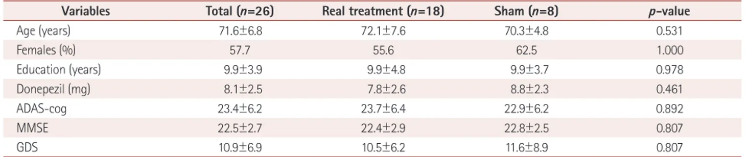
No significant differences in the baseline characteristics such as age, gender, duration of education, and neuropsychological assessments (ADAS-cog, MMSE, and GDS scores) were de- tected between these two groups (Table 1).
Primary outcomes
Comparison of the changes in ADAS-cog score between groups revealed no significant group-by-time interaction (p=0.208), although the improvement in the ADAS-cog score was much greater in the treatment group than in the sham group (Fig. 1).
The patients in the treatment group exhibited an improve- ment of 4.28 points on the ADAS-cog score immediately after the end of rTMS-COG treatment (6 weeks) from baseline (p=0.018). The effect of rTMS-COG treatment remained steady or was even enhanced at 6 weeks after the end of rTMS-COG treatment (12 weeks), as demonstrated the 5.39-point im- provement in ADAS-cog score in the treatment group from baseline at that time point (p=0.002). The patients in the sham group also improved slightly, by 1.75 and 2.88 points imme- diately and 6 weeks after treatment, respectively, although dif- ference in interactions follow-up and two group (treatment and sham group) was not statistically significant (Fig. 2, Table Table 1. Baseline characteristics of the participants
Variables Total (n=26) Real treatment (n=18) Sham (n=8) p-value
Age (years) 71.6±6.8 72.1±7.6 70.3±4.8 0.531
Females (%) 57.7 55.6 62.5 1.000
Education (years) 9.9±3.9 9.9±4.8 9.9±3.7 0.978
Donepezil (mg) 8.1±2.5 7.8±2.6 8.8±2.3 0.461
ADAS-cog 23.4±6.2 23.7±6.4 22.9±6.2 0.892
MMSE 22.5±2.7 22.4±2.9 22.8±2.5 0.807
GDS 10.9±6.9 10.5±6.2 11.6±8.9 0.807
Values denote means±SD unless specified otherwise.
ADAS-cog: Alzheimer’s disease assessment scale-cognitive subscale, GDS: Geriatric Depression Scale, MMSE: Mini-Mental Statue Examination.
Fig. 1. Differences in ADAS-cog score at each measurement time point (immediate after, 6 weeks after treatment) from baseline. There was no significant time×group interaction, although significant im- provements were found in the treatment group. The solid and dotted lines indicate the treatment and sham-treated groups, respectively.
ADAS-cog: Alzheimer’s disease Assessment Scale-cognitive subscale.
Baseline Immediate after
treatment 6 weeks after treatment -10
-6
-8 -4 2
0
-2
Changes of ADAS-cog
Sham Treatment
Lee J et al.
JCN
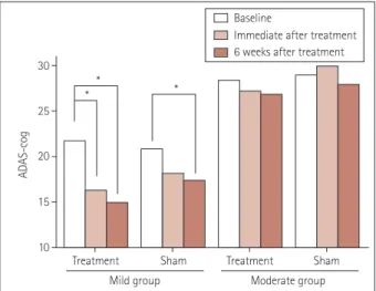
2). The ADAS-cog score improved much more significantly from baseline in the mild group (MMSE score=21–26) than in the moderate group (Fig. 3).
Secondary outcomes MMSE score
The average MMSE score improved from 22.39 (baseline) to 23.89 (immediate after) and 24.39 (6 weeks after) in the treatment group as a whole; these differences were not statis- tically significant. However, the score improved significantly between baseline and 6 weeks after treatment in the mild AD group (p=0.015) (Table 2).
GDS score
The GDS score did not improve significantly in the treatment group; however, a significant improvement at the end of the study was observed in the sham group (p=0.02) (Table 2).
CGIC score
The average CGIC scores in the treatment and sham groups were 2.4 and 3.5 immediately after treatment, respectively (p=0.009), and 2.6 and 3.3 at 6 weeks after the end of treatment (p=0.196).
Cognitive domains
Improvements in the memory and language cognitive do- mains were observed following rTMS-COG treatment; these Fig. 2. Significantly improved ADAS-cog scores were found follow-
ing treatment compared with baseline (immediately after treatment, improved by 4.28 points, p=0.014; 6 weeks after treatment, improved by 5.39 points, p=0.002). *p<0.05. ADAS-cog: Alzheimer’s disease As- sessment Scale-cognitive subscale.
Treatment
Group Sham
10 15 20 30
* * 25
ADAS-cog
Baseline
Immediate after treatment 6 weeks after treatment
Table 2. Neuropsychological test scores at each time points with all participants and in each mild and moderate group Neuropsychological
assessments Group (n)
Scores (mean±SD) p value
Baseline [B] Immediate after [6]
6 weeks after
[12] [B] vs. [6] [B] vs. [12] Time* group
ADAS
Treatment (18) 23.61 (6.40) 19.33 (8.30) 18.22 (8.85) 0.018* 0.002*
0.208
Sham (8) 22.88 (6.20) 21.12 (7.66) 20.00 (9.49) 0.238 0.46
Mild treatment (13) 21.77 (5.09) 16.31 (6.40) 14.92 (7.43) 0.035* 0.005*
0.111
Mild sham (6) 20.83 (3.76) 18.17 (4.54) 17.33 (4.93) 0.396 0.056
Moderate treatment (5) 28.4 (7.54) 27.2 (7.95) 26.8 (6.30) 1 0.264
0.966
Moderate sham (2) 29.0 (9.90) 30.0 (9.90) 28.0 (18.39) NA NA
MMSE
Treatment (18) 22.39±2.87 23.89±4.44 24.39±4.57 0.139 0.058
0.729
Sham (8) 22.75±2.49 24.50±4.90 25.75±4.56 0.769 0.213
Mild treatment (13) 23.77±2.01 25.62±3.33 26.46±2.93 0.058 0.015*
0.785
Mild sham (6) 23.83±1.72 26.67±2.16 27.5±2.51 0.461 0.204
Moderate treatment (5) 8.8±0.84 19.4±3.98 19.0±3.54 1 1
0.784
Moderate sham (2) 19.5±0.71 18.0±5.66 20.5±6.36 NA NA
GDS
Treatment (18) 10.50±6.14 7.89±5.71 7.50±6.44 0.635 0.452
Sham (8) 11.63±8.93 9.38±6.99 8.00±6.97 0.656 0.020* 0.77
Mild treatment (13) 11.62±4.82 9.92±5.25 10.00±5.85 1 1
0.754
Mild sham (6) 11.83±8.45 9.17±6.97 7.50±6.16 1 0.148
Moderate treatment (5) 7.60±8.71 2.60±2.70 1.00±0.71 1 1
0.484
Moderate sham (2) 11.00±14.14 10.00±9.90 9.50±12.02 NA NA
Repeatitive measures ANCOVA adjusted with age, sex, duration of education and post-hoc analysis with Bonferroni comparison. As moderate sham group included only two participants, analysis was impossible within group.
*p value<0.05.
ADAS: Alzheimer’s disease Assessment Scale-cognitive subscale, GDS: Geriatric Depression Scale, MMSE: Mini-Mental State Examination, NA: not ap- plicable.
Treatment of AD with rTMS and Cognitive Training
JCN
results were more significant in the mild-AD treatment group (Table 3). ADAS-cog subdomains such as word recall, word recognition, orientation, naming objects, and fingers and com- mands were improved in the treatment group. No statistically significant change in cognitive function was detected among the moderate-AD treatment group (Table 3).
DISCUSSION
A significant improvement in cognition was observed among the AD patients in this study after rTMS-COG treatment, although the differences between the treatment and sham groups were not significant. In addition, the mean ADAS-cog scores among those with mild AD improved by 5.46 points after rTMS-
COG treatment. These results were remarkable compared with the treatment effects of cholinesterase inhibitors, which resulted in an average improvement of 2.7 points over 6 months4 and 1.8 points over 12 weeks.24
While the mechanisms underlying the beneficial effects of rTMS are not fully understood, more efficient processing due to the direct modulation of cortical areas or networks has been proposed as an underlying mechanism.25 The synaptic neuro- nal activities involved in long-term potentiation (LTP) might be related to memory and learning processes based on the Hebbian theory of changes in synaptic strength via coactivation of input neurons, and such neural coactivation might be facil- itated by TMS.25,26 Given the activation of Hebbian and LTP- like mechanisms, TMS has the potential to accelerate learning skill by targeting a cortical area that is essential to performing or learning the skill, especially when TMS is applied in con- junction with training or exercise of the skill.27 High-frequency rTMS was applied to multiple cortical sites coincident with as- sociated cognitive training in the present study. Thus, rTMS- COG may increase the probability of cortical plasticity by apply- ing rTMS and subsequently performing cognitive training to the targeted cortical areas.
Cotelli et al.16,17,28 demonstrated improvements in language and auditory sentence comprehension after rTMS, and Devi et al.29 reported improvements in certain cognitive parameters after four sessions of rTMS in AD patients, primarily in their verbal and nonverbal agility. The performance in the language and memory domains was also significantly improved in the treatment group in the present study.
The cognitive outcome in the sham group was slightly better than in previous studies, which could have been due to our presentation of peripheral auditory clicking sounds from the Table 3. Changes of cognitive subdomains in the rTMS-COG treatment group
Group Cognitive domains
Changes of scores, mean (SD) p value
Δ immediate after treatment
Δ 6 weeks
after treatment [B] vs. [6] [B] vs. [12] Time
Treatment
Memory 2.00 (2.81) 2.56 (3.05) 0.040* 0.006* 0.054
Language 1.28 (1.53) 1.44 (1.92) 0.004* 0.003* 0.009*
Executive function 0.67 (1.24) 0.56 (1.50) 0.117 0.413 0.995
Mild treatment
Memory 1.77 (3.09) 2.69 (3.17) 0.275 0.031* 0.178
Language 1.23 (1.79) 1.23 (1.96) 0.04* 0.019* 0.002*
Executive function 0.92 (1.12) 0.92 (1.50) 0.031* 0.093 0.2
Moderate treatment
Memory 2.60 (2.07) 2.20 (3.03) 0.627 0.688 0.397
Language 1.40 (0.55) 2.00 (1.87) 0.243 1.000 0.947
Executive function 0.00 (1.41) 0.40 (1.14) 1.000 0.969 0.084
Repeatitive measures analysis of covariance including age, gender and duration of education as covariates with multiple comparisons with Bonferro- ni correction.
*p value<0.05.
rTMS-COG: repetitive transcranial magnetic stimulation with cognitive training, Δ: differences from baseline to at each point, [B]: baseline, [6]: imme- diately after the end of treatment, [12]: 6 weeks after the end of treatment.
Fig. 3. Significant improvements of ADAS-cog scores compared with baseline were observed in the mild-AD treatment group. *p<0.005 AD: Alzheimer’s disease, ADAS-cog: AD Assessment Scale-cognitive subscale.
Treatment Treatment
Mild group Moderate group
Sham Sham
10 15 20 30
* *
* 25
ADAS-cog
Baseline
Immediate after treatment 6 weeks after treatment
Lee J et al.
JCN
rTMS coil without cortical magnetic stimulation, thereby evok- ing intersensory facilitation.30 However, a definite effect of this intersensory facilitation phenomenon was not detected in pre- vious rTMS studies. Placebo responses are psychological con- structs related to treatment and expected outcomes, and the emotional valence attached to placebo responses include goal- seeking and optimism regarding the treatment.31 However, it cannot be concluded definitively that placebo effects were the only contribution to the improvement of cognitive function in sham participants. Improvement of depression as measured by the GDS was detected exclusively in the sham group, which might have been due to the close attention of the caregivers to the patients during this study positively influencing their psy- chological stability.
This study was subject to some limitations. First, the num- ber of patients was small, especially in moderate-AD sham group (n=2). Second, this study had only two arms: rTMS- COG treatment and sham treatment; it would have been ben- eficial to also include a group of patients who received only rTMS without cognitive training. However, the effect of rTMS has been studied previously, and the aim of the present study was to compare the effects of cognitive training with rTMS ver- sus sham. Furthermore, it was envisaged that significant prob- lems would be encountered when attempting to enroll sufficient participants for a three-arm design. Despite these limitations, the present findings demonstrate an effect of rTMS-COG treat- ment among AD patients.
This is the first study to compare rTMS-COG treatment in AD patients with a sham-treated group in Korea. Analysis of ADAS-cog subdomains and comparison of mild- and moder- ate-AD patients have not been performed previously in stud- ies with similar protocols. This study provides new evidence regarding the effects of rTMS-COG on patients with AD, es- pecially during the mild stage, which suggest that rTMS-COG represents a useful adjuvant therapy with cholinesterase in- hibitors for the treatment of mild AD.
Acknowledgements
This search was supported by Chungnam National University Hospital Research Fund, 2012.
Conflicts of Interest
The authors have no financial conflicts of interest.
REFERENCES
1. Sosa-Ortiz AL, Acosta-Castillo I, Prince MJ. Epidemiology of de- mentias and Alzheimer’s disease. Arch Med Res 2012;43:600-608.
2. Wisniewski T, Konietzko U. Amyloid-beta immunisation for Al- zheimer’s disease. Lancet Neurol 2008;7:805-811.
3. Holmes C, Boche D, Wilkinson D, Yadegarfar G, Hopkins V, Bayer A, et al. Long-term effects of Abeta42 immunisation in Alzheimer’s disease: follow-up of a randomised, placebo-controlled phase I trial.
Lancet 2008;372:216-223.
4. Birks J. Cholinesterase inhibitors for Alzheimer’s disease. Cochrane Database Syst Rev 2006:CD005593.
5. Park KW, Kim HS, Cheon SM, Cha JK, Kim SH, Kim JW. Dementia with Lewy bodies versus Alzheimer’s disease and Parkinson’s disease dementia: a comparison of cognitive profiles. J Clin Neurol 2011;7:
19-24.
6. Kobayashi M, Pascual-Leone A. Transcranial magnetic stimulation in neurology. Lancet Neurol 2003;2:145-156.
7. Rossini PM, Rossi S. Transcranial magnetic stimulation: diagnostic, therapeutic, and research potential. Neurology 2007;68:484-488.
8. Pilato F, Profice P, Ranieri F, Capone F, Di Iorio R, Florio L, et al. Syn- aptic plasticity in neurodegenerative diseases evaluated and modulated by in vivo neurophysiological techniques. Mol Neurobiol 2012;46:563- 9. Tokay T, Holl N, Kirschstein T, Zschorlich V, Köhling R. High-fre-571.
quency magnetic stimulation induces long-term potentiation in rat hippocampal slices. Neurosci Lett 2009;461:150-154.
10. Burt T, Lisanby SH, Sackeim HA. Neuropsychiatric applications of transcranial magnetic stimulation: a meta analysis. Int J Neuropsy- chopharmacol 2002;5:73-103.
11. Jahanshahi M, Rothwell J. Transcranial magnetic stimulation studies of cognition: an emerging field. Exp Brain Res 2000;131:1-9.
12. George MS, Lisanby SH, Avery D, McDonald WM, Durkalski V, Pavlicova M, et al. Daily left prefrontal transcranial magnetic stimu- lation therapy for major depressive disorder: a sham-controlled ran- domized trial. Arch Gen Psychiatry 2010;67:507-516.
13. Luber B, McClintock SM, Lisanby SH. Applications of transcranial magnetic stimulation and magnetic seizure therapy in the study and treatment of disorders related to cerebral aging. Dialogues Clin Neuro- sci 2013;15:87-98.
14. Anderkova L, Rektorova I. Cognitive effects of repetitive transcranial magnetic stimulation in patients with neurodegenerative diseases- clinician’s perspective. J Neurol Sci 2014;339:15-25.
15. Nardone R, Tezzon F, Höller Y, Golaszewski S, Trinka E, Brigo F. Tran- scranial magnetic stimulation (TMS)/repetitive TMS in mild cognitive impairment and Alzheimer’s disease. Acta Neurol Scand 2014;129:351- 16. Cotelli M, Manenti R, Cappa SF, Geroldi C, Zanetti O, Rossini PM, 366.
et al. Effect of transcranial magnetic stimulation on action naming in patients with Alzheimer disease. Arch Neurol 2006;63:1602-1604.
17. Cotelli M, Manenti R, Cappa SF, Zanetti O, Miniussi C. Transcranial magnetic stimulation improves naming in Alzheimer disease patients at different stages of cognitive decline. Eur J Neurol 2008;15:1286- 1292.
18. Haffen E, Chopard G, Pretalli JB, Magnin E, Nicolier M, Monnin J, et al. A case report of daily left prefrontal repetitive transcranial mag- netic stimulation (rTMS) as an adjunctive treatment for Alzheimer disease. Brain Stimul 2012;5:264-266.
19. Rosen WG, Mohs RC, Davis KL. A new rating scale for Alzheimer’s disease. Am J Psychiatry 1984;141:1356-1364.
20. Ahmed MA, Darwish ES, Khedr EM, El Serogy YM, Ali AM. Effects of low versus high frequencies of repetitive transcranial magnetic stimulation on cognitive function and cortical excitability in Alzheim- er’s dementia. J Neurol 2012;259:83-92.
21. Bentwich J, Dobronevsky E, Aichenbaum S, Shorer R, Peretz R, Khai- grekht M, et al. Beneficial effect of repetitive transcranial magnetic stimulation combined with cognitive training for the treatment of Al- zheimer’s disease: a proof of concept study. J Neural Transm 2011;118:
463-471.
22. Rabey JM, Dobronevsky E, Aichenbaum S, Gonen O, Marton RG, Khaigrekht M. Repetitive transcranial magnetic stimulation combined with cognitive training is a safe and effective modality for the treat- ment of Alzheimer’s disease: a randomized, double-blind study. J Neu-
Treatment of AD with rTMS and Cognitive Training
JCN
ral Transm 2013;120:813-819.
23. Rossi S, Hallett M, Rossini PM, Pascual-Leone A; Safety of TMS Con- sensus Group. Safety, ethical considerations, and application guide- lines for the use of transcranial magnetic stimulation in clinical prac- tice and research. Clin Neurophysiol 2009;120:2008-2039.
24. Lee JH, Hong YJ, Bae HJ, Kim BJ, Na DL, Han SH, et al. The effects of galantamine treatment on attention and its relationship with cog- nition and activities of daily living in patients with mild to moderate Alzheimer’s disease. J Clin Neurol 2015;11:66-72.
25. Luber B, Lisanby SH. Enhancement of human cognitive performance using transcranial magnetic stimulation (TMS). Neuroimage 2014;85 Pt 3:961-970.
26. Tegenthoff M, Ragert P, Pleger B, Schwenkreis P, Förster AF, Nicolas V, et al. Improvement of tactile discrimination performance and en- largement of cortical somatosensory maps after 5 Hz rTMS. PLoS Biol 2005;3:e362.
27. Thickbroom GW. Transcranial magnetic stimulation and synaptic
plasticity: experimental framework and human models. Exp Brain Res 2007;180:583-593.
28. Cotelli M, Calabria M, Manenti R, Rosini S, Zanetti O, Cappa SF, et al.
Improved language performance in Alzheimer disease following brain stimulation. J Neurol Neurosurg Psychiatry 2011;82:794-797.
29. Devi G, Voss HU, Levine D, Abrassart D, Heier L, Halper J, et al.
Open-label, short-term, repetitive transcranial magnetic stimulation in patients with Alzheimer’s disease with functional imaging correlates and literature review. Am J Alzheimers Dis Other Demen 2014;29:248- 30. Terao Y, Ugawa Y, Suzuki M, Sakai K, Hanajima R, Gemba-Shimizu K, 255.
et al. Shortening of simple reaction time by peripheral electrical and submotor-threshold magnetic cortical stimulation. Exp Brain Res 1997;
115:541-545.
31. Horing B, Weimer K, Muth ER, Enck P. Prediction of placebo respons- es: a systematic review of the literature. Front Psychol 2014;5:1079.