ISSN 2234-3806 • eISSN 2234-3814
https://doi.org/10.3343/alm.2018.38.4.316
Determining Genotypic Drug Resistance by Ion
Semiconductor Sequencing With the Ion AmpliSeq™
TB Panel in Multidrug-Resistant Mycobacterium tuberculosis Isolates
Joonhong Park, M.D.1*, So Youn Shin, M.D.2*, Kyungjong Kim, Ph.D.2, Kuhn Park, M.D.3, Soyoung Shin, M.D.1, and Chunhwa Ihm, M.D.4
Department of Laboratory Medicine1, College of Medicine, The Catholic University of Korea, Seoul; Korean Institute of Tuberculosis2, Cheongju; Department of Thoracic and Cardiovascular Surgery3, College of Medicine, The Catholic University of Korea, Seoul; Department of Laboratory Medicine4, Eulji University Hospital, Daejeon, Korea
Background: We examined the feasibility of a full-length gene analysis for the drug resis- tance-related genes inhA, katG, rpoB, pncA, rpsL, embB, eis, and gyrA using ion semi- conductor next-generation sequencing (NGS) and compared the results with those ob- tained from conventional phenotypic drug susceptibility testing (DST) in multidrug-resis- tant Mycobacterium tuberculosis (MDR-TB) isolates.
Methods: We extracted genomic DNA from 30 pure MDR-TB isolates with antibiotic sus- ceptibility profiles confirmed by phenotypic DST for isoniazid (INH), rifampin (RIF), eth- ambutol (EMB), pyrazinamide (PZA), amikacin (AMK), kanamycin (KM), streptomycin (SM), and fluoroquinolones (FQs) including ofloxacin, moxifloxacin, and levofloxacin. En- riched ion spheres were loaded onto Ion PI Chip v3, with 30 samples on a chip per se- quencing run, and Ion Torrent sequencing was conducted using the Ion AmpliSeq TB panel (Life Technologies, USA).
Results: The genotypic DST results revealed good agreement with the phenotypic DST re- sults for EMB (Kappa 0.8), PZA (0.734), SM (0.769), and FQ (0.783). Agreements for INH, RIF, and AMK+KM were not estimated because all isolates were phenotypically re- sistant to INH and RIF, and all isolates were phenotypically and genotypically susceptible to AMK+KM. Moreover, 17 novel variants were identified: six (p.Gly169Ser, p.Ala256Thr, p.Ser383Pro, p.Gln439Arg, p.Tyr597Cys, p.Thr625Ala) in katG, one (p.Tyr113Phe) in inhA, five (p.Val170Phe, p.Thr400Ala, p.Met434Val, p.Glu812Gly, p.Phe971Leu) in rpoB, two (p.Tyr319Asp and p.His1002Arg) in embB, and three (p.Cys14Gly, p.Asp63Ala, p.Gly162Ser) in pncA.
Conclusions: Ion semiconductor NGS could detect reported and novel amino acid changes in full coding regions of eight drug resistance-related genes. However, genotypic DST should be complemented and validated by phenotypic DSTs.
Key Words: Ion semiconductor sequencing, Ion AmpliSeq TB panel, Multidrug-resistant, Mutations, Mycobacterium tuberculosis
Received: July 30, 2017
Revision received: December 11, 2017 Accepted: February 13, 2018 Corresponding author: Chunhwa Ihm Department of Laboratory Medicine, Eulji University Hospital, Eulji University School of Medicine, 95 Dunsanseo-ro, Seo-gu, Daejeon 35233, Korea
Tel: +82-42-611-3478 Fax: +82-42-611-3464 E-mail: [email protected]
* These authors contributed equally to this work.
© Korean Society for Laboratory Medicine This is an Open Access article distributed under the terms of the Creative Commons Attribution Non-Commercial License (http://creativecom- mons.org/licenses/by-nc/4.0) which permits unrestricted non-commercial use, distribution, and reproduction in any medium, provided the original work is properly cited.
2017-03-16 https://crossmark-cdn.crossref.org/widget/v2.0/logos/CROSSMARK_Color_square.svg
INTRODUCTION
Mycobacterium tuberculosis (MTB), an obligate pathogenic bac- terial species that causes tuberculosis (TB), is a highly transmis- sible agent with significant mortality and morbidity. It has long been recognized that elderly people are more vulnerable to TB, and the TB treatment outcomes in elderly adults are often rela- tively poor owing to delayed diagnosis, more drug-related ad- verse effects, frequent co-morbidity, and overarching poverty [1]. Thus, targeted efforts to improve TB detection and treat- ment prognosis in the elderly population are needed to achieve global TB control [2].
Currently, the reference method for determining multidrug-re- sistant (MDR) or extensively drug-resistant (XDR) TB is culture- based drug susceptibility testing (DST). However, this method is labor-intensive, time-consuming (taking weeks to months to ob- tain results), expensive, and technically challenging, especially in resource-limited countries [3]. Moreover, this methodology requires handling viable and potentially infectious cultures, spe- cialized laboratory facilities, expensive consumables, and instru- ment maintenance [4]. There are currently several commercially available molecular assays to rapidly and reliably detect com- mon mutations related to isoniazid (INH) and rifampin (RIF) re- sistance as potential alternatives to phenotypic DST, including the GenoType MTBDRplus line probe (LPA) [5] and GeneXpert MTB/RIF assays [6]. However, the absence of wild-type bands related to a specific gene on the LPA interpreted as resistant to the test drug may not be supported by a phenotypic assay [7].
Furthermore, one of the main current limitations of nucleic acid- based assays is that they do not accommodate all the mutations conferring resistance to anti-TB drugs.
Routinely, liquid acid-fast bacilli (AFB) cultures are sent to medical laboratories for phenotypic DST when they are culture- positive (usually one to three weeks after inoculation), and the results are reported after an additional two to three weeks ac- cording to breakpoints to determine if the cultures are suscepti- ble or resistant to anti-TB drugs. Thus, clinically crucial informa- tion to initiate the most appropriate treatment is only available 4 to 6 weeks after a patient has been diagnosed with TB.
Next-generation sequencing (NGS) has emerged as a tech- nique that can significantly contribute to TB control and treat- ment owing to its capacity to rapidly identify multiple gene tar- gets associated with drug resistance-determining regions. Ion semiconductor sequencing is one of the available NGS platforms, which uses a small chip to detect released hydrogen ions emit- ted during DNA polymerization when deoxynucleotide triphos-
phates are incorporated into a DNA strand growing on a semi- conductor device [8]. Several studies have described the clini- cal utility of Ion Torrent sequencing to characterize known mu- tations and discover novel variants linked to molecular-based drug resistance [9-11]. We examined the feasibility of full-length gene analysis using ion semiconductor NGS with the Ion Am- pliSeq™ TB panel (Life Technologies, Carlsbad, CA, USA) through comparison with the results of conventional phenotypic DST in 30 MDR-TB isolates using eight drug resistance-related genes.
METHODS
1. Sample preparation
The study protocol was approved by the Institutional Review Board of The Catholic University of Korea (DC16EISI0099). A total of 30 pure MDR-TB isolates were randomly selected and provided by the Korean Institute of Tuberculosis, Cheongju, Ko- rea. Samples were requested for routine MTB culture and phe- notypic DST from January to December 2016. Remaining sam- ples revealing RIF and INH resistance in the phenotypic DST were sub-cultured again. Individual MTB colonies grown on the slants of 3% Ogawa solid medium within eight weeks in an in- cubator at 37°C were carefully isolated. Mycobacterial genomic DNA was extracted from the pure MDR-TB isolates using a QIA- amp DNA Mini Kit (Qiagen, Hamburg, Germany) according to the manufacturer’s protocol. Purified genomic DNA was stored at –20°C until analysis.
2. Phenotypic DST
Phenotypic drug resistance for first- and second-line drugs was determined using conventional DST (absolute concentration method with Löwenstein-Jensen medium) at the Korean Insti- tute of Tuberculosis. The critical concentrations for each anti-TB drug tested were as follows: first-line drugs INH 0.2 μg/mL, RIF 40 μg/mL, ethambutol (EMB) 2.0 μg/mL, and second-line drugs streptomycin (SM) 10 μg/mL, amikacin (AMK) 30 μg/mL, kana- mycin (KM) 30 μg/mL, and fluoroquinolones (FQs), including ofloxacin (OFX) 4.0 μg/mL, moxifloxacin (MXF) 2.0 μg/mL, and levofloxacin (LEV) 2.0 μg/mL. Pyrazinamide (PZA) susceptibility was estimated using a pyrazinamidase activity test. Besides INH and RIF resistance, the phenotypic DST results revealed 15 iso- lates resistant to EMB (50%), 10 isolates resistant to SM (33%), seven isolates resistant to PZA (23%), three isolates resistant to OFX (10%), two isolates resistant to LEV (7%), and one isolate resistant to MXF (3%); however, all 30 MDR-TB isolates were phenotypically susceptible to AMK and KM.
3. Ion semiconductor sequencing with the Ion AmpliSeq TB panel
Nucleic acid quality and quantity were assessed with the Broad- Range Qubit DNA kit and a Qubit 2.0 fluorometer (Life Technol- ogies) followed by agarose gel electrophoresis. To identify gene variants correlated with drug resistance in genomic DNA extracted from the pure MDR-TB isolates, we generated AmpliSeq librar- ies using the Ion AmpliSeq Library Kit 2.0 and the Ion AmpliSeq TB Research Panel (Life Technologies). This panel amplifies 109 amplicons (two pools) from coding sequences of eight genes re- lated to MTB drug resistance: inhA, katG, rpoB, pncA, rpsL embB, eis, and gyrA.
4. PCR amplification
The MDR-related genes were amplified through multiplex PCRs using 10 ng of genomic DNA with a premixed primer pool and the Ion AmpliSeq HiFi master mix (Ion AmpliSeq Library Kit 2.0) at 99°C for 2 minutes 18 times (99°C for 15 seconds and 60°C for 4 minutes), followed by a hold at 10°C. We treated the ob- tained PCR amplicons with 2 µL FuPa reagent to partially digest the primer sequences, and phosphorylated the amplicons at 50°C for 10 minutes, followed by 55°C for 10 minutes and 60°C for 20 minutes. We ligated the amplicons to adapters with diluted bar codes from the Ion Xpress Barcode Adapters kit (Life Tech- nologies) for 30 minutes at 22°C and then 72°C for 20 minutes.
We purified adapter-ligated amplicons (library) using Agencourt AMPure XP reagent (Beckman Coulter, Brea, CA, USA). The li- brary was quantified using a Tapestation High Sensitivity DNA Assay kit (Agilent Technologies Inc., Santa Clara, CA, USA) and an Ion Library Quantification kit (Life Technologies) for quantita- tive PCR.
5. Emulsion PCR
Clonal amplification of the bar-coded DNA library onto ion sphe- res (ISPs) was conducted using emulsion PCR, and the DNA of the ISPs was subsequently isolated using an Ion PI HIQ OT2 200 kit and Ion OneTouch ES (Life Technologies) according to the manufacturer’s instructions. We determined the polyclonal percentages and quality of enriched, template-positive ISPs us- ing an Ion Sphere Quality Control kit (Life Technologies). We then sequenced samples with polyclonal percentages <30%
and enriched, template-positive ISPs >80% on the Ion Torrent PROTON system (Life Technologies).
6. Sequencing using Ion Torrent Ion PROTON
We loaded the enriched ISPs onto Ion PI Chip v3, using 30 sam-
ples on a chip per sequencing run with the Ion PI HIQ Sequenc- ing 200 kit (Life Technologies). For sequencing, we used 520 flow runs that generated approximately 200-bp reads. Ion Tor- rent sequencing was performed using the Ion PI HIQ Sequenc- ing 200 kit, following the standard protocol.
7. Bioinformatics analysis
The raw signal data from sequencing runs were automatically transferred from Ion Torrent Ion PROTON to the Torrent Server hosted in the Torrent Suite software to process the raw voltage semiconductor sequencing data into DNA base calls. We aligned the sequences to the MTB H37Rv reference genome (NC_000 962.3), and the base calling was conducted using Torrent Suite. Variants were identified with the Ion Torrent Vari- ant Caller plug-in and Ion Reporter software (Life Technolo- gies), and coverage maps were generated using a coverage analysis plug-in. Following data analysis, single nucleotide sub- stitutions, insertions, and deletions were annotated with Ion Reporter software (Life Technologies). We then defined annota- tions of variants using snpEff v4.1 with upstream, coding, and downstream region information [12]. Amino acid changes such as missense, nonsense, or frame-shift variants were further evaluated. In addition, the sequence data were visually con- firmed with Integrative Genomics Viewer (http://software.broadin- stitute.org/software/igv/), and sequence, alignment, or variant call error artifacts were discarded.
For the rapid diagnosis of drug-resistant TB using sequence- based diagnostic methods, we searched the TB Drug Resistance Mutation Database (https://tbdreamdb.ki.se/Info/), which is a comprehensive list of genetic polymorphisms associated with first- and second-line anti-TB drug resistance, especially the most common mutations found for the major groups of anti-TB drugs in clinical MTB isolates detected worldwide [13].
8. Statistical analyses
To evaluate the agreement between genotypic DST using the Ion Ampliseq TB panel and phenotypic DST, we calculated the inter-rater agreement statistic (kappa) and 95% confidence in- terval (CI) for kappa, and interpreted each according to Altman’s guidelines: poor, <0.2; fair, 0.21–0.40; moderate, 0.41–0.60;
good, 0.61–0.80; and very good, 0.81–1.00 [14]. We then con- ducted statistical analyses using MedCalc ver. 17.6 (MedCalc Software, Mariakerke, Belgium), considering P <0.05 as statisti- cally significant.
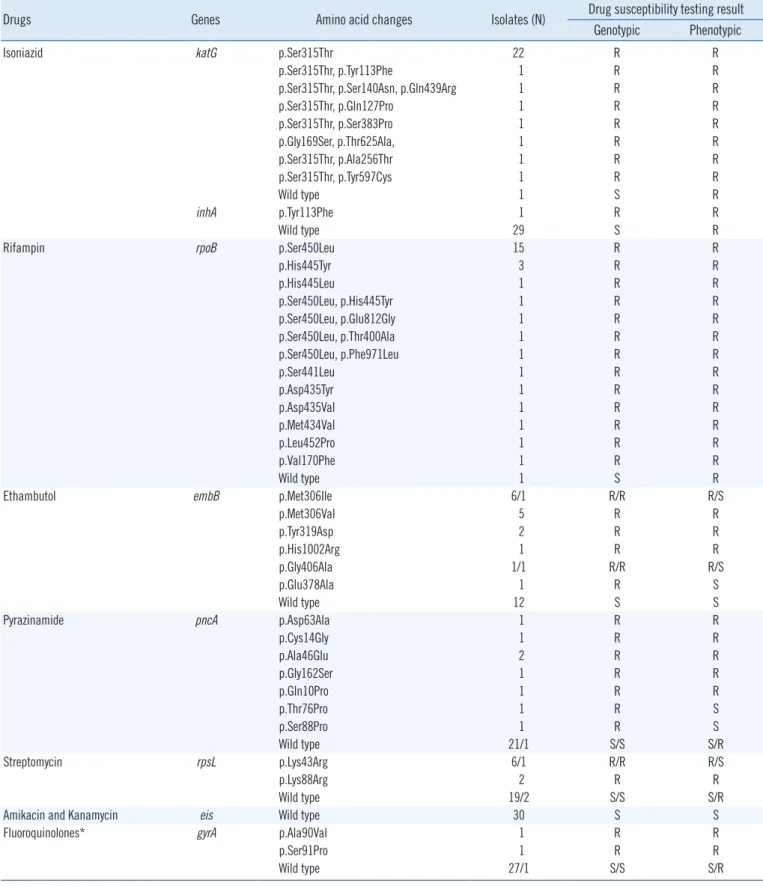
Table 1. Results of genotypic drug susceptibility testing by ion semiconductor sequencing in 30 multidrug-resistant Mycobacterium tuber- culosis isolates
Drugs Genes Amino acid changes Isolates (N) Drug susceptibility testing result
Genotypic Phenotypic
Isoniazid katG p.Ser315Thr 22 R R
p.Ser315Thr, p.Tyr113Phe 1 R R
p.Ser315Thr, p.Ser140Asn, p.Gln439Arg 1 R R
p.Ser315Thr, p.Gln127Pro 1 R R
p.Ser315Thr, p.Ser383Pro 1 R R
p.Gly169Ser, p.Thr625Ala, 1 R R
p.Ser315Thr, p.Ala256Thr 1 R R
p.Ser315Thr, p.Tyr597Cys 1 R R
Wild type 1 S R
inhA p.Tyr113Phe 1 R R
Wild type 29 S R
Rifampin rpoB p.Ser450Leu 15 R R
p.His445Tyr 3 R R
p.His445Leu 1 R R
p.Ser450Leu, p.His445Tyr 1 R R
p.Ser450Leu, p.Glu812Gly 1 R R
p.Ser450Leu, p.Thr400Ala 1 R R
p.Ser450Leu, p.Phe971Leu 1 R R
p.Ser441Leu 1 R R
p.Asp435Tyr 1 R R
p.Asp435Val 1 R R
p.Met434Val 1 R R
p.Leu452Pro 1 R R
p.Val170Phe 1 R R
Wild type 1 S R
Ethambutol embB p.Met306Ile 6/1 R/R R/S
p.Met306Val 5 R R
p.Tyr319Asp 2 R R
p.His1002Arg 1 R R
p.Gly406Ala 1/1 R/R R/S
p.Glu378Ala 1 R S
Wild type 12 S S
Pyrazinamide pncA p.Asp63Ala 1 R R
p.Cys14Gly 1 R R
p.Ala46Glu 2 R R
p.Gly162Ser 1 R R
p.Gln10Pro 1 R R
p.Thr76Pro 1 R S
p.Ser88Pro 1 R S
Wild type 21/1 S/S S/R
Streptomycin rpsL p.Lys43Arg 6/1 R/R R/S
p.Lys88Arg 2 R R
Wild type 19/2 S/S S/R
Amikacin and Kanamycin eis Wild type 30 S S
Fluoroquinolones* gyrA p.Ala90Val 1 R R
p.Ser91Pro 1 R R
Wild type 27/1 S/S S/R
*Fluoroquinolones include ofloxacin, moxifloxacin, and levofloxacin.
Abbreviations: R, resistant; S, susceptible.
RESULTS
1. Phenotypic and genotypic DST
Table 1 summarizes the detected amino acid changes in the 30 MDR-TB isolates using Ion Torrent semiconductor sequencing with Ion Ampliseq TB panels for inhA, katG, rpoB, pncA, rpsL embB, eis, and gyrA and compares the genotypic and pheno- typic DST results. Overall, we identified 17 novel variants in the isolates: six (p.Gly169Ser, p.Ala256Thr, p.Ser383Pro, p.Gln439- Arg, p.Tyr597Cys, p.Thr625Ala) in katG, one (p.Tyr113Phe) in inhA, five (p.Val170Phe, p.Thr400Ala, p.Met434Val, p.Glu812Gly, p.Phe971Leu) in rpoB, two (p.Tyr319Asp and p.His1002Arg) in embB, and three (p.Cys14Gly, p.Asp63Ala, p.Gly162Ser) in pncA.
Because the Ion Ampliseq TB panel only targets coding sequen- ces for these eight genes, we could not analyze the presence of
common promoter mutations of inhA [15], eis [16], and pncA [17].
2. Genotypic DST using the Ion AmpliSeq TB panel as a predictor of phenotypic DST
The concordance between the Ion AmpliSeq TB and phenotypic DST results varied for each of the eight anti-TB drugs tested: for INH, RIF, EMB, PZA, SM, AMK+KM, and FQs, the concordance rates were 97%, 97%, 90%, 90%, 90%, 100%, and 97%, re- spectively, with agreement (kappa values) ranging from 0.734 to 0.800 (Table 2). The agreement rates for INH, RIF, and AMK+
KM could not be estimated, because all isolates were phenotypi- cally resistant to both INH and RIF, and all isolates were pheno- typically and genotypically susceptible to AMK+KM. Overall, the genotypic DST results revealed “good” agreement with the phe- notypic DST results for EMB, PZA, SM, and FQ. The overall ma- jor error rate, defined as phenotypic resistance with no mutation found, was 11% (4/35). Individual major error rates ranged from 0 to 33%: 0% for EMB (0/15), 14% for PZA (1/7), 20% for SM (2/10), and 33% for FQs (1/3). The overall major error rate de- fined as phenotypically susceptible with a high-confidence mu- tation related to drug resistance was 7% (6/85), whereas the in- dividual major error rates ranged from 0 to 20%: 0% for FQ (0/27), 5% for SM (1/20), 9% for PZA (2/23), and 20% for EMB (3/15).
DISCUSSION
We evaluated the feasibility of genotypic DST using ion semicon- ductor NGS with the Ion AmpliSeq TB panel by characterizing 30 MDR-TB strains. Currently available commercial molecular methods offer limited detection capabilities, particularly when uncommon or new amino acid changes are contained within common drug-resistance regions or when undetected amino acid mutations impact drug resistance [11]. This targeted NGS approach can characterize known mutations and facilitate the discovery of novel variants in the entire coding regions of eight full-length genes that have been previously implicated in MDR- TB resistance.
The katG gene encodes catalase peroxidase, an enzyme that converts INH into the active form. The majority of cases of INH resistance are associated with the katG codon corresponding to amino acid 315 (p.Ser315Thr) [18], although mutations in the ahpC and inhA promoter regions have also been reported to con- tribute to resistance [19]. Among the 30 strains we assessed, 28 (93%) contained the common amino acid change of serine Table 2. Agreement between phenotypic and genotypic drug sus-
ceptibility testing by ion semiconductor sequencing Genotypic DST results
Phenotypic DST
results (N) Agreement [Kappa (95% confidence
interval)]
Resistant Susceptible Isoniazid*
Mutated (N=29) 29 0 Not estimated
Unmutated (N=1) 1 0
Rifampin
Mutated (N=29) 29 0 Not estimated
Unmutated (N=1) 1 0
Ethambutol
Mutated (N=18) 15 3 Good [0.800 (0.590–1)]
Unmutated (N=12) 0 12
Pyrazinamide
Mutated (N=8) 6 2 Good [0.734 (0.452–1)]
Unmutated (N=22) 1 21
Streptomycin
Mutated (N=9) 8 1 Good [0.769 (0.523–1)]
Unmutated (N=21) 2 19
Amikacin and Kanamycin
Mutated (N=0) 0 0 Not estimated
Unmutated (N=30) 0 30
Fluoroquinolones†
Mutated (N=2) 2 0 Good [0.783 (0.374–1)]
Unmutated (N=28) 1 27
*Results of agreement are for katG mutation and phenotypic INH resistance only; †Fluoroquinolones include ofloxacin, moxifloxacin, and levofloxacin.
Abbreviation: DST, drug susceptibility testing.
to threonine at position 315 (p.Ser315Thr) that confers INH re- sistance. Novel double variants, namely p.Thr625Ala and p.Gly- 169Ser, in one MDR-TB strain were detected in a phenotypically INH-resistant strain.
Mutations within an 81-bp RIF-resistance determining region (RRDR) of rpoB are responsible for approximately 95% of all cases of RIF resistance in MTB strains [20]. Three of these mu- tations that yield substitutions at positions p.Asp435Val, p.His- 445Asp/Tyr, and p.Ser450Leu, respectively, constitute most of the mutations within this region. Of the 30 RIF-resistant strains characterized, 19 (63%) carried p.Ser450Leu, two (7%) con- tained an amino acid change at position 435, and five (17%) contained a mutation at position 445 of the encoded rpoB prod- uct. Interestingly, ion semiconductor sequencing revealed that five of the 29 rpoB-mutated strains contained a novel variant.
We identified an uncommon amino acid change, p.Met434Val, at position 434 in the rpoB product, in which the most prevalent mutation was a change from methionine to isoleucine (p.Met- 434Ile) [21].
EMB is a first-line anti-TB agent used to prevent the emer- gence of drug resistance and is included in most MDR-TB regi- mens when susceptibility is demonstrated [22]. EMB interferes with mycobacterial cell wall synthesis and integrity by inhibiting arabinosyl transferases encoded by embC, embA, and embB [23]. The most common mutations in the EMB resistance-de- termining region (ERDR) are found in embB codons 306, 406, or 497; the remaining are located outside of these three posi- tions between codons 296 and 426 or correspond to mutations in the embC-embA intergenic region [24]. Three of the MDR- TB isolates with embB mutations p.Met306Ile, p.Glu378Ala, or p.Gly406Ala were found to be phenotypically susceptible to EMB, in line with previous findings [25]. It has recently been proposed that the conventional phenotypic DST result for EMB should not be used in the design of individualized treatment for MDR-TB.
The broth dilution method is more closely correlated with embB mutations than the proportion method in MDR-TB [26].
PZA, a synthetic derivative of nicotinamide, is used as a first- line drug, and PZA resistance is attributed to mutations in pncA encoding a pyrazinamidase [27]. However, these resistance-con- ferring mutations are variable and widely dispersed [28]. The difficulty in assessing PZA phenotypes and the sparse distribu- tion of mutations along pncA highlight the useful application of NGS to assess mutations in this hypervariable gene.
SM, an aminocyclitol glycoside, interferes with translation proof- reading, thereby inhibiting protein synthesis [29]. Various muta- tions in three genes, rrs, rpsL, and gidB, are involved in confer-
ring resistance to SM [30]. We identified common mutations converting Lys to Arg in rpsL at codons 43 and 88 in eight of the 10 SM-resistant isolates, which is in line with previous reports [31-33].
The main target of FQs is DNA gyrase, a type II topoisomer- ase composed of subunits A and B encoded by gyrA and gyrB, respectively [34]. Amino acid changes located within a short re- gion of gyrA known as the quinolone-resistance determining re- gion (QRDR) account for the majority of known FQ-resistant strains [35]. Two missense mutations, p.Ala90Val and p.Ser91Pro, were identified in the QRDR in two of three FQ-resistant isolates. Al- though gyrA and gyrB mutations can lead to ofloxacin resistance, there were no significant differences among strains with muta- tion locations in gyrA and gyrB, as well as levels of ofloxacin re- sistance [36].
From this perspective, ion semiconductor NGS with the Ion AmpliSeq TB panel has the potential to provide useful informa- tion several weeks before phenotypic DST results are available, even when the genotypic DST is conducted directly from culture isolates. In contrast to most Sanger sequencers, Ion Torrent se- quencing does not require lasers, which are expensive and in- volve substantial maintenance, and typically require modified fluorescence-based, light-sensitive chemistry; thus, this platform offers a broader testing range at lower cost, without compromis- ing assay performance or turn-around times in clinical settings [10]. We do not suggest that phenotypic DST should be replaced completely; however, for critical cases, there is clinical significance associated with the delay in obtaining broader DST results, to which genotypic DST can provide a complementary and more rapid result. In particular, genotypic DST can play a major role in determining resistance to the drugs EMB and PZA, for which phenotypic DST is known to be relatively less reliable [37]. Given that PZA is associated with a highly adverse effect of hepatotox- icity, especially in older patients, a genotypic DST for pncA may support phenotypic DST results and enable clinicians to man- age and initiate anti-TB treatment earlier.
One of the main concerns related to performing full-gene NGS using only MDR-TB isolates is the discovery of novel mutations that may have no impact on drug susceptibility; conversely, this approach could reveal rare variants outside of known resistance sites that may be responsible for partial or complete resistance when accompanied by other mutations within either the same or other genes. The main limitation is that the Ion AmpliSeq TB panel is designed to cover the coding sequences of only eight genes. Thus, the panel should be revised to provide a primer that extends to promoter regions so as to cover the frequent pro-
moter mutations associated with drug resistance, including inhA c.-15C>T [38], eis c.-14C>T, c.-12C>T, c.-10G>C/A [16], and pncA c.-11A>G, c-15A>C [17, 39]. In addition, the rrs gene encoding 16S rRNA should be included in the panel, which is associated with high-level resistance to second-line injectable drugs, such as AMK and KM, used to treat MDR-TB [40].
In conclusion, ion semiconductor NGS could provide the op- portunity to detect reported or novel amino acid changes in the full coding regions of these eight genes. However, genotypic drug resistance profiles should be complemented and validated by subsequent phenotypic DST studies.
Authors’ Disclosures of Potential Conflicts of Interest
No potential conflict of interest relevant to this article was re- ported.
Acknowledgment
This research was supported by the Bio & Medical Technology Development Program of the NRF funded by the Korean gov- ernment, MSIP (No. 2016M3A9B694241).
REFERENCES
1. Rajagopalan S. Tuberculosis in older adults. Clin Geriatr Med 2016;32:
479-91.
2. Negin J, Abimbola S, Marais BJ. Tuberculosis among older adults--time to take notice. Int J Infect Dis 2015;32:135-7.
3. Cegielski JP, Kurbatova E, van der Walt M, Brand J, Ershova J, Tupasi T, et al. Multidrug-resistant tuberculosis treatment outcomes in relation to treatment and initial versus acquired second-line drug resistance. Clin Infect Dis 2016;62:418-30.
4. Rodrigues C, Jani J, Shenai S, Thakkar P, Siddiqi S, Mehta A. Drug sus- ceptibility testing of Mycobacterium tuberculosis against second-line drugs using the Bactec MGIT 960 System. Int J Tuberc Lung Dis 2008;
12:1449-55.
5. Meaza A, Kebede A, Yaregal Z, Dagne Z, Moga S, Yenew B, et al. Evalu- ation of genotype MTBDRplus VER 2.0 line probe assay for the detec- tion of MDR-TB in smear positive and negative sputum samples. BMC Infect Dis 2017;17:280.
6. Geleta DA, Megerssa YC, Gudeta AN, Akalu GT, Debele MT, Tulu KD.
Xpert MTB/RIF assay for diagnosis of pulmonary tuberculosis in sputum specimens in remote health care facility. BMC Microbiol 2015;15:220.
7. Cirillo DM, Miotto P, Tagliani E. Reaching consensus on drug resistance conferring mutations. Int J Mycobacteriol 2016;5(S1):S33.
8. Rothberg JM, Hinz W, Rearick TM, Schultz J, Mileski W, Davey M, et al.
An integrated semiconductor device enabling non-optical genome se- quencing. Nature 2011;475:348-52.
9. Colman RE, Anderson J, Lemmer D, Lehmkuhl E, Georghiou SB, Hea- ton H, et al. Rapid drug susceptibility testing of drug-resistant Mycobac-
terium tuberculosis isolates directly from clinical samples by use of am- plicon sequencing: a proof-of-concept study. J Clin Microbiol 2016;54:
2058-67.
10. Daum LT, Fischer GW, Sromek J, Khubbar M, Hunter P, Gradus MS, et al. Characterization of multi-drug resistant Mycobacterium tuberculosis from immigrants residing in the USA using ion torrent full-gene sequenc- ing. Epidemiol Infect 2014;142:1328-33.
11. Daum LT, Rodriguez JD, Worthy SA, Ismail NA, Omar SV, Dreyer AW, et al. Next-generation ion torrent sequencing of drug resistance mutations in Mycobacterium tuberculosis strains. J Clin Microbiol 2012;50:3831-7.
12. Cingolani P, Platts A, Wang le L, Coon M, Nguyen T, Wang L, et al. A program for annotating and predicting the effects of single nucleotide polymorphisms, SnpEff: SNPs in the genome of Drosophila melanogas- ter strain w1118; iso-2; iso-3. Fly (Austin) 2012;6:80-92.
13. Sandgren A, Strong M, Muthukrishnan P, Weiner BK, Church GM, Mur- ray MB. Tuberculosis drug resistance mutation database. PLoS Med 2009;6:e2.
14. Bland JM and Altman DG. Statistical methods for assessing agreement between two methods of clinical measurement. Lancet 1986;1:307-10.
15. Jagielski T, Bakula Z, Roeske K, Kaminski M, Napiorkowska A, Augus- tynowicz-Kopec E, et al. Mutation profiling for detection of isoniazid re- sistance in Mycobacterium tuberculosis clinical isolates. J Antimicrob Chemother 2015;70:3214-21.
16. Kambli P, Ajbani K, Nikam C, Sadani M, Shetty A, Udwadia Z, et al. Cor- relating rrs and eis promoter mutations in clinical isolates of Mycobacte- rium tuberculosis with phenotypic susceptibility levels to the second-line injectables. Int J Mycobacteriol 2016;5:1-6.
17. Ramirez-Busby SM and Valafar F. Systematic review of mutations in pyr- azinamidase associated with pyrazinamide resistance in Mycobacterium tuberculosis clinical isolates. Antimicrob Agents Chemother 2015;59:
5267-77.
18. Kim SY, Park YJ, Kim WI, Lee SH, Ludgerus Chang C, Kang SJ, et al.
Molecular analysis of isoniazid resistance in Mycobacterium tuberculo- sis isolates recovered from South Korea. Diagn Microbiol Infect Dis 2003;
47:497-502.
19. Nieto RL, Mehaffy C, Creissen E, Troudt J, Troy A, Bielefeldt-Ohmann H, et al. Virulence of Mycobacterium tuberculosis after acquisition of isoni- azid resistance: individual nature of katG mutants and the possible role of AhpC. PLoS One 2016;11:e0166807.
20. Telenti A, Imboden P, Marchesi F, Lowrie D, Cole S, Colston MJ, et al.
Detection of rifampicin-resistance mutations in Mycobacterium tuber- culosis. Lancet 1993;341:647-50.
21. Sajduda A, Brzostek A, Poplawska M, Augustynowicz-Kopec E, Zwolska Z, Niemann S, et al. Molecular characterization of rifampin- and isonia- zid-resistant Mycobacterium tuberculosis strains isolated in Poland. J Clin Microbiol 2004;42:2425-31.
22. Alcaide F, Pfyffer GE, Telenti A. Role of embB in natural and acquired resistance to ethambutol in mycobacteria. Antimicrob Agents Chemoth- er 1997;41:2270-3.
23. Belanger AE, Besra GS, Ford ME, Mikusova K, Belisle JT, Brennan PJ, et al. The embAB genes of Mycobacterium avium encode an arabinosyl transferase involved in cell wall arabinan biosynthesis that is the target for the antimycobacterial drug ethambutol. Proc Natl Acad Sci U S A 1996;93:11919-24.
24. Brossier F, Sougakoff W, Bernard C, Petrou M, Adeyema K, Pham A, et al. Molecular analysis of the embCAB locus and embR gene involved in ethambutol resistance in clinical isolates of Mycobacterium tuberculosis in France. Antimicrob Agents Chemother 2015;59:4800-8.
25. Shi D, Li L, Zhao Y, Jia Q, Li H, Coulter C, et al. Characteristics of embB mutations in multidrug-resistant Mycobacterium tuberculosis isolates in
Henan, China. J Antimicrob Chemother 2011;66:2240-7.
26. Zhang Z, Wang Y, Pang Y, Kam KM. Ethambutol resistance as deter- mined by broth dilution method correlates better than sequencing re- sults with embB mutations in multidrug-resistant Mycobacterium tuber- culosis isolates. J Clin Microbiol 2014;52:638-41.
27. Sengstake S, Bergval IL, Schuitema AR, de Beer JL, Phelan J, de Zwaan R, et al. Pyrazinamide resistance-conferring mutations in pncA and the transmission of multidrug resistant TB in Georgia. BMC Infect Dis 2017;
17:491.
28. Mphahlele M, Syre H, Valvatne H, Stavrum R, Mannsaker T, Muthivhi T, et al. Pyrazinamide resistance among South African multidrug-resistant Mycobacterium tuberculosis isolates. J Clin Microbiol 2008;46:3459- 64.
29. Sreevatsan S, Pan X, Stockbauer KE, Williams DL, Kreiswirth BN, Muss- er JM. Characterization of rpsL and rrs mutations in streptomycin-resis- tant Mycobacterium tuberculosis isolates from diverse geographic local- ities. Antimicrob Agents Chemother 1996;40:1024-6.
30. Jagielski T, Ignatowska H, Bakula Z, Dziewit L, Napiorkowska A, Augus- tynowicz-Kopec E, et al. Screening for streptomycin resistance-confer- ring mutations in Mycobacterium tuberculosis clinical isolates from Po- land. PLoS One 2014;9:e100078.
31. Finken M, Kirschner P, Meier A, Wrede A, Bottger EC. Molecular basis of streptomycin resistance in Mycobacterium tuberculosis: alterations of the ribosomal protein S12 gene and point mutations within a functional 16S ribosomal RNA pseudoknot. Mol Microbiol 1993;9:1239-46.
32. Nair J, Rouse DA, Bai GH, Morris SL. The rpsL gene and streptomycin resistance in single and multiple drug-resistant strains of Mycobacteri- um tuberculosis. Mol Microbiol 1993;10:521-7.
33. Rezaei F, Haeili M, Imani Fooladi A, Azari Garmjan GA, Feizabadi MM.
Screening for streptomycin resistance conferring mutations in Myco- bacterium tuberculosis isolates from Iran. J Chemother 2017;29:14-8.
34. Aubry A, Pan XS, Fisher LM, Jarlier V, Cambau E. Mycobacterium tu- berculosis DNA gyrase: interaction with quinolones and correlation with antimycobacterial drug activity. Antimicrob Agents Chemother 2004;48:
1281-8.
35. Maruri F, Sterling TR, Kaiga AW, Blackman A, van der Heijden YF, May- er C, et al. A systematic review of gyrase mutations associated with fluo- roquinolone-resistant Mycobacterium tuberculosis and a proposed gy- rase numbering system. J Antimicrob Chemother 2012;67:819-31.
36. Cui Z, Wang J, Lu J, Huang X, Hu Z. Association of mutation patterns in gyrA/B genes and ofloxacin resistance levels in Mycobacterium tuber- culosis isolates from East China in 2009. BMC Infect Dis 2011;11:78.
37. Falzon D, Jaramillo E, Schunemann HJ, Arentz M, Bauer M, Bayona J, et al. WHO guidelines for the programmatic management of drug-resis- tant tuberculosis: 2011 update. Eur Respir J 2011;38:516-28.
38. Niehaus AJ, Mlisana K, Gandhi NR, Mathema B, Brust JC. High preva- lence of inhA promoter mutations among patients with drug-resistant tu- berculosis in KwaZulu-Natal, South Africa. PLoS One 2015;10:e0135003.
39. Rahman A, Ferdous SS, Ahmed S, Rahman SMM, Uddin MKM, Phol- wat S, et al. Pyrazinamide susceptibility and pncA mutation profiles of Mycobacterium tuberculosis among multidrug-resistant tuberculosis pa- tients in Bangladesh. Antimicrob Agents Chemother 2017;61:e00511-7.
40. Sowajassatakul A, Prammananan T, Chaiprasert A, Phunpruch S. Mo- lecular characterization of amikacin, kanamycin and capreomycin re- sistance in M/XDR-TB strains isolated in Thailand. BMC Microbiol 2014;
14:165.