Picosecond 755-nm Alexandrite Laser in Asians
Vol. 29, No. 6, 2017 779
Received September 5, 2016, Revised December 21, 2016, Accepted for publication January 13, 2017
Corresponding author: Sung-Eun Chang, Department of Dermatology, Asan Medical Center, 88 Olympic-ro 43-gil, Songpa-gu, Seoul 05505, Korea. Tel:
82-2-3010-3460, Fax: 82-2-486-7831, E-mail: changse2016@gmail.com This is an Open Access article distributed under the terms of the Creative Commons Attribution Non-Commercial License (http://creativecommons.
org/licenses/by-nc/4.0) which permits unrestricted non-commercial use, distribution, and reproduction in any medium, provided the original work is properly cited.
Copyright © The Korean Dermatological Association and The Korean Society for Investigative Dermatology
pISSN 1013-9087ㆍeISSN 2005-3894
Ann Dermatol Vol. 29, No. 6, 2017 https://doi.org/10.5021/ad.2017.29.6.779
CASE REPORT
Treatment of Melasma and Post-Inflammatory
Hyperpigmentation by a Picosecond 755-nm Alexandrite Laser in Asian Patients
Ye Jin Lee, Ho Jeong Shin, Tai-Kyung Noh, Kwang-Ho Choi1, Sung-Eun Chang
Department of Dermatology, Asan Medical Center, University of Ulsan College of Medicine, 1Chois Dermatology Clinic, Seoul, Korea
The picosecond lasers have shown to effectively treat tattoo pigments that are intractable to previous multiple Q-switch- ed (QS) laser treatments. Therefore we hypothesized that a picosecond laser would show better efficacy with minimal adverse events in the treatment of melasma and post-in- flammatory hyperpigmentation (PIH) that are difficult to treat with conventional QS lasers. Two patients with melasma and one patient with PIH were treated with a Picosecond 755-nm Alexandrite Laser (Cyanosure, USA). All patients were Korean with skin type IV and no longer responding to QS la- ser treatments. Laser treatment was well tolerated in all the patients. Adverse events such as PIH were not reported dur- ing 8 weeks of follow up period. After the multiple treatment sessions, one patient reported fair improvement and two pa- tients reported good improvement. Consistent with the clin- ical results, ex vivo skin model irradiated with a Picosecond 755-nm Alexandrite Laser also showed decreased epidermal keratinocyte necrosis compared with the 532-nm QS Neodymium-Doped Yttrium Aluminium Garnet Laser (Lutronic, Korea) yet decreased melanin content. In con- clusion, the Picosecond 755-nm Alexandrite Laser may be useful for effective treatment of intractable melasma and PIH with fewer adverse events in dark Asian skin. (Ann Dermatol
29(6) 779∼781, 2017) -Keywords-
Melanosis, Picosecond laser, Postinflammatory hyper- pigmentation
INTRODUCTION
Q-switched (QS) lasers, which emit high-energy pulses in the nanosecond range at varying wavelengths, have been used with good efficacy for treatment of pigmented lesions.
Use of a low-fluence QS neodymium-doped yttrium alu- minium garnet (QSNY) laser or ‘laser toning’ has shown efficacy in treating various pigment disorders, including melasma and post-inflammatory hyperpigmentation (PIH) in Asian countries1. However, treatment outcomes are in- consistent, and adverse events such as rebound hyper- pigmentation and mottled hypopigmentation have been reported, especially in darker-skinned patients1.
The picosecond laser was introduced in the 1990s, and several studies have shown improved efficacy in clearing tattoo pigments compared with QS lasers2. Moreover, tat- too pigments that are intractable to multiple QS laser treat- ments have responded to picosecond lasers3. Chesnut et al.4 reported successful treatment of a recalcitrant nevus of Ota with Picosecond 755-nm Alexandrite Lasers. Therefore, we hypothesized that a picosecond laser would show bet- ter efficacy with minimal adverse events in the treatment of melasma and PIH that are difficult to treat with conven- tional QS lasers.
YJ Lee, et al
780 Ann Dermatol
Fig. 1. Melasma in a 53-year-old female. (A) Findings at baseline.
(B) Findings after six treatments of 0.57 J/cm2 with a 6-mm spot size using a Picosecond 755-nm Alexandrite Laser.
Fig. 2. Melasma in a 45-year-old female. (A) Findings at baseline.
(B) Findings after 14 treatments of 0.57 J/cm2 with a 6-mm spot size using a Picosecond 755-nm Alexandrite Laser.
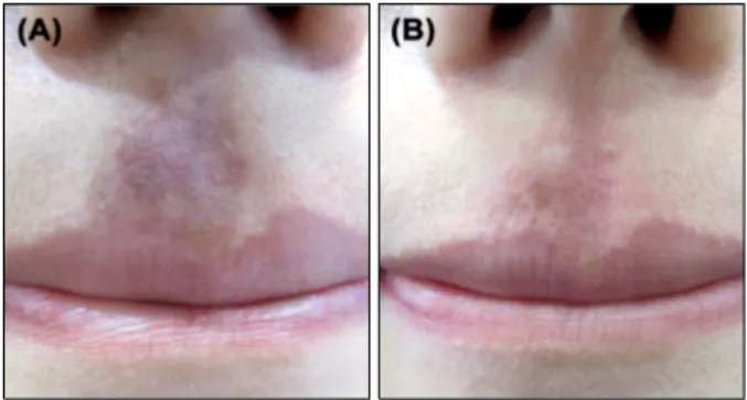
Fig. 3. Post-inflammatory hyperpigmentation in a 20-year-old female. (A) Findings at baseline. (B) Findings after seven treatments of 5.25 J/cm2 with a 2-mm spot size using a Picosecond 755-nm Alexandrite Laser.
CASE REPORT
Two patients with melasma and one patient with PIH were treated with a 750-picosecond pulse using a 755-nm Alexandrite Laser (Cyanosure, Westford, MA, USA). All patients were Korean women with skin type IV. All pa- tients had multiple previous low-fluence QSNY laser treat- ments but were no longer responding to such treatments.
Informed consent was obtained for all patients. Two fe- male patients, 53 and 45 years old (patient 1 and 2), had inhomogeneous pigmentation on their cheeks, noses, and temples, consistent with melasma and were treated with a spot size of 6 mm (0.57 J/cm2) for 6 and 14 sessions, re- spectively, with two-week intervals in between treatments (Fig. 1, 2). One 20-year-old female patient (patient 3) had an ill-defined, 2-cm, brownish patch on her philtrum for more than 3 years that was diagnosed as PIH (Fig. 3). She was treated seven times, two weeks apart, with a 2-mm (5.25 J/cm2) spot size. Laser treatment was well tolerated with minimal downtime. Post-laser erythema was not evident. Neither blistering nor petechia was reported. No patients had developed PIH at 8 weeks after laser treatment.
The melasma lesions showed significant improvement at this time (Fig. 1∼3). Patient 1 showed fair improvement and patients 2 and 3 showed good improvement.
An ex vivo skin model from the abdomen of a Korean fe- male was irradiated with a Picosecond 755-nm Alexandrite Laser and a QSNY laser at 532 nm and 1,064 nm. The la- ser parameters were 1.26 J/cm2 with a 4.5-mm spot size, 1 J/cm2, with a 7-mm spot size, and 3 J/cm2 with a 7-mm spot size, respectively. Immunohistochemical staining us- ing nitro blue tetrazolium was performed 4 hours after the laser treatment to see immediate effect of laser irradiation
and detect viable cells. Remaining melanin pigments were quantitatively measured by Fontana-Masson staining 7 days after laser treatment using Image J (National Institutes of Health, Bethesda, MA, USA). Ex vivo experiments re- sults were consistent with clinical outcomes. Four hours after the irradiation, the Picosecond 755-nm Alexandrite Laser led to decreased epidermal keratinocyte necrosis compared with the 532-nm QSNY Laser (Lutronic, Goyang, Korea) (Fig. 4). Quantitative measurements showed that the melanin content was decreased by both the Picosecond 755-nm Alexandrite Laser and the 1,064-nm QSNY Laser (Lutronic) (Fig. 4).
DISCUSSION
The Picosecond 755-nm Alexandrite Laser, which has a pulse duration that is much shorter than the thermal relax- ation time of melanosomes, enabled us to selectively and effectively destroy melanosomes while causing minimal damage to surrounding tissues such as vessel hemoglobin
Picosecond 755-nm Alexandrite Laser in Asians
Vol. 29, No. 6, 2017 781 Fig. 4. (A∼D) Photomicrographs of a skin model stained for nitro blue tetrazolium after laser treatment: (A) control, (B) 1,064-nm Q-switched Neodymium-Doped Yttrium Aluminium Garnet (QSNY) Laser, (C) 532-nm QSNY Laser, (D) Picosecond 755-nm Alexandrite Laser; A∼D, ×200). (E) Relative value of melanin index seven days after treatment. Quantitative measurement of melanin pigments was performed using Image J analysis following Fontana-Masson staining.
and epidermis. Low-fluence 1,064-nm QSNY Laser treat- ments of melasma or PIH often reach a steady state after multiple treatment sessions. Energy delivery in the pico- second range may achieve more selective photothermolysis of fragmented melanin granules from previous repetitive laser treatments5. With a picosecond laser, lower fluence can be used, which should decrease adverse effects while keeping the peak energy substantially higher than that typ- ically produced by QS lasers. Except its high cost, the pi- cosecond laser has a more favorable effect and safety pro- file to the QS nanosecond lasers.
In conclusion, the Picosecond 755-nm Alexandrite Laser may be useful for treating melasma and PIH that are in- tractable to conventional laser toning or residual lesions after QS lasers and shows high efficacy and fewer adverse events in dark Asian skin.
ACKNOWLEDGMENT
This work was funded by the Industrial Core Technology Development Program of the Korean Ministry of Trade, Industry and Energy (No. 10048690).
CONFLICTS OF INTEREST
The authors have nothing to disclose.
REFERENCES
1. Kim BW, Lee MH, Chang SE, Yun WJ, Won CH, Lee MW, et al. Clinical efficacy of the dual-pulsed Q-switched neodymium:yttrium-aluminum-garnet laser: comparison with conservative mode. J Cosmet Laser Ther 2013;15:340-341.
2. Ross V, Naseef G, Lin G, Kelly M, Michaud N, Flotte TJ, et al. Comparison of responses of tattoos to picosecond and nanosecond Q-switched neodymium: YAG lasers. Arch Dermatol 1998;134:167-171.
3. Saedi N, Metelitsa A, Petrell K, Arndt KA, Dover JS.
Treatment of tattoos with a picosecond alexandrite laser: a prospective trial. Arch Dermatol 2012;148:1360-1363.
4. Chesnut C, Diehl J, Lask G. Treatment of nevus of ota with a picosecond 755-nm alexandrite laser. Dermatol Surg 2015;41:508-510.
5. Kim JE, Chang SE, Yeo UC, Haw S, Kim IH. Histo- pathological study of the treatment of melasma lesions using a low-fluence Q-switched 1064-nm neodymium:
yttrium-aluminium-garnet laser. Clin Exp Dermatol 2013;
38:167-171.