Received July 22, 2021, Revised August 31, 2021, Accepted September 3, 2021 Corresponding author: Sujung Yeo
Department of Meridian & Acupoint, College of Korean Medicine, Sangji University, 83 Sangjidae-gil, Wonju 26339, Korea Tel: +82-33-738-7506, E-mail: pinkteeth@naver.com
This research was supported by Sangji University Research Fund, 2019.
CCThis is an open access article distributed under the terms of the Creative Commons Attribution Non-Commercial License (http://creativecommons.org/licenses/ by-nc/4.0) which permits unrestricted non-commercial use, distribution, and reproduction in any medium, provided the original work is properly cited.
파킨슨병 동물 모델에서 도파민세포의 감소와 관련된 Slc6a4 발현의 증가
여수정1,2
1상지대학교 한의과대학 경혈학교실, 2상지대학교 한의학연구소
Increased Slc6a4 Expression Associated with Decreased Dopaminergic Neurons in an MPTP
Induced Parkinsonism Mouse Model
Sujung Yeo1,2
1Department of Meridian & Acupoint, College of Korean Medicine, Sangji University,
2Research Institute of Korean Medicine, Sangji Univeristy
Objectives : Parkinson’s disease is a neurodegenerative disease caused by a decrease in the dopaminergic neurons in the sub- stantia nigra. The abnormal expression of solute carrier family 6 member 4 (Slc6a4) has been reported in patients with Parkinson’s disease. Methods : In this study, we used MPTP to examine the changes in the expression of Slc6a4 in the brain of mice with Parkinson’s disease and investigate its effect on dopaminergic neuronal cell death. Results : In the examination of the Slc6a4 expression in the substantia nigra of MPTP-treated mice for 4 weeks. The gene expression was increased compared to the normal group. To investigate the relationship between Slc6a4 and dopaminergic neurons, we performed a study using siRNA of Slc6a4 in the dopaminergic neuronal cell line SH-SY5Y. Using the siRNA of Slc6a4 to evaluate gene expression, it revealed that the tyrosine hydroxylase (TH) expression increases when Slc6a4 decreases. Moreover, this confirms its effects on the dopaminergic neurons. Additionally, through the evaluation of factors related to apoptosis, in particular, it was established that the value of bax/bcl2 decreased and was affected. These results suggest that a decreased Slc6a4 expression induces an increase in TH expression, providing a mechanism of action for dopaminergic neurons regulated by Slc6a4 expression. Conclusions : Slc6a4 is deemed to be involved in the regulation of dopaminergic neurons, suggesting that an increased Slc6a4 expression induced by MPTP may influence a reduction of dopaminergic neurons.
Key words : Parkinson’s disease, slc6a4, MPTP, MPP+
서 론
인간의 평균 수명이 증가함에 따라 퇴행성 질환의 발병률이 증 가하고 있다1,2). 이러한 질병 중 파킨슨병은 진전, 무동증, 자세의 불안정, 경직 등의 운동기능과 관련된 증상을 주요 특징으로 하는
신경계의 만성 진행성 퇴행성 뇌질환으로써 알쯔하이머병에 이어 두 번째로 많은 수를 차지한다2). 파킨슨병은 대뇌 흑질의 도파민성 신경세포가 손실된 결과로 발생하는데1,2), 도파민 세포의 수가 감 소하는 근본적인 이유는 아직 밝혀지지 않았다2-4).
파킨슨병과 관련이 있는 인자들을 확인하기 위하여 유전자 배열
분석을 통하여 파킨슨병 동물모델에서 유전자의 발현 변화를 살펴 본 결과 파킨슨병을 유발한 동물모델에서 Solute Carrier Family 6 (Neurotransmitter Transporter, Serotonin), Member 4 (Slc6a4) 유전자 발현이 증가하는 것으로 나타났다. Slc6a4는 SERT, Sero- tonin Transporter 1, 5HT Transporter, 그리고 5-HTT 등으로도 알려져 있는데, 이 유전자는 신경 전달 물질인 세로토닌을 시냅스 공간에서 시냅스 전 뉴런으로 재흡수하는 수송체인 막 단백질을 암호화한다5). 인체에서 세로토닌의 수준은 주로 Slc6a4에 의해 암 호화 된 세로토닌 재흡수 수송체에 의해 조절된다6,7). 이러한 세로 토닌 재흡수 수송체의 이상이 파킨슨병과 밀접한 관련성이 있다고 보고된 바 있으며, 이를 통해 각각의 신경전달물질들이 상호 영향 을 주는 것으로 생각되고 있다8,9).
중국인 파킨슨병 환자를 대상으로 진행한 연구에서 안정 시 진 전, 우울증 등의 증상이 Slc6a4 발현 이상과 관련되어 있다고 보고 하였다10). 또 이탈리아인을 대상으로 한 연구에서 Slc6a4의 발현 이상이 파킨슨병의 유전적 위험요소가 될 수 있다고 보고했다11). 그리고 Slc6a4에 의해 발현되는 물질을 포함하는 세로토닌 시스템 은 파킨슨병의 병태 생리학에서 중요한 역할을 할 것으로 보고되 고 있다12).
이들 앞선 연구결과들을 통하여 Slc6a4와 파킨슨병과의 연관성 을 살펴 볼 수 있었는데, 대다수의 논문들이 임상연구 결과로써, Slc6a4이 파킨슨병에 미치는 기전에 대하여는 아직 연구된 바가 없다. 본 연구에서는 MPTP로 유도된 파킨슨병 동물모델의 뇌 흑 질에서 Slc6a4 발현 변화를 조사하고, SH-SY5Y 세포주에서 짧은 간섭 RNA (siRNA)를 활용하여 파킨슨병의 주요 요소인 도파민성 신경세포와 관련된 Slc6a4의 작용에 대하여 탐색하여 파킨슨병에 미치는 기전에 대하여 살펴 보았다.
재료 및 방법
1. 동물모델
6주령 수컷 C57BL/6 마우스(체중, 20∼22 g, DBL, 한국)를 대 조군(CTL)과 MPTP 치료군(MPTP)으로 나누어 1주일간 적응 기 간을 가진 후 실험을 진행하였다. CTL 그룹의 마우스는 0.9%
(100 μl) 식염수를 4주 동안 매일 1회 복강 주사하고 MPTP 그룹 의 마우스는 0.9% MPTP-HCl (20 mg/kg)을 복강 내 주사했다.
최종 MPTP를 처리한 다음 날, 마우스를 Alfaxan (JUROX, 호주)을 사용하여 마취하고 저온 0.05 M 인산 나트륨 완충액(PBS)으로 관 류하여 면역 조직 화학적 평가를 수행했다. 본 연구에 사용된 모든
동물 프로토콜은 상지대학교 동물 실험위원회의 승인을 받았다 (No. 2016-07). 연구에 사용된 모든 시약은 달리 언급하지 않는 한 Sigma (미국) 제품을 사용했다.
2. RNA 추출 및 마이크로 어레이 분석
RNeasy Plus Mini kit (QIAGEN, 미국)를 사용하여 각 그룹의 양측 흑질(SN) 조직에서 총 RNA를 추출했다. 분리된 RNA의 품질 은 NanoDrop ND-1000 (NanoDrop Technologies, 미국)을 사 용하여 정량했다. 전체 RNA 300 ng을 GeneChip Whole Transcript Sense에 따라 Affymetrix GeneChipⓇ Mouse Gene 1.0 ST Array (Affymetrix, 미국)에 적용했다. Expression Console 소프트웨어 (Affymetrix, 미국)를 사용하여 스캔하고 얻은 Microarray 데이터 는 GenPlex ver. 3.0 (ISTECH, 한국)을 이용하여 분석하였다.
Student t-test를 시행하여 유의성 기준 (p<0.05)을 충족하는 유 전자를 대상으로 DEG-finding 모듈을 사용하여 분석했다.
3. 세포배양
본 실험에 사용된 세포주는 human neuroblastoma인 SH- SY5Y세포이며(한국세포주은행, 한국), 10% FBS (Welgen, 한국) 에서 100 U/ml의 페니실린, 0.1 mM의 비필수아미노산 및 100 mg/ml의 스트렙토 마이신을 포함하는 MEM (Welgen, 한국)에서 표준 세포 배양 조건(5% CO2, 37℃) 하에 배양되었다.
4. 짧은 간섭 RNA (siRNA)을 이용한 유전자 발현억제 Slc6a4에 대한 siRNA (5’- UUG UUG AAC UUG UUG UAG CUA -3’) 및 음성 대조군(Slc6a4에 대한 스크램블 된 siRNA 5’- UUC UCC GAA CGU GUC ACG UTT -3’)는 Bioneer Inc (한국) 에서 구입했다.
5. 면역 조직 화학 염색
마우스의 뇌를 4% 파라 포름 알데히드를 함유하는 4℃의 0.05M PBS에 12시간 동안 고정하고 30% 수크로스 용액에 48 시 간 동안 탈수시켰다. 뇌는 40μm 두께로 −20℃에서 마이크로톰 을 사용하여 절단했다. ABC 키트와 Mouse on Mouse (M.O.M) 면역 검출 키트(Vector Laboratories, CA)를 사용하여 avidin- biotin-peroxidase 방법을 사용하여 면역 조직 화학적 분석을 수 행했다. 절편된 조직을 3% H2O2를 포함하는 PBS에서 처리하고 1% bovine serum albumin 및 10% horse serum이 들어있는 PBS로 1시간 동안 처리했다. 후에 실온에서 1시간 동안 M.O.M mouse Ig-blocking reagent (Vector Laboratories, CA)로 처리
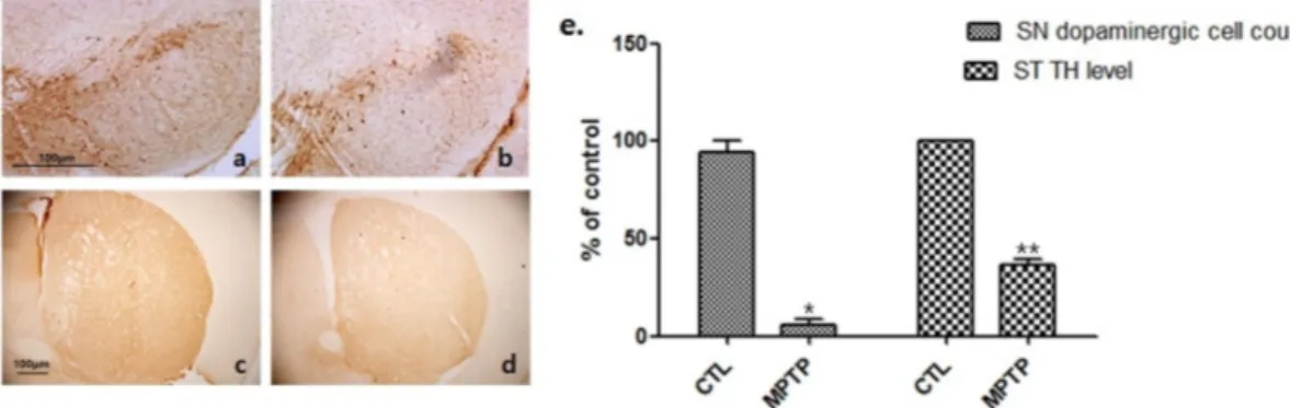
Fig. 1. Dopaminergic cell expression decrease in a MPTP-induced chronic parkinsonism mouse model.
The substantia nigra (SN) and striatum (ST) were immune-stained by an anti-tyrosine hydroxylase (TH; a, b, 100×; c, d, 40×) antibody.
Histogram (e) shows that the TH expression decreased in the MPTP groups; these were statistically significant in comparison with the MPTP group normalized to 100% of the control group. CTL : saline control (n=3), MPTP : MPTP treated (n=3). *p<0.05 and **p<0.01, compared to CTL. Analyzed by Student’s t-test.
하고, 각 섹션을 anti-TH 항체(1:2000; Santa Cruz Biotechnology, USA)로 4℃에서 밤새 염색하여 도파민성 뉴런을 확인했다. 그런 다음 절편을 비오틴화 된 a biotinylated anti-mouse IgG에 이어 avidin-biotin-peroxidase complex로 순차적으로 처리하고 dia- minobenzidine-hydrogen peroxide에 반응시켰다. 사진은 Nikon X-cite 시리즈 120 Q 현미경(Nikon, 일본)을 사용하여 촬영했다.
6. 면역 형광 염색
세포를 4℃에서 밤새 anti-SERT (1:2000)로 처리했다. 각 그룹 은 비오틴화 된 항 마우스 IgG로 처리 한 후 Rhodamine Avidin D (Vector Laboratories, CA)와 함께 배양했다. Nikon X-cite 시 리즈 120 Q 현미경(Nikon, 일본)을 사용하여 촬영했으며 노출 매 개 변수는 CTL 및 MPP+ 그룹에서 동일했다.
7. 웨스턴 블롯
양측 ST 및 SN 영역을 얼음에서 30분 동안 lysis buffer로 균질 화한 후 4℃에서 20분 동안 원심 분리(12,000 rpm)하여 상층액을 동일한 단백질 농도(총 단백질 30 μg)로 정량하였다. 샘플을 12%
sodium dodecyl sulfate-polyacrylamide gel 전기 영동(SDS- PAGE)을 사용하여 분리 한 다음 polyvinylidene difluoride (PVDF, Bio-Rad, USA) 막으로 이동시켰다. 단백질이 옮겨진 PVDF는 5%
탈지유를 포함하는 0.1% Tris- 완충 식염수(TBS; 20-mM Tris- HCl (pH 7.5) 및 0.1% Tween-20을 함유하는 150-mM NaCl, TBST)를 사용하여 실온에서 1시간 동안 처리하고, anti-TH (1:
2,000), anti-SERT (1:2000; Cloud-clone corp., USA), anti-bax (1:2000, abcam), anti-bcl2 (1:2000, abcam) 및 anti-β actin (1:5000; Santa Cruz Biotechnology, 미국) 항체와 함께 24시간
배양되었다. 그 후, 막을 0.1% TBST로 세척하고 anti-mouse IgG-peroxidase 항체(1:2,000, Bio-Rad)와 함께 배양 한 후 Pierce ECL western blotting substrate (Thermo Scientific, 미국)을 사 용하여 현상했다.
8. 통계 분석
통계분석은 Student’s t-test 또는 분산 분석(ANOVA)을 수행했 다. 모든 값은 평균±표준 오차(SE)로 표현되었으며 유의수준은 p<0.05로 지정했다.
결 과
1. 만성 MPTP 유발 파킨슨병 마우스 모델
만성 MPTP 유발 파킨슨병 마우스 모델의 확립을 확인하기 위 해 먼저 뇌의 SN의 도파민세포와 ST에서 TH의 발현 변화를 평가 했다. CTL 그룹(Fig. 1a, c)과 비교하여 MPTP 그룹(Fig. 1b, d)의 SN 영역에서 도파민세포 및 ST 영역에서 TH의 발현의 현저한 감 소가 관찰되었다. 이는 통계적으로 유의하게 나타났다(*p<0.05 와 **p<0.01, Fig. 1e).
2. MPTP 유발 만성 파킨슨증 마우스 모델에서 Slc6a4 발현의 변화
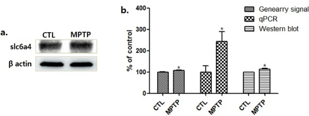
MPTP 그룹의 SN에서 Slc6a4 발현의 증가가 나타났다(Fig. 2a).
마이크로 어레이를 이용한 유전자 발현 분석에서 MPTP 그룹의 Slc6a4증가를 나타냈으며, 이를 정량하기 위한 qPCR과 면역 블롯 분석에서도 통계적으로 유의하게 증가하는 것으로 확인되었다
Fig. 2. Solute carrier family 6 member 4 (Slc6a4) expression caused by MPTP treatment increased in a MPTP induced chronic parkinsonism model.
(a) The results of the immunoblot assay are shown. (b) Histograms of the microarray, qPCR and the western-blot analyses represent that Slc6a4 expression significantly increased in the MPTP group, compared to the control normalized to 100%. CTL : saline control (n=3), MPTP : MPTP treated (n=3). *p<0.05, compared to CTL. Analyzed by Student’s t-test.
Fig. 3. Western blot analysis (a) demonstrating that the administration of a solute carrier family 6 member 4 (Slc6a4) short interfering RNA (siRNA) led to an increase in the tyrosine hydroxylase (TH) expression in SH-SY5Y cells. (b) Histograms of TH, bax, bcl2 and bax/bcl2 expressions are shown in comparison of the control (CTL) group, siRNA 10, and siRNA 100.
CTL and C, control siRNA treatment (100 nM for 2 days, n=3); 10, Slc6a4 siRNA treatment (10 nM for 2 days, n=3); 100, Slc6a4 siRNA treatment (100 nM for 2 days, n=3). *p<0.05 and **p<0.01, compared to CTL. Analyzed by Student’s t-test.
(*p<0.05, Fig. 2b).
3. siRNA를 사용하여 Slc6a를 감소시킨 SH-SY5Y 세포 에서 TH 발현 증가
Slc6a4와 도파민성 신경세포와의 관계를 조사하기 위해 Slc6a4 siRNA를 사용하여 Slc6a4의 유전자 발현을 감소시킨 SH-SY5Y 세포에서 실험을 수행했다. 웨스턴 면역 블롯 분석은 Slc6a4 유전 자의 발현을 감소시킨 SH-SY5Y 세포에서 TH 발현이 증가되는 결과를 보여주었다(*p<0.05, Fig. 3a). 이와 관련된 기전을 살펴 보기 위해 apoptosis와 연관된 bax와 bcl2를 살펴본 결과 bax/bcl2의 값이 감소하여 apoptosis 감소 경향을 나타내는 것으 로 확인되었다(*p<0.05 와 **p<0.01, Fig. 3b). 이 결과는 Slc6a4 이 도파민성 신경세포의 apoptosis를 조절하는 기전에 대한 증거 를 제공하고 있다.
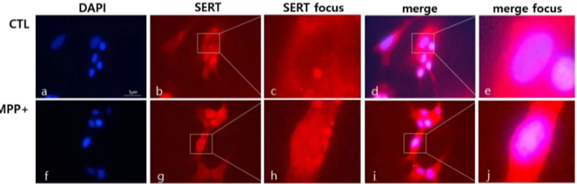
4. 면역형광염색으로 살펴본 MPP+를 처리한 SH-SY5Y 세포에서 Slc6a4의 발현 변화
SH-SY5Y 세포를 면역형광염색을 시행하여 MPP+에 의하여 Slc6a4의 발현이 변화되는 양상을 관찰했다. Slc6a4의 발현은 MPP+ 그룹에서 증가하였으며, 특징적으로 중심 부분에서의 발현 이 MPP+그룹에서 증가된 양상으로 나타났다(Fig. 4).
고 찰
기존의 파킨슨병 동물모델을 사용한 연구결과들에서 도파민세 포의 감소와 이에 따라 나타나는 변화를 TH의 감소를 통하여 확인 하였다13). 본 실험에서도 만성 MPTP 유발 파킨슨병 마우스 모델 을 확인하기 위해 뇌의 SN에서의 도파민세포의 발현과 ST에서 TH 의 발현 변화를 평가했다. CTL 그룹과 비교하여 MPTP 그룹의 SN
Fig. 4. Solute carrier family 6 member 4 (Slc6a4) expressions in SH-SY5Y cells treated with 1-methyl-4-phenylpyridinium (MPP+).
The cells were immunofluorescence labeled with dapi (a, f; blue) and anti-Slc6a4 antibodies by using Rhodamine Avidin (b, c, g, h;
red). Slc6a4 focus (c, h) are of magnified images of squares in Slc6a4 (b, g). The merge panels (d, i) show merged images of the left (a, f) and middle (b, g) panels, respectively. The merge focus panels (e, J) are of magnified images of squares in merge panels (d, i). Note the purple-colored regions indicating the co-localization of the Slc6a4 and nucleus. CTL, Control : treatment none, MPP+ : 500 μM MPP+ treatment.
부위에서 도파민세포의 발현이 현저히 감소한 것을 확인하였으며, ST부위에서 TH의 발현의 현저한 감소를 통하여 파킨슨병 모델이 잘 확립된 것을 확인하였다(Fig. 1). 이 모델의 SN 부위에서 마이크 로 어레이를 이용한 유전자 발현 분석을 수행한 결과 MPTP 그룹 에서 Slc6a4의 현저한 증가를 나타냈다. Slc6a4와 도파민성 신경 세포와의 관계를 조사하기 위해 Slc6a4 siRNA를 사용하여 Slc6a4 의 유전자 발현을 감소시킨 SH-SY5Y 세포에서 TH 발현 증가를 관찰하였다. 그리고 apoptosis와 연관된 bax와 bcl2를 살펴본 결 과, Slc6a4 siRNA에 의하여 bax와 bcl2 둘 다 증가하는 것으로 나타났는데, bax/bcl2의 값이 감소하여 apoptosis 감소 경향을 나 타내는 것을 확인했다(Fig. 3). 이러한 결과는 Slc6a4이 도파민성 신경세포의 apoptosis에 영향을 줄 수 있는 가능성에 대한 증거를 제공하고 있다. 특히 Slc6a4 감소가 TH의 증가를 나타낸 결과를 통하여 MPTP 파킨슨병 동물모델에서 나타난 Slc6a4의 증가는 파 킨슨병을 악화시킬 것으로 생각된다. MPP+로 처리한 SH-SY5Y 세포의 면역형광염색 결과에서는, 특징적으로 세포의 중심부분에 서 Slc6a4의 발현이 증가된 양상으로 나타났다(Fig. 4). 대조군의 경우에는 중심 부분이 다른 부분에 비하여 연하게 염색된 것에 반 하여 MPP+로 처리한 SH-SY5Y 세포에서는 중심 부분이 다른 세 포영역에 비하여 좀 더 진하게 염색되어 MPTP 파킨슨병 동물모델 에서 나타난 Slc6a4의 증가가 세포의 중심부분에서 나타나는 것으 로 생각되었다. 또한 병증화와 관련하여 이 부위의 Slc6a4의 증가 가 영향을 줄 것으로 예상되었다.
세로토닌 재흡수 수송체의 이상이 파킨슨병과 밀접한 관련성이 있다고 보고되고 있는데8,9), 특히 파킨슨병의 전구증상으로 나타나 는 불면증의 경우 세로토닌 재흡수 수송체와 관련되어 있는 것으 로 보고되었다14). PET을 사용하여 세로토닌 수송체 표지자를 관찰
한 연구에서 파킨슨병의 운동 증상 발현 전에 세로토닌 변화가 발 생할 수 있음을 시사한다고 보고하였다15). 이들의 연구를 통하여, 병의 초기에 세로토닌 재흡수 수송체에 변화가 생기면서 파킨슨병 의 병리학적인 변화를 일으킬 수 있을 것으로 생각된다.
파킨슨병 환자를 대상으로 진행한 연구에서 파킨슨병의 운동 증상 중의 하나인 진전과 Slc6a4 발현 이상이 관련되어 있다는 보 고를 통하여 도파민성 신경세포에 Slc6a4이 영향을 주어 증상이 나타날 수 있을 것으로 생각되었다10). 또한 Slc6a4에 의해 발현되 는 물질을 포함하는 세로토닌 시스템은 파킨슨병의 병태 생리학에 서 중요한 역할을 할 것으로 보고되고 있는데12), 특히 Slc6a4발현 변화가 도파민성세포에서의 apoptosis를 조절하는 것으로 나타난 결과도 이를 뒷받침하고 있다.
본 연구에서는 Microarray를 사용하여 실험하였으나 Slc6a4의 유전자 발현 변화에 대한 결과만을 제시하고 있다. 따라서 다른 변화 인자나 각 인자들 간의 pathway 분석에 관한 연구가 더 진행 되어야 할 것으로 생각된다.
결론적으로 본 실험에서는 파킨슨병 동물모델의 뇌 흑질에서 Slc6a4의 증가를 확인하였으며, 도파민성 신경 세포의 조절에 관 여하는 Slc6a4의 영향에 대해 살펴 보았다. 이를 통하여 아직 정확 한 원인을 알 수 없는 파킨슨병에서 도파민성 신경세포의 감소에 대한 원인을 찾기 위한 근거로써 사용할 수 있을 것으로 생각된다.
Acknowledgement
연구비 지원기관: 상지대학교
연구비 지원기관은 연구의 설계, 자료 수집과 분석, 출판결정,
원고 작성에 어떠한 영향도 없었음을 밝힌다.
Funding
This research was supported by Sangji University Research Fund, 2019.
Data availability
The authors can provide upon reasonable request.
Conflicts of interest
저자들은 아무런 이해 상충이 없음을 밝힌다.
References
1. Elkouzi A, Vedam-Mai V, Eisinger RS, Okun MS. Emerging therapies in Parkinson disease - repurposed drugs and new approaches. Nat Rev Neurol. 2019 ; 15(4) : 204-23. https://doi.
org/10.1038/s41582-019-0155-7
2. Tysnes OB, Storstein A. Epidemiology of Parkinson’s disease. J Neural Transm (Vienna). 2017 ; 124(8) : 901-5. https://doi.org/
10.1007/s00702-017-1686-y
3. Del Rey NL, Quiroga-Varela A, Garbayo E, Carballo-Carbajal I, Fernandez-Santiago R, Monje MHG, et al. Advances in Parkinson’s Disease: 200 Years Later. Front Neuroanat. 2018 ; 12 : 113.
https://doi.org/10.3389/fnana.2018.00113
4. Cacciatore I, Ciulla M, Marinelli L, Eusepi P, Di Stefano A.
Advances in prodrug design for Parkinson’s disease. Expert Opin Drug Discov. 2018 ; 13(4) : 295-305. https://doi.org/10.1080/
17460441.2018.1429400
5. Murphy DL, Fox MA, Timpano KR, Moya PR, Ren-Patterson R, Andrews AM, et al. How the serotonin story is being rewritten by new gene-based discoveries principally related to SLC6A4, the serotonin transporter gene, which functions to influence all cel-
lular serotonin systems. Neuropharmacology. 2008 ; 55(6) : 932- 60. https://doi.org/10.1016/j.neuropharm.2008.08.034 6. Montanez S, Daws LC, Gould GG, Frazer A. Serotonin (5-HT)
transporter (SERT) function after graded destruction of seroto- nergic neurons. J Neurochem. 2003 ; 87(4) : 861-7. https://doi.
org/10.1046/j.1471-4159.2003.02032.x
7. Martel F, Monteiro R, Lemos C. Uptake of serotonin at the apical and basolateral membranes of human intestinal epithelial (Caco-2) cells occurs through the neuronal serotonin transporter (SERT). J Pharmacol Exp Ther. 2003 ; 306(1) : 355-62.
https://doi.org/10.1124/jpet.103.049668
8. Koren G, Ornoy A. Clinical implications of selective serotonin reuptake inhibitors-selective serotonin norepinephrine reup- take inhibitors pharmacogenetics during pregnancy and lactation.
Pharmacogenomics. 2018 ; 19(14) : 1139-45. https://doi.org/10.
2217/pgs-2018-0076
9. Zahavi AY, Sabbagh MA, Washburn D, Mazurka R, Bagby RM, Strauss J, et al. Serotonin and Dopamine Gene Variation and Theory of Mind Decoding Accuracy in Major Depression: A Preliminary Investigation. PLoS One. 2016 ; 11(3) : e0150872.
https://doi.org/10.1371/journal.pone.0150872
10. Wang JY, Fan QY, He JH, Zhu SG, Huang CP, Zhang X, et al.
SLC6A4 Repeat and Single-Nucleotide Polymorphisms Are Associated With Depression and Rest Tremor in Parkinson’s Disease: An Exploratory Study. Front Neurol. 2019 ; 10 : 333.
https://doi.org/10.3389/fneur.2019.00333
11. Albani D, Vittori A, Batelli S, Polito L, De Mauro S, Galimberti D, et al.
Serotonin transporter gene polymorphic element 5-HTTLPR in- creases the risk of sporadic Parkinson’s disease in Italy. Eur Neurol.
2009 ; 62(2) : 120-3. https://doi.org/10.1159/000222784 12. Mossner R, Schmitt A, Syagailo Y, Gerlach M, Riederer P, Lesch
KP. The serotonin transporter in Alzheimer’s and Parkinson’s disease. J Neural Transm Suppl. 2000 ; (60) : 345-50.
13. Choi YG, Lim S. N(varepsilon)-(carboxymethyl)lysine linkage to alpha-synuclein and involvement of advanced glycation end products in alpha-synuclein deposits in an MPTP-intoxicated mouse model. Biochimie. 2010 ; 92(10) : 1379-86. https://doi.
org/10.1016/j.biochi.2010.06.025
14. Paus S, Seeger G, Brecht HM, Koster J, El-Faddagh M, Nothen MM, et al. Association study of dopamine D2, D3, D4 receptor
and serotonin transporter gene polymorphisms with sleep at- tacks in Parkinson’s disease. Mov Disord. 2004 ; 19(6) : 705-7.
https://doi.org/10.1002/mds.20134
15. Wile DJ, Agarwal PA, Schulzer M, Mak E, Dinelle K, Shahinfard
E, et al. Serotonin and dopamine transporter PET changes in the premotor phase of LRRK2 parkinsonism: cross-sectional studies. Lancet Neurol. 2017 ; 16(5) : 351-9. https://doi.org/
10.1016/s1474-4422(17)30056-x