Distribution of HCV Genotypes in Chronic Korean HCV Patients
Kyung-Ok Lee, Su-Jin Jeong, Ji-Young Byun, Ae-Sug Shim, Hye-Soon Seong, and Kyung-Tae Kim
Genome Research Center, Neodin Medical Institute, Seoul 133-847, Korea
HCV is a single-stranded RNA virus and more than 1 million new cases are reported annually worldwide.
The six major HCV genotypes and numerous subtypes vary in their geographic distribution. It is thought that genetic heterogeneity of HCV may account for some of the differences in disease outcome and response to treatment observed in HCV infected persons. In this study, we determined HCV genotypes among chronic Korean HCV patients and evaluated direct sequence PCR protocols developed. For the study, 232 chronic HCV patient sera were used. HCV RNA was extracted and two pairs of consensus PCR primers were selected in 5’UTR region for amplification of HCV RNA. Amplification products obtained from the HCV positive cases were subjected to automatic sequencing. Sequences were compared with those in GenBank by using the BLAST program. From this study, five HCV genotypes, 1b, 2a, 2b, 2c and 3a were found. HCV genotypes 4, 5 and 6 were not determined. HCV genotype 1b (53.9%, 125/232) and 2a (35.8%, 83/232) were most frequently found. This group was followed by 2b (3.9%, 9/232), 3a (3.4%, 8/232) and 2c (3.0%, 7/232).
The data presented here suggest a complex distribution of HCV types and they were well correlated with other reports on Koreans and will be helpful for type-specific follow-up of Korean HCV patients. This study showed that 5’UTR direct sequence analysis is a sensitive and rapid method to identify HCV genotypes.
Key words : HCV genotyping, PCR-direct sequencing
I. INTRODUCTION
1)The Hepatitis C virus (HCV) is chronically infecting at least 1% of the world's population and is believed to be more prevalent than the hepatitis B virus infection (Cooreman et al, 1996). The HCV genome is a positive-sense single- stranded RNA genome approximately 10 kb long. It has marked similarities to those of members of the genera Pestivirus and Flavivirus (Zein et al, 1996). After the complete HCV genome was
Corresponding Author : Kyung-Ok Lee
Genome Research Center, Neodin Medical Institute 2-3 Yongdap-Dong, Sungdong-Gu, Seoul 133-847, Korea Tel : 82-2-2244-6500 Fax : 82-2-2214-5809
E-mail : [email protected]
determined by Choo et al (1991), several HCV isolates from different parts of the world were obtained and sequenced (Delisse et al, 1991; Kato et al, 1991; Li et al, 1991; Chen et al, 1992). Different HCV isolates from around the world show substantial nucleotide sequence variability throughout the viral genome (Choo et al, 1991).
It is important to consider the role of HCV genotypes in liver disease progression or response to interferon therapy and clinical utility of genotyping. Sequence variability is distributed equally throughout the viral genome, apart from the highly conserved 5'UTR and core regions and the hypervariable envelope (E) region (Kata et al, 1990).
HCV is classified on the basis of the similarity of nucleotide sequence into major genetic groups designated
-341 0 915 1491 2769 3420 5477 6258 7601 9375
Structural Nonstructural
5'UTR
C E1 E2 NS2 NS3 NS4 NS5
3'UTR
Core Envelope
Glycoproteins Metalloprotease Protease
& Helicase
RNA Polymerase
C : Core, E : Envelope, NS : Nonstructural, UTR : untranslated
Fig. 1. Genomic organization of HCV. HCV specific primer pairs were selected in 5'UTR region of HCV genome.
genotypes. Based on the identification of these genomic differences, HCV has been classified into multiple strains.
HCV displays a remarkable degree of genomic diversity, with the six major genotypes and twenty four subtypes differing in geographic distribution. It is thought that genetic heterogeneity of HCV may account for some of the differences in disease outcome and response to treatment observed in HCV-infected persons. HCV genotyping can be particularly useful in studying worldwide and local evolutions of the HCV endemics, since the epidemiology of HCV is changing rapidly.
Several studies have reported that that HCV genotype 1b helps the rapid progression of cirrhosis, and hepatocellular carcinoma. It also shows resistance to interferon treatment, and poor prognosis following liver transplantation (Zein et al, 1996). In the present study, we examined the distribution of HCV genotypes in Korean HCV patients and evaluated the efficiency of homebrew PCR-direct sequencing PCR protocols based on 5’UTR analysis.
II. MATERIALS AND METHODS
Materials
In this study, 232 Korean HCV patient samples that were referred to a reference laboratory for HCV genotyping were analyzed. For the evaluation of HCV genotyping method developed by this study, reference panels of HCV genotypes (Acrometrix Co., USA) and quality control materials from CAP (College of American Pathologist, USA) were used for this study.
Methods
HCV-RNA was extracted using Viral Gene-spinTM Viral DNA/RNA Extraction kit (Intron Co., Korea). For the RT-PCR of HCV RNA, PCR was performed with HCV specific primers described by Okamoto et al (1991) (Fig.
1). For the reverse transcription of HCV RNA and cDNA amplification was performed using of 11 μL of HCV RNA, 0.5 μL of each primer (10 μM), and 8 μL of One step RT Mixture (Intron Co., Korea) in a final volume of 20 μL. The amplification conditions included a reverse transcription for 30 min at 45℃ and an initial denaturation for 5 min at 94℃, 30 cycles of amplification with denaturation at 94℃ for 1 min, annealing at 50℃ for 1 min, extension at 72℃ for 45 sec, followed by final extension at 72℃ for 10 min. For the second round PCR, amplifications were performed using of 1.5 μL of
first-round PCR product, 0.5 μM of primer set, 2 mM of MgCl2, 25 mM of KCl, 30 mM of Tris-HCl (pH 9.0), 0.5 mM of dNTPs, and 1 Unit of Taq DNA polymerase (Intron Co., Korea) in a final volume of 20 μL. The amplification conditions included an initial denaturation for 5 min at 94℃, 30 cycles of amplification with denaturation at 94℃ for 30 sec, annealing at 50℃ for 30 sec, extension at 72℃ for 30 sec, followed by final extension at 72℃ for 7 min. The amplified product (215bps) of HCV RNA was detected by 2% agarose gel electrophoresis. PCR products were neutralized with HCl and purified using columns of Qiaquick spin PCR purification kit (Qiagen, USA) to eliminate excess deoxyribonucleotides and amplification primers before dye terminator cycle sequencing. Sequencing was accomplished in both directions by use of the ABI PRISM BigDye Terminator Cycle Sequencing Kit and an ABI 377 DNA Automatic Sequencer (Perkin Elmer, USA). Sequences were compared with those of GenBank by using the BLAST program.
III. RESULT
In this study, the HCV genotyping method by PCR-direct sequencing was developed in 5'UTR region. It was evaluated with materials of reference HCV panels (Acrometrix USA) and quality control survey materials of CAP (College of American Pathologist, USA). The results perfectly matched the ones described in the kit insert and survey reports (data now shown). The PCR-direct sequencing procedure developed was rapid and accurate for discrimination of HCV genotypes. Moreover the sequencing results were clear to analyze the HCV genotype. When HCV genotyping was performed using PCR-direct sequencing method developed by this study among 232 chronic Korean HCV patients, five HCV genotypes, including 1b, 2a, 2b, 2c and 3a were determined. HCV genotype 1a, 3b, 4, 5 and 6 were not found in this study.
HCV subtype 1b was dominant (53.9%, 125/232) in chronic Korean HCV patients. Eighty three individuals (35.8, 83/232) were infected by the subtype 2a. HCV subtypes 1b and 2a were responsible for more than 90%
of infections in chronic Korean HCV patients. This group was followed by 2b (3.9%, 9/232), 3a (3.4%, 8/232) and 2c (3.0%, 7/232) in descending order (Table 1).
HCV genotype Number %
1a 0
1b 125 53.9
2a 83 35.8
2b 9 3.9
2c 7 3.0
3a 8 3.4
4 0
5 0
6 0
Total 232
Table 1. Genotype frequencies of HCV in chronic Korean HCV patients
IV. DISCUSSION
Chronically infected individuals with HCV are at increased risk of developing liver cirrhosis and hepatocellular carcinoma. HCV genotype has been identified by some investigators as an important factor in the progression of liver disease. Amoroso et al (1998) specifically investigated the role of HCV genotypes in persistence of HCV infection following an acute exposure. HCV genotype may potentially play an important role in the development of chronic infection following acute exposure to HCV and is one of the factors that influences response to therapy. The rate of evolution to chronicity after acute exposure to HCV was 92% in patients exposed to HCV genotype 1b infection. Moreover, infection with genotype 1b has been associated with more advanced liver disease, cirrhosis, and hepatocellular carcinoma than infection with other
genotypeHCV This study
Korea Lee et al
12)*
Korea Park et al
17)*
Korea Kim et al
29)*
Japan Takada et al
25)*
Bangladesh Ali et al.
2)*
Zein et alUSA 28)*
France Tamalet et al
27)*
Brazil Pradal et al
20)*
1a 1b 2a 2b 2c 3a 4 5 6
53.9%
35.8%
3.9%
3.0%
3.4%
57.7%
32.1%
3.0%
47.3%
42.6%
2.4%
0.6%
39.9%
38.2%
1.1%
77.7%
16.5%
5.0%
10%
16%
20%
30%
58%
21%
4%
13%
5%
1%
33%
26%
7%
22%
10.7%
27%
38%
2%
33%
* means reference number quoted in this study.
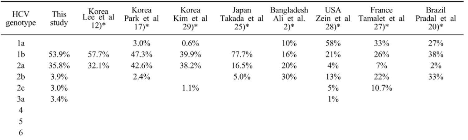
Table 2. HCV genotype distributions in different ethnic groups genotypes (Takahashi et al, 1993; Pistello et al, 1994;
Nousbaum et al, 1995; Pozzato et al, 1995).
At least six major genotypes of HCV, each comprising multiple subtypes, have been identified worldwide. HCV genotypes 1, 2 and 3 are responsible for more than 90%
of infections in America (Zein et al, 1996), Europe (Nousbaum et al, 1995) and Korea (Lee et al, 1998; Park et al, 1998). In Japan, subtype 1b caused more than 70%
of HCV infections (Takada et al, 1993). HCV genotype 4 appeared to be prevalent in North Africa and the Middle East (Chamberlain et al, 1997; Abdulkarim et al, 1998), and genotypes 5 and 6 seemed to be confined to South Africa and HongKong, respectively (Zein et al, 1996).
The present data showed that HCV genotypes 1b and 2a were predominant in chronic Korean HCV patients. It was well correlated with previous reports on Korean patients (Lee et al, 1998; Park et al, 1998; Kim et al, 2002). Park et al (1998) and Kim et al (2002) reported that HCV subtype 1a was found in 3% and 0.6%, respectively.
However it was not found in this study. Frequency of HCV 1b (39.9%) by Kim et al (2002) was lower than that of other Korean studies including this study (Table 2). It would be due to two factors, one is the high rate of unclassified cases (20.2%) depending on their analytical method and another is the study subjects. Their study samples were from anti-HCV ELISA-positive cases who
had undergone a periodic health examination. However subjects of most Korean studies (Lee et al, 1998; Park et al, 1998; this study) used were samples of chronic HCV patients. Therefore some differences of HCV genotype frequencies could be occurred between studies. When HCV genotype distributions of chronic Korean HCV patients was compared with other ethnic groups, it was similar with Japan, but different from those of Caucasian.
HCV subtype 3a was found more in than 30% of the infections in Bangladesh and Brazil, however it was very rare in Korea, Japan and the United States (Table 2). The geographic distribution and diversity of HCV genotypes may provide clues about the historical origin of HCV (Smith et al, 1997).
Because disease outcome and response to therapy among HCV genotypes have been suggested, reliable methods for determining the HCV genotype may become important clinical tests (Frederic et al, 2001). Several molecular methods, including direct sequencing, PCR-SSP (sequence specific primers)(Okamoto et al, 1991), PCR-SSOP (sequence specific oligonucleotide probes) (Li et al, 1991; Qu et al, 1994) or Restriction Fragment Length Polymorphism (RFLP) (McOmish et al, 1994;
Park et al, 1998) have been used for determination of HCV genotype. HCV typing using PCR-SSP was first introduced by Okamoto et al (1991) and a commercial kit
for HCV genotyping using PCR-SSOP has been introduced by Innogenetics and is based on hybridization of 5'UTR amplification products with genotype specific probes (Stuyver et al, 1993). Although all these methods are able to identify correctly the major genotypic groups, the direct nucleotide sequencing method which is sequencing of a specific PCR-amplified portion of the HCV genome, is the reference standard and the most definitive method (Abdulkarim et al, 1998). It is efficient in discriminating among HCV subtypes and remains the gold standard (Simmonds et al, 1995). However sequencing of amplified DNA does not usually identify mixed infections with two different HCV genotypes.
Multiple genotypes of HCV have been isolated throughout the world. It is clear that HCV genotypes are important epidemiologic markers and may alter the sensitivity and specificity of diagnostic assays for the detection of HCV.
Although not efficient by itself, HCV genotyping in combination with other markers, such as quantitative evaluation of HCV RNA, may be beneficial in the routine pretreatment evaluation of chronic HCV infection and in the selection of candidates for interferon treatment. In conclusion, the PCR-direct sequencing method developed by this study, could be useful for clinical laboratories to identify the HCV genotypes. Moreover this data could be helpful for epidemiologic studies of HCV in Koreans.
REFERENCES
1. Abdulkarim AS, Zein NN, Germer JJ, Kolbert CP, Kabbani L, Krajnik KL, Hola A, Agha MN, Tourogman M, Persing DH. Hepatitis C virus genotypes and hepatitis G virus in hemodialysis patients from Syria: identification of two novel hepatitis C virus subtypes. Am J Trop Med Hyg 59:571-576, 1998.
2. Ali MS, Chaudhury HS, Ahmed M, Rahman MM, Martnatunnessa M, Kabir M, Ali ME, Huda Q.
Hepatitic C virus genotype among HCV seropositive patients in Bangladesh. AACC abstract 2006.
3. Amoroso P, Rapicetta M, Tosti ME, Mele A, Spada E, Buonocore S, Chionne P, Ciccaglione AR, Sagliocca L. Correlation between virus genotype and chronicity rate in acute hepatitis C. J Hepatol 38:939-944, 1998.
4. Chamberlain RW, Adams N, Saed AA, Simmonds P, Elliott RM. Complete nucleotide sequence of type 4 hepatitis C virus variant, the predominant genotype in the Middle East. J Gen Virol 78:1341-1347, 1997.
5. Chen PJ, Lin MH, Tae KF, Liu PC, Lin CJ, Chen DS.
The Taiwanese hepatitis C virus genome: sequence determination and mapping the 5’ termini of viral genomic and antigenomic RNA. Virology 188:102-113, 1992.
6. Choo QL, Richman KH, Han JH, Berger K, Lee C, Dong C, Gallegos C, Coit D, Medina-Selby R, Barr PJ, Weiner AJ, Bradley DW, Kuo G, Houghton M.
Genetic organization and diversity of the hepatitis C virus. Proc Natl Acad Sci USA 88:2451-2455, 1991.
7. Cooreman MP and Schoondermark-Van de VE.
Hepatitis C virus: biological and clinical consequences of genetic heterogeneity. Scand J Gastroenterol Suppl 218:106-115, 1996.
8. Delisse AM, Descurieux M, Rutgers T, D’Hondt E, De WM, Arima T, Barrera-Sala JM, Ercilla MG, Ruelle JL, Cabezon T. Sequence analysis of the putative structural genes of hepatitis C virus from Japanese and European origin. J Hepatol 13:20-23, 1991.
9. Frederic SN. Hepatitis C virus genotyping: clinical implications and methods. Mol diag 6:265-277, 2001.
10. Kata N, Higikata M, Oosuyama Y, Nakagawa M, Ohkoshi S, Sugimura T, Shimotohno K. Molecular cloning of the human hepatitis C virus genome from Japanese patients with non-A, non-B hepatitis. Proc Natl Acad Sci USA 87:9524-9528, 1990.
11. Kato N, Ootsuyama Y, Ohkoshi S, Nakazawa T, Mori S, Hujikata M, Shimotohno K. Distribution of pleural HCV types in Japan. Biochem Biophys Res Commun
181:279-285, 1991.
12. Lee YS, Chung YH, Min YI, Moon DH, Na DS, Suh DJ. Hepatitis C virus genotyping of 100 consecutive anti-HCV positive case with PCR using type-specific primers. J Kor Liver Dis 4:235-243, 1998.
13. Li JS, Tong SP, Vitvitski L, Lepot D, Trepo C. Two French genotypes of hepatitis C virus: homology of the predominant genotype with the prototype American strain. Gene 105:167-172, 1991.
14. McOmish F, Yap PL, Dow BC, Follett EA, Seed C, Keller AJ, Cobain TJ, Krusius T, Kolho E, Naukkarinen R, Lin C, Lai C, Leong S, Medgyesi GA, Hejjas M, Kiyokawa H, Fukada K, Cuypers T, Saeed AA, Al-Rasheed AM, Lin M, Simmonds P.
Geographical distribution of hepatitis C virus genotypes in blood donors: an international coll- aborative survey. J Clin Microbiol 32:884-892, 1994.
15. Nousbaum JB, Pol S, Nalpas B, Landais P, Berthelot P, Brechot C. The collaborative study group. Hepatitis C virus type 1b (II) infection in France and Italy. Ann Intern Med 122:161-168, 1995.
16. Okamoto H, Okada S, Sugiyama Y, Kurai K, Akahane Y, Sugai Y, Tanaka T, Sato K, Tsuda F, Miyakawa Y, Mayumi M. Nucleotide sequence of the genomic RNA of hepatitis C virus isolated from a human carrier:
comparison with reported isolates for conserved and divergent regions. J Gen Virol 72:2697- 2704, 1991.
17. Park YS, Lee KO, Oh MJ, Cha YG. Distribution of genotypes in the 5'translated region of hepatitis C virus in Korea. J Med Micobiol 47:643-647, 1998.
18. Pistello M, Maggi F, Vatteroni L, Cecconi N, Panicucci F, Bresci GP, Gambardella L, Taddei M, Bionda A, Tuoni M, Bendinelli M. Prevalence of hepatitis C virus genotypes in Italy. J Clin Microbiol 32:232-234, 1994.
19. Pozzato G, Moretti M, Croce LS, Sasso F, Kaneko S, Unoura M, Kobayashi K, Crovatto M, Santini G, Tiribelli C. Interferon therapy in chronic hepatitis C virus: evidence of different outcome with respect to
different viral strains. J Med Virol 45:445-450, 1995.
20. Pradal MG, Carloni VN, Silbiger RD. Genotyping diversity of hepatitis C virus in Brazilian patients.
AACC abstract 2006.
21. Qu D, Li JS, Vitvitski L, Mechai S, Berby F, Tong SP, Bailly F, Wang QS, Martin JL, Trepo C. Hepatitis C virus genotypes in France: Comparison of clinical features of patients infected with HCV type I and type II. J Hepatol 21:70-75, 1994.
22. Smith DB and Simmonds P. Molecular epidemiology of hepatitis C virus. J Gastroenterol Hepatol 12:522- 527, 1997.
23. Simmonds P. Variability of hepatitis C virus.
Hepatology 21:570-583, 1995.
24. Stuyver L, Rossau R, Wyseur A, Duhamel M, Vanderborght B, Maertens G. Typing of hepatitis C virus isolates and characterization of new subtypes using a line probe assay. J Gen Virol 74:1093-1102, 1993.
25. Takada N, Takase S, Takada A, Date T. Differences in the hepatitis C virus genotypes in different countries. J Hepatol 17:277-283, 1993.
26. Takahashi M, Yamada G, Miyamoto R, Doi T, Endo H, Tsuji T. Natural course of chronic hepatitis C. Am J Gastroenterol 88:240-243, 1993.
27. Tamalet C, Colson P, Tissot-Dupont H, Henry M, Tourres C, Tivoli N, Botta D, Ravaux I, Pozot-Martin I, Yahi N. Genomic and phylogenetic analysis of hepatitis C virus isolates: a survey of 535 strains circula- ting in southern France. J Med Virol 71:391-398, 2003.
28. Zein NN, Rakela J, Krawitt EL, Reddy KR, Tominaga T, Persing DH. The collaborative study group.
Hepatitis C virus genotypes in the United States:
epidemiology, pathogenicity, and response to interferon therapy. Ann Intern Med 125:634-639, 1996.
29. Kim YS, Ahn YO, Lee HS. Risk factors for Hepatitis C virus infection among Koreans according to the Hepatitis C virus genotype. J Korean Med Sci 17:187-92, 2002.
국문 초록
HCV는 single stranded RNA 바이러스로서 감염 시에는 만성간염 및 간경화 간암으로 진행될 수 있는 가능성이 높다. HCV는 6종의 주된 genotype과 그에 따른 많은 종류의 subtype이 보고되고 있으며, 세계 각 지역별로 그 분포는 매우 다양하다. 여러 가지 HCV genotype 중에서 1b 형에 감염되었을 경우 간경화나 간암으로 진행할 가능성이 높으며 치료효과도 떨어진다는 보고가 있어, 최근 HCV 환자의 치료에 있어서 HCV 바이러스 정량검사와 함께 HCV genotyping 검사의 임상적 활용이 높아지고 있다. 본 연구에서는 PCR-direct sequencing을 이용한 HCV genotyping 검사방법을 이용하여, 한국인 만성 HCV 간염환자에서 HCV genotype의 분포를 조사하였다. 검체로는 232명의 한국인 만성간염환자의 혈청을 사용하였으며, HCV 5'UTR 영역에서 선택한 2쌍의 primer로 nested PCR을 실시하였다. 증폭된 PCR산물 (215 bps)은 2% agrose gel로 전기영동을 하고 sequencing을 실시한 후 GeneBank의 BLAST 프로그램을 사용하여 HCV genotype을 분석하였다. HCV genotyping을 실시한 232명에서 5종류의 genotype, HCV 1b, 2a, 2b, 2c, 3a, 이 발견되었 으며, HCV genotype 4, 5, 6 은 검출되지 않았다. 발견된 HCV genotype 중에서 HCV 1b의 검출률이 53.9%
로 가장 높았고, 다음은 HCV 2a가 35.8%로 높게 나타나, 위 두 가지 HCV genotype을 합하면 거의 90%였 다. 다음으로 HCV genotype 2b가 3.9%, 3a가 3.4% 그리고 2c가 3.0%의 순서로 검출되었다. 본 결과는 한국 인 만성 HCV간염 환자의 치료 및 예후관리에 참고가 될 것으로 사료된다. 또한 PCR-direct sequencing을 이용한 HCV genotyping 검사는 간편하고 분명하게 결과를 판독할 수 있어 임상실험실에서 유용하게 사용 될 수 있을 것으로 판단된다.