Received on September 11, 2013. Revised on September 23, 2013. Accepted on September 27, 2013.
CC This is an open access article distributed under the terms of the Creative Commons Attribution Non-Commercial License (http://creativecommons.org/licenses/by-nc/3.0) which permits unrestricted non-commercial use, distribu- tion, and reproduction in any medium, provided the original work is properly cited.
*Corresponding Author. Inhak Choi, Department of Microbiology and Immunology, Advanced Research Center for Multiple Myeloma, Inje University College of Medicine, 75, Bokji-ro, Busanjin-gu, Busan, Korea. Tel: 82-51-890-6724;
Fax: 82-51-891-6004; E-mail: [email protected]
Keywords: Co-signaling molecule, B7 family, Co-stimulation, Co-inhibition
Abbreviations: MHC, major histocompatibility; CTLA-4, cytotoxic T lymphocyte antigen-4; PD-1, programmed death-1;
ICOS, inducible T-cell costimulator; BTLA, B- and T-lymphocyte attenuator; IL-2, interleukin-2; IFN-γ, interferon-γ;
GM-CSF, granulocycte-monocyte colony-stimulating factor; LPS, lipopolysaccharide; VSITA, V-domain Ig suppressor of T cell activation; BMDC, bone marrow-derived dendritic cell; OVA, ovalbumin
Emerging Co-signaling Networks in T Cell Immune Regulation
Keunok Jung and Inhak Choi*
Department of Microbiology and Immunology, Advanced Cancer Research of Multiple Myeloma, Inje University College of Medicine, Busan 614-735, Korea
Co-signaling molecules are surface glycoproteins that pos- itively or negatively regulate the T cell response to antigen.
Co-signaling ligands and receptors crosstalk between the surfaces of antigen-presenting cells (APCs) and T cells, and modulate the ultimate magnitude and quality of T cell re- ceptor (TCR) signaling. In the past 10 years, the field of co-signaling research has been advanced by the under- standing of underlying mechanisms of the immune modu- lation led by newly identified co-signaling molecules and the successful preclinical and clinical trials targeting co-inhibitory molecules called immune checkpoints in the treatment of au- toimmune diseases and cancers. In this review, we briefly de- scribe the characteristics of well-known B7 co-signaling fam- ily members regarding the expression, functions and ther- apeutic implications and to introduce newly identified B7 members such as B7-H5, B7-H6, and B7-H7.
[Immune Network 2013;13(5):184-193]
INTRODUCTION
The most essential role of the immune system in the human body is to kill invading pathogens by inducing a protective immunity and not to harm the host by inducing tolerance to self-tissues. This is achieved through a fine tuning of T cell activities in initiation, differentiation and effector phase of the immune response (1). T cell response is initiated by specific
recognition of cognate peptide presented by MHC proteins on antigen-presenting cell (APC) through T cell receptors (TCRs), which are referred to as a first signal for T cell activation. However, the ultimate magnitude and quality of T cell response is determined by a balance between co-stim- ulatory and co-inhibitory signals (collectively called co-sig- nals) that are transduced into T cells, which is referred to as a second signal (2,3). Following TCR engagement by cognate peptide-MHC complexes, co-signaling receptors are often mo- bilized and colocalized with TCRs, forming the immunological synapse between APCs and T cells. This synaptic interface is the place where the crosstalk between co-signaling ligands and receptors synergize or antagonize with TCR signaling, rendering T cells activated or inhibited (4).
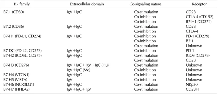
The B7 family is a group of surface glycoproteins that share structural features with immunoglobulin (Ig), whose ex- tracellular domains bear homology to IgV and IgC domains of Ig (Table I). A hallmark of the B7 family molecules is their capability to co-stimulate or co-inhibit T cell responses in the presence of peptide/MHC complex-mediated TCR signaling (4). B7 family members primarily bind to the members of CD28 family including CD28, CTLA-4, PD-1, ICOS, and BTLA that transmit co-signals into T cells (Fig. 1). The B7 co-stim- ulatory ligands are important for full activation of naïve T cells in the lymphoid organs, in which APCs, particularly den- dritic cells (DCs), are the primary cellular source providing
Figure 1. Schematic diagram of B7 co-signaling family network.
Table I. Structure, co-signaling nature, and receptors of B7 family members
B7 family Extracellular domain Co-signaling nature Receptor
B7.1 (CD80) IgV+IgC Co-stimulation CD28
Co-inhibition CTLA-4 (CD152)
Co-inhibition B7-H1 (CD274)
B7.2 (CD86) IgV+IgC Co-stimulation CD28
Co-inhibition CTLA-4
B7-H1 (PD-L1, CD274) IgV+IgC Co-inhibition PD-1 (CD279)
Co-inhibition B7.1
Co-stimulation Unknown
B7-DC (PD-L2, CD273) IgV+IgC Co-inhibition PD-1
B7-H2 (ICOSL, CD275) IgV+IgC Co-stimulation ICOS (CD278)
Co-stimulation CD28
B7-H3 (CD276) IgV+IgC+IgV+IgC (Hu) Co-stimulation Unknown
IgV+IgC (Mo) Co-inhibition Unknown
B7-H4 (VTCN1) IgV+IgC Co-inhibition Unknown
B7-H5 (VISTA) IgV Co-inhibition Unknown
B7-H6 (NCR3LG1) IgV+IgC Co-stimulation NKp30
B7-H7 (HHLA2) IgV+IgC+IgV Co-stimulation CD28H
the ligands including B7.1 and B7.2. In contrast, B7 co-in- hibitory ligands are crucial for the termination of over- activated T cell response, maintenance of self-tolerance, and protection of tissues from damage induced by invading pathogens. In addition, co-inhibitory ligands expressed on
pathological microenvironment such as tumor microenviron- ment actively inhibit the effector functions of cytotoxic T lym- phocytes (CTLs) or induce the generation of regulatory T cells (2,3), thus, playing as important immune checkpoint proteins that are involved in the immune resistance of cancer cells.
In this review, we briefly describe the characteristics of well-known B7 family members regarding the expression, functions and therapeutic implications. We also introduce newly identified B7 members such as B7-H5, B7-H6, and B7-H7.
B7.1 AND B7.2 (CD80 AND CD86)
The B7.1 and B7.2, best-characterized co-signaling ligands, are expressed on APCs including DCs, macrophages, and B cells. The B7.1 and B7.2 deliver a co-stimulatory signal through CD28 constitutively expressed on naïve T cells, whose cytoplasmic tail contains three signaling motif. The first motif containing YMNM sequence serves as a binding site for the SH2-containg proteins, p85, growth factor receptor 2 (Grb2) and GADS. The second motif contains PRRP sequence and interacts with the SH3 domain of IL-2-inducible T cell kinase (Itk) of which involvement in CD28-mediated activa- tion is still controversial. The third motif containing PYAP se- quence associates with the SH3 domain of Grb2, GADS, lym- phocyte cell-specific protein-tyrosine kinase (Lck), and fila- min-A. Particularly, the association of Grb2 to both the YMNM and PYAP motifs is critically involved in the recruit- ment of protein kinase Cθ (PKCθ) and RAS guanyl nucleo- tide-releasing protein (RASGRP) to the immunological syn- apse and its subsequent activation (5-7). Therefore, B7.1/
B7.2-CD28 pathway lowers the threshold for optimal T cell activation, subsequently resulting in T cell proliferation, upre- gulating anti-apoptotic proteins including Bcl-xL, and increas- ing IL-2 production (8-10). Recently, the B7-CD28 pathway was also shown to play an essential role in both suppressive function and survival of regulatory T (Treg) cells (11).
In addition, B7.1 and B7.2 deliver a co-inhibitory signal in- to activated T cells through CTLA-4 whose cytoplasmic tail contains the immunoreceptor tyrosine inhibitory motif (ITIM) recruiting Src homology 2 domain-containing phosphatase (SHP)-1 and SHP-2 (12,13). Due to its high affinity to B7.1/
B7.2, CTLA-4 competes out CD28 for binding to B7.1 and B7.2, conferring signaling-independent T cell suppression by sequestering B7.1/B7.2 from the APC surface (14). In contrast to the CD28 pathway, the B7-CTLA-4 pathway sets the thresh- old high enough so that T cell activation can be reduced and finally terminated. Since CTLA-4 is constitutively expressed on Treg cells, CTLA-4 engagement by B7 molecules also enhan- ces their immunosuppressive activity with upregulation of FOXP3 gene transcription (15,16). A great deal of mouse tu-
mor models revealed that blockade of the B7-CTLA-4 path- way could be a potential therapeutic target for cancer immun- otherapy. Specifically, ipilimumab, a CTLA-4 blocking anti- body, was the first FDA-approved antibody therapeutic for melanoma, adopting the notion that immune checkpoint block- ade might enhance endogenous anti-tumor immunity (3).
Interestingly, B7.1 and B7.2 are believed to deliver bidirec- tional signalings. Specifically, B7 molecules expressed on T cells act as a counter receptor that receives an inhibitory sig- nal from Treg cells probably through CTLA-4, an important in vivo T-T interaction for T cell homeostasis (17). Another reverse signaling through B7.1 and B7.2 was reported in T cell-APC interaction (18-20). B7 molecules expressed on DCs can transmit a suppressive signal into DCs through CTLA-4 on Treg cells or CTLA-4.Ig by enhancing IFN-γ production of DCs, which, in turn, induces indoleamine 2,3-dioxygenase (IDO), an enzyme metabolizing tryptophan into kynurenine, an immunosuppressive metabolite, in autocrine or paracrine mode (18). Thus, the bidirectional B7-CTLA-4 pathway ap- pears to be critical for the downregulation of T cell response and induction of T cell tolerance.
B7-H1 (CD274, PD-L1) AND B7-DC (CD273, PD-L2)
B7-H1 is constitutively expressed not only on APCs but also in a wide range of normal somatic tissues including the heart, lung, placenta and liver (21,22). In contrast, B7-DC ex- pression is largely restricted to APCs such as DCs and macrophages. Both molecules share the PD-1 receptor that is expressed on T cells, Treg cells, B cells, activated mono- cytes, DCs, NK cells, and NKT cells (23,24). The cytoplasmic tail of the PD-1 receptor contains ITIM and the immunor- eceptor switch motif (ITSM) that binds to SHP-1 and SHP-2, transmitting a co-inhibitory signal into T cells and down- regulating Bcl-xL expression, which leads to T cell functional impairment and apoptosis (21).
Due to its ectopic expression on non-hematopoietic tissues including normal peripheral organs and cancer tissues, B7-H1 is believed to limit T cell activation in peripheral organs, lead- ing to peripheral tolerance. Thus, the B7-H1-PD-1 pathway in tumor microenvironment can be a critical immune escape mechanism (25-27), indicating that B7-H1 and PD-1 can act as key immune checkpoint proteins. Many clinical observat- ions further support the immune checkpoint activities of the B7-H1-PD-1 pathway by demonstrating the correlation be-
tween B7-H1 expression levels in cancer tissues and patients’
survival. Specifically, high levels of cancer B7-H1 expression is associated with poor prognosis in patients with kidney, lung, pancreas and urothelial cancers (28-32). In addition, chronic exposure to antigens that are derived from cancers and chronic viral infections can upregulate the PD-1 ex- pression on antigen-specific effector T cells, which leads to a state of functional exhaustion or anergy, one of the critical immune evasion mechanisms for cancers and persistently in- fecting viruses (33-35).
In an effort to discover another possible receptor for B7-H1, recent studies revealed that B7-H1 binds to B7.1 that acts as a counter receptor delivering a co-inhibitory signal.
Interestingly, B7-H1 can also act as a counter receptor for B7.1 and transmit a co-inhibitory signal, indicating that the B7-H1-B7.1 pathway regulates immune functions through co-inhibitory bidirectional interaction (22). Furthermore, there are some in vitro and in vivo evidence indicating that both B7-H1 and B7-DC bind to a yet-unknown co-stimulatory re- ceptor that is involved in T cell activation (36,37). Similar to the B7-CTLA-4 pathway, B7-H1-PD-1 pathway is also im- plicated in expansion and suppressive activity of Treg cells (38).
It has been well known that IFN-γ is a key cytokine to induce B7-H1 in non-hematopoietic cells such as cancer cells.
Therefore, B7-H1 induction in tumor microenvironment by PD-1+ CTLs that infiltrated and produced IFN-γ may repre- sent an adaptive immune resistance, leading to immune es- cape of cancer cells in the presence of anti-tumor responses (3,27,39). Many preclinical and clinical trials have been un- derway using B-H1 or PD-1 blocking antibodies. Early clinical studies have shown the promise in the treatment of patients with advanced cancers such as colon, renal, and lung cancers (40). Preclinical models also demonstrate a powerful synergy between tumor vaccines and blockade of the B7-H1-PD-1 pathway (41,42).
B7-H2 (ICOSL, CD275)
B7-H2 is a co-stimulatory ligand that binds only to ICOS, a co-stimulating receptor. B7-H2 is detected on the surface of APCs including B cells, DCs, and macrophages and a subset of CD3+ T cells. It is also expressed on the surface of non-lymphoid cells such as endothelial cells and some epi- thelial cells (43-48). B7-H2 mRNA is constitutively expressed in non-hematopoietic tissues including the kidney, liver, lung
and testes (43-45,49,50). Anatomically, B7-H2 is expressed in B cell areas and follicles of lymphoid organs and ICOS is de- tected on T cells in the germinal centers and T cell areas (51-53). B7-H2 delivers a co-stimulatory signal through ICOS, whose cytoplasmic tail contains the YMFM motif that binds to the p85 subunit of PI3K, but not Grb2 (54). The B7-H2- ICOS pathway augments T cell effector function, but not pro- liferation of naïve T cells, which leads to enhancement of Th1 and Th2 cytokine production (55-57). The B7-H2-ICOS path- way also regulates humoral immune responses by enabling the germinal center T cells to provide critical helper signals to B cells, leading to the formation of germinal centers and antibody maturation. The fact that the B7-H2-ICOS pathway stimulates IL-10 production suggests that this signaling path- way may contribute to the regulation of Treg cell function, T cell tolerance, and autoimmunity (58-60). It was recently discovered that only human B7-H2, but not mouse B7-H2, binds to CD28 and CTLA-4, and that the B7-H2-CD28 path- way delivers co-stimulatory signals into T cells. Thus, ICOS, CD28 and CTLA-4 may compete for a similar binding site on human B7-H2 (61). However, the questions about the physio- logic role of the B7-H2-CD28 and B7-H2-CTLA-4 pathway in vivo still remain unanswered.
B7-H3 (CD276)
B7-H3 differs in extracellular domain composition between humans and mice; human B7-H3 has a tandem repeat of IgV and IgC domains giving rise to IgV-IgC-IgV-IgC, but mouse B7-H3 contain set of IgV and IgC domains (62,63). However, the functional significance remains unclear between the 2 structures. Although human and mouse B7-H3 mRNA are de- tectable in lymphoid and non-lymphoid organs, human B7-H3 protein is not expressed on resting monocytes, B cells, T cells, or NK cells, but it can be induced on these cell types in response to GM-CSF, LPS, or phorbol myristate acetate (PMA) and ionomycin (62-64). Aberrant expression of B7-H3 has been reported in a wide range of solid cancer tissues in- cluding brain, lung, and pancreatic cancers (65-67). In some diseases including cancers, sepsis, and meningitis, B7-H3 can be secreted from the cells, and soluble B7-H3 levels are pos- itively correlated with disease states (68-70).
The receptor for B7-H3 has not yet been identified, al- though a previous study suggested the triggering receptor ex- pressed on myeloid cell (TREM)-like transcript 2 (TLT-2) as a possible receptor for mouse B7-H3, which is not confirmed
in human B7-H3 (71). Initial studies indicated that B7-H3 is a co-stimulatory molecule as evidenced by the facts that hu- man B7-H3.Ig fusion protein co-stimulates T cell proliferation in an in vitro assay and the B7-H3-overexpressing tumor cells are completely regressed in the tumor-bearing mouse. How- ever, some in vivo experiments using B7-H3 knockout (KO) mice suggested that B7-H3 is a co-inhibitory player, which is supported by the observation that B7-H3 KO mice showed an increased alloreactive T cell expansion in mixed lympho- cyte reaction (MLR), and developed more severe airway in- flammation, experimental allergic encephalomyelitis (EAE) (5). In addition, tumor B7-H3 expression is often correlated with increased tumor size, the decreased number of tumor-in- filtrating lymphocytes, and suppression of anti-tumor im- munity (72-74). Thus, dual activity of B7-H3 may depend on the presence of putative co-stimulatory or co-inhibitory re- ceptors on T cells in tissue microenvironment.
B7-H4 (B7-S1, B7x, VTCN1)
Although the protein structure of B7-H4 is predicted as a type 1 transmembrane protein, partial sensitivity to cleavage by phosphatidylinositol-phospholipase C (PI-PLC) suggested that B7-H4 is a GPI-anchored molecule, which makes B7-H4 dif- ferent from other members of the B7-family in terms of top- ology (75,76). B7-H4 mRNA is expressed in lymphoid and non-lymphoid organs, but B7-H4 protein is not detectable in normal lymphoid and non-lymphoid organs except for acti- vated APCs and breast ductal and lobular epithelia (75,77).
Like B7-H1 and B7-H3, B7-H4 is ectopically expressed in a wide range of solid cancers including lung, thyroid, breast, ovary, esophageal cancers, and its expression in cancer tis- sues is correlated with poor prognosis (78-81). It is believed that immunosuppressive cytokines in tumor microenviron- ment such as IL-6 and IL-10 induce B7-H4 expression on tu- mor infiltrating macrophages, but not on cancer cells (82).
There is still no report on which factors are responsible for B7-H4 induction in cancer cells. Although the receptor for B7-H4 is still unknown, it functions as a co-inhibitory factor whose engagement suppresses T cell expansion, cytokine production, and arrest cell cycle at the G0/G1 phase (75).
A recent study using B7-H4 KO mice demonstrated that B7-H4 exerts an inhibitory effect on neutrophil expansion by inhibiting the growth of bone marrow-derived neutrophil pro- genitor (83), indicating that B7-H4 can also regulate innate immunity.
Recently, there have been several reports on an un- expected activity of B7-H4 in cancer progression in addition to its involvement in immune escape mechanisms. Unlike B7-H1, B7-H4 is expressed not only on the cell surface but also the inside of cancer cells which is an observation that attracts attention to its role in cancer cell biology. Specifically, many in vitro studies using siRNA for B7-H4 knockdown showed that downregulation of cancer B7-H4 inhibits pro- liferation, colony formation, and migration of cancer cells (84-86) and that intracellular B7-H4 may act as a cellular regu- lator promoting cancer cell proliferation and metastasis. How- ever, molecular mechanisms behind B7-H4-mediated cancer progression remain unclear.
B7-H5 (PD-1H, VISTA, GI24, DIES1)
B7-H5, also known as VISTA, Gi24, or Dies1 has recently been identified as a co-inhibitory ligand bearing homology to B7-H1. Unlike B7-H1, B7-H5 contains a single IgV domain which has 3 additional cysteine residues, a unique structural feature different from other B7 family members (87,88). The cytoplasmic tail of B7-H5 does not contain any signaling motifs. B7-H5 is preferentially expressed on mature myeloid APCs including peritoneal macrophages, mature BMDC, neu- trophils and CD11c+ DCs, and to a less extent on T cells and activated Treg cells. B7-H5 expression is inducible on T cells in response to PMA/ionomycin and on the myeloid cell population in response to OVA immunization with adjuvants such as complete Freund’s adjuvant (CFA), indicating that B7-H5 expression is inducible during inflammatory response (87).
B7-H5 functions as a co-inhibitory ligand through an un- known receptor by inhibiting T cell proliferation and cytokine production and by arresting cell cycle (87). The co-inhibitory activity of B7-H5 is further supported by the finding that B7-H5-expressing MCA105 tumor cells grow vigorously in vaccinated hosts, whereas the control tumors do not. This re- sult suggests that B7-H5 may inhibit a protective antitumor immunity in the host (87). Interestingly, consistent with can- cer-associated B7- H4 showing non-immune activity, B7-H5 expressed on cancer cells enhances tumor-invasive growth by augmenting membrane type 1 matrix metalloproteinase (MMP) (89). In addition, B7-H5 is also required for proper differentiation of mouse embryonic cells via the bone mor- phogenetic protein 4 (BMP4) pathway (90).
B7-H6 (NCR3LG1)
B7-H6, also known as NCR3LG1, is a newly identified B7 family member, acting as a co-stimulatory ligand that delivers a stimulatory signal to NK cells through the receptor NKp30 (91). B7-H6 consists of an IgV-IgC extracellular domain, a transmembrane region and a long cytoplasmic tail that con- tains signal transducing motifs such as an ITIM, a Src homol- ogy 2 (SH2)-binding domain, and an SH3-binding motif.
However, there have been few reports on the receptor activ- ity of B7-H6. B7-H6 mRNA and protein are not detected in normal tissues. In contrast, B7-H6 is expressed on primary blood and bone marrow cells from hematological malig- nancies, including acute lymphoblastic leukemia, acute non- lymphoblastic leukemia, and non-Hodgkin’s lymphoma (91).
B7-H6 expression is regulated by class I histone deacetylases (HDACs) 2 and 3, so that knockdown of HDAC3 down-regu- lates B7-H6 surface expression (92).
NKp30 is a NK cell surface receptor that delivers an activat- ing signal into NK cells. Since NKp30 consists of a single IgV domain in its extracellular region, a unique structural feature of the CD28 family, it belongs to CD28 family. NKp30 also has another ligand, BAT3 which is a nuclear protein involved in the interaction with P53 and apoptosis induction after DNA damage-induced cellular stress (93,94). NKp30 has been shown to mediate anti-tumor effects in gastrointestinal stromal tumors and lymphoid leukemia (95,96). Many studies indicate that the B7-H6-NKp30 pathway is implicated in anti-tumor ac- tivity by stimulating primary NK cells to produce IL-2 and IFN-γ and to enhance the cytotoxic activity (91,92). Further studies are needed to investigate the physiological role of B7-H6 in T cell immunity.
B7-H7
B7-H7 was previously known as human endogenous retro- virus-H long terminal repeat associating 2 (HHLA2 (97)) with unidentified function. Recently, B7-H7 has been identified as a specific ligand for human CD28H. Notably, mice and rats do not have the ortholog for human B7-H7 that consists of 3 signature of extracellular domains (IgV-IgC-IgV) (98).
Interestingly, the first IgV domain of B7-H7, which presum- ably binds to a putative receptor, shows the highest homol- ogy to other B7 family members. B7-H7 mRNA is expressed in APCs, and non-lymphoid organs including the testis, colon, lung, kidney and pancreas while its transcript was not de-
tected in T cells and a little in the small intestine, liver and skeletal muscle. Unlike the transcript expression pattern, cell surface B7-H7 expression is not detected on T cells, B cells, NK cells or monocytes freshly isolated from human PBMCs, but induced by poly (I:C), suggesting that B7-H7 is largely an inducible molecule on APCs in response to proinflam- matory stimuli (99).
The B7-H7-CD28H pathway strongly promoted CD4+
T-cell proliferation and cytokine production (IL-2, IFN-γ, TNF-α and IL-10) via an AKT-dependent signaling cascade in the presence of TCR signaling, suggesting B7-H7 comprises a new co-stimulatory pathway (99). Co-stimulation via the B7-H7-CD28H pathway appears to be dependent on endoge- nous B7- CD28 interaction because addition of CTLA-4.Ig blocking B7-CD28 interactions greatly inhibited T-cell pro- liferation and cytokine production, even in the presence of agonistic CD28H mAb. The potential involvement of B7-H7 in human diseases has not yet been reported. However, it is worth while to further investigate the B7-H7-CD28H path- way in patients with autoimmune diseases or cancer, which will shed insight on a new potential therapeutic target in immunotherapy.
CONCLUSION
Co-signaling molecules comprise a complex molecular net- work that positively or negatively modulates T cell responses.
Thanks to the improvement of in silico cloning technology and the development of high throughput screening method, more co-signaling ligands and receptors have been dis- covered, which establishes much more complicated co-signal- ing interactions. Due to the nature of suppressive activity, co-inhibitory molecules act as immune checkpoints that main- tain self-tolerance and regulate the ultimate magnitude and quality of normal physiological immune response. Recently, promising clinical outcomes has come from several clinical trials that have used immune checkpoint blockade strategies for the treatment of autoimmune diseases and cancers. It is quite clear that successful clinical trials targeting immune checkpoints must be preceded by understanding fundamental mechanisms of immune regulations led by co-signaling mole- cules in normal and pathological microenvironment.
ACKNOWLEDGEMENTS
This study was supported by the 2005 Inje University research
grant.
CONFLICTS OF INTEREST
The authors have no financial conflict of interest.
REFERENCES
1. Van Parijs, L. and A. K. Abbas. 1998. Homeostasis and self-tolerance in the immune system: turning lymphocytes off.
Science 280: 243-248.
2. Chen, L. and D. B. Flies. 2013. Molecular mechanisms of T cell co-stimulation and co-inhibition. Nat. Rev. Immunol. 13:
227-242.
3. Pardoll, D. M. 2012. The blockade of immune checkpoints in cancer immunotherapy. Nat. Rev. Cancer 12: 252-264.
4. Saito, T., T. Yokosuka, and A. Hashimoto-Tane. 2010.
Dynamic regulation of T cell activation and co-stimulation through TCR-microclusters. FEBS Lett. 584: 4865-4871.
5. Greenwald, R. J., G. J. Freeman, and A. H. Sharpe. 2005.
The B7 family revisited. Annu. Rev. Immunol. 23: 515-548.
6. Isakov, N. and A. Altman. 2012. PKC-theta-mediated signal delivery from the TCR/CD28 surface receptors. Front.
Immunol. 3: 273.
7. Rudd, C. E. and H. Schneider. 2003. Unifying concepts in CD28, ICOS and CTLA4 co-receptor signalling. Nat. Rev.
Immunol. 3: 544-556.
8. Viola, A. and A. Lanzavecchia. 1996. T cell activation de- termined by T cell receptor number and tunable thresholds.
Science 273: 104-106.
9. Boise, L. H., A. J. Minn, P. J. Noel, C. H. June, M. A.
Accavitti, T. Lindsten, and C. B. Thompson. 1995. CD28 cos- timulation can promote T cell survival by enhancing the ex- pression of Bcl-XL. Immunity 3: 87-98.
10. Rulifson, I. C., A. I. Sperling, P. E. Fields, F. W. Fitch, and J. A. Bluestone. 1997. CD28 costimulation promotes the pro- duction of Th2 cytokines. J. Immunol. 158: 658-665.
11. Zhang, R., A. Huynh, G. Whitcher, J. Chang, J. S. Maltzman, and L. A. Turka. 2013. An obligate cell-intrinsic function for CD28 in Tregs. J. Clin. Invest. 123: 580-593.
12. Walunas, T. L., D. J. Lenschow, C. Y. Bakker, P. S. Linsley, G. J. Freeman, J. M. Green, C. B. Thompson, and J. A.
Bluestone. 1994. CTLA-4 can function as a negative regulator of T cell activation. Immunity 1: 405-413.
13. Salama, A. K. and F. S. Hodi. 2011. Cytotoxic T-lympho- cyte-associated antigen-4. Clin. Cancer Res. 17: 4622-4628.
14. Qureshi, O. S., Y. Zheng, K. Nakamura, K. Attridge, C.
Manzotti, E. M. Schmidt, J. Baker, L. E. Jeffery, S. Kaur, Z.
Briggs, T. Z. Hou, C. E. Futter, G. Anderson, L. S. Walker, and D. M. Sansom. 2011. Trans-endocytosis of CD80 and CD86: a molecular basis for the cell-extrinsic function of CTLA-4. Science 332: 600-603.
15. Hori, S., T. Nomura, and S. Sakaguchi. 2003. Control of regu- latory T cell development by the transcription factor Foxp3.
Science 299: 1057-1061.
16. Fontenot, J. D., M. A. Gavin, and A. Y. Rudensky. 2003. Foxp3
programs the development and function of CD4+CD25+ regu- latory T cells. Nat. Immunol. 4: 330-336.
17. Paust, S., L. Lu, N. McCarty, and H. Cantor. 2004. Engage- ment of B7 on effector T cells by regulatory T cells prevents autoimmune disease. Proc. Natl. Acad. Sci. USA 101: 10398- 10403.
18. Grohmann, U., C. Orabona, F. Fallarino, C. Vacca, F.
Calcinaro, A. Falorni, P. Candeloro, M. L. Belladonna, R.
Bianchi, M. C. Fioretti, and P. Puccetti. 2002. CTLA-4-Ig regu- lates tryptophan catabolism in vivo. Nat. Immunol. 3:
1097-1101.
19. Fallarino, F., U. Grohmann, K. W. Hwang, C. Orabona, C.
Vacca, R. Bianchi, M. L. Belladonna, M. C. Fioretti, M. L.
Alegre, and P. Puccetti. 2003. Modulation of tryptophan ca- tabolism by regulatory T cells. Nat. Immunol. 4: 1206-1212.
20. Munn, D. H., M. D. Sharma, and A. L. Mellor. 2004. Ligation of B7-1/B7-2 by human CD4+ T cells triggers indoleamine 2,3-dioxygenase activity in dendritic cells. J. Immunol. 172:
4100-4110.
21. Keir, M. E., M. J. Butte, G. J. Freeman, and A. H. Sharpe.
2008. PD-1 and its ligands in tolerance and immunity. Annu.
Rev. Immunol. 26: 677-704.
22. Butte, M. J., M. E. Keir, T. B. Phamduy, A. H. Sharpe, and G. J. Freeman. 2007. Programmed death-1 ligand 1 interacts specifically with the B7-1 costimulatory molecule to inhibit T cell responses. Immunity 27: 111-122.
23. Shlapatska, L. M., S. V. Mikhalap, A. G. Berdova, O. M.
Zelensky, T. J. Yun, K. E. Nichols, E. A. Clark, and S. P.
Sidorenko. 2001. CD150 association with either the SH2-con- taining inositol phosphatase or the SH2-containing protein ty- rosine phosphatase is regulated by the adaptor protein SH2D1A. J. Immunol. 166: 5480-5487.
24. Agata, Y., A. Kawasaki, H. Nishimura, Y. Ishida, T. Tsubata, H. Yagita, and T. Honjo. 1996. Expression of the PD-1 anti- gen on the surface of stimulated mouse T and B lymphocytes.
Int. Immunol. 8: 765-772.
25. Dong, H., S. E. Strome, D. R. Salomao, H. Tamura, F.
Hirano, D. B. Flies, P. C. Roche, J. Lu, G. Zhu, K. Tamada, V. A. Lennon, E. Celis, and L. Chen. 2002. Tumor-associated B7-H1 promotes T-cell apoptosis: a potential mechanism of immune evasion. Nat. Med. 8: 793-800.
26. Blank, C., I. Brown, A. C. Peterson, M. Spiotto, Y. Iwai, T.
Honjo, and T. F. Gajewski. 2004. PD-L1/B7H-1 inhibits the effector phase of tumor rejection by T cell receptor (TCR) transgenic CD8+ T cells. Cancer Res. 64: 1140-1145.
27. Taube, J. M., R. A. Anders, G. D. Young, H. Xu, R. Sharma, T. L. McMiller, S. Chen, A. P. Klein, D. M. Pardoll, S. L.
Topalian, and L. Chen. 2012. Colocalization of inflammatory response with B7-h1 expression in human melanocytic le- sions supports an adaptive resistance mechanism of immune escape. Sci. Transl. Med. 4: 127ra137.
28. Thompson, R. H., S. M. Kuntz, B. C. Leibovich, H. Dong, C. M. Lohse, W. S. Webster, S. Sengupta, I. Frank, A. S.
Parker, H. Zincke, M. L. Blute, T. J. Sebo, J. C. Cheville, and E. D. Kwon. 2006. Tumor B7-H1 is associated with poor prognosis in renal cell carcinoma patients with long-term fol- low-up. Cancer Res. 66: 3381-3385.
29. Boland, J. M., E. D. Kwon, S. M. Harrington, J. A. Wampfler,
H. Tang, P. Yang, and M. C. Aubry. 2013. Tumor B7-H1 and B7-H3 expression in squamous cell carcinoma of the lung.
Clin. Lung Cancer 14: 157-163.
30. Mu, C. Y., J. A. Huang, Y. Chen, C. Chen, and X. G. Zhang.
2011. High expression of PD-L1 in lung cancer may contrib- ute to poor prognosis and tumor cells immune escape through suppressing tumor infiltrating dendritic cells matura- tion. Med. Oncol. 28: 682-688.
31. Wang, L., Q. Ma, X. Chen, K. Guo, J. Li, and M. Zhang.
2010. Clinical significance of B7-H1 and B7-1 expressions in pancreatic carcinoma. World J. Surg. 34: 1059-1065.
32. Nakanishi, J., Y. Wada, K. Matsumoto, M. Azuma, K. Kikuchi, and S. Ueda. 2007. Overexpression of B7-H1 (PD-L1) sig- nificantly associates with tumor grade and postoperative prog- nosis in human urothelial cancers. Cancer Immunol. Immun- other. 56: 1173-1182.
33. Barber, D. L., E. J. Wherry, D. Masopust, B. Zhu, J. P.
Allison, A. H. Sharpe, G. J. Freeman, and R. Ahmed. 2006.
Restoring function in exhausted CD8 T cells during chronic viral infection. Nature 439: 682-687.
34. Day, C. L., D. E. Kaufmann, P. Kiepiela, J. A. Brown, E.
S. Moodley, S. Reddy, E. W. Mackey, J. D. Miller, A. J. Leslie, C. DePierres, Z. Mncube, J. Duraiswamy, B. Zhu, Q.
Eichbaum, M. Altfeld, E. J. Wherry, H. M. Coovadia, P. J.
Goulder, P. Klenerman, R. Ahmed, G. J. Freeman, and B.
D. Walker. 2006. PD-1 expression on HIV-specific T cells is associated with T-cell exhaustion and disease progression.
Nature 443: 350-354.
35. West, E. E., H. T. Jin, A. U. Rasheed, P. Penaloza-Macmaster, S. J. Ha, W. G. Tan, B. Youngblood, G. J. Freeman, K. A.
Smith, and R. Ahmed. 2013. PD-L1 blockade synergizes with IL-2 therapy in reinvigorating exhausted T cells. J. Clin.
Invest. 123: 2604-2615.
36. Shin, T., K. Yoshimura, T. Shin, E. B. Crafton, H. Tsuchiya, F. Housseau, H. Koseki, R. D. Schulick, L. Chen, and D. M.
Pardoll. 2005. In vivo costimulatory role of B7-DC in tuning T helper cell 1 and cytotoxic T lymphocyte responses. J. Exp.
Med. 201: 1531-1541.
37. Dong, H., G. Zhu, K. Tamada, and L. Chen. 1999. B7-H1, a third member of the B7 family, co-stimulates T-cell pro- liferation and interleukin-10 secretion. Nat. Med. 5: 1365- 1369.
38. Francisco, L. M., V. H. Salinas, K. E. Brown, V. K. Vanguri, G. J. Freeman, V. K. Kuchroo, and A. H. Sharpe. 2009.
PD-L1 regulates the development, maintenance, and function of induced regulatory T cells. J. Exp. Med. 206: 3015-3029.
39. Gajewski, T. F., J. Louahed, and V. G. Brichard. 2010. Gene signature in melanoma associated with clinical activity: a po- tential clue to unlock cancer immunotherapy. Cancer J. 16:
399-403.
40. Brahmer, J. R., C. G. Drake, I. Wollner, J. D. Powderly, J.
Picus, W. H. Sharfman, E. Stankevich, A. Pons, T. M. Salay, T. L. McMiller, M. M. Gilson, C. Wang, M. Selby, J. M.
Taube, R. Anders, L. Chen, A. J. Korman, D. M. Pardoll, I.
Lowy, and S. L. Topalian. 2010. Phase I study of single-agent anti-programmed death-1 (MDX-1106) in refractory solid tu- mors: safety, clinical activity, pharmacodynamics, and im- munologic correlates. J. Clin. Oncol. 28: 3167-3175.
41. van Elsas, A., A. A. Hurwitz, and J. P. Allison. 1999. Combi- nation immunotherapy of B16 melanoma using anti-cytotoxic T lymphocyte-associated antigen 4 (CTLA-4) and granulo- cyte/macrophage colony-stimulating factor (GM-CSF)-produc- ing vaccines induces rejection of subcutaneous and metastatic tumors accompanied by autoimmune depigmentation. J. Exp.
Med. 190: 355-366.
42. Li, B., M. VanRoey, C. Wang, T. H. Chen, A. Korman, and K. Jooss. 2009. Anti-programmed death-1 synergizes with granulocyte macrophage colony-stimulating factor--secreting tumor cell immunotherapy providing therapeutic benefit to mice with established tumors. Clin. Cancer Res. 15: 1623- 1634.
43. Swallow, M. M., J. J. Wallin, and W. C. Sha. 1999. B7h, a novel costimulatory homolog of B7.1 and B7.2, is induced by TNFalpha. Immunity 11: 423-432.
44. Ling, V., P. W. Wu, H. F. Finnerty, K. M. Bean, V. Spaulding, L. A. Fouser, J. P. Leonard, S. E. Hunter, R. Zollner, J. L.
Thomas, J. S. Miyashiro, K. A. Jacobs, and M. Collins. 2000.
Cutting edge: identification of GL50, a novel B7-like protein that functionally binds to ICOS receptor. J. Immunol. 164:
1653-1657.
45. Yoshinaga, S. K., J. S. Whoriskey, S. D. Khare, U. Sarmiento, J. Guo, T. Horan, G. Shih, M. Zhang, M. A. Coccia, T.
Kohno, A. Tafuri-Bladt, D. Brankow, P. Campbell, D. Chang, L. Chiu, T. Dai, G. Duncan, G. S. Elliott, A. Hui, S. M.
McCabe, S. Scully, A. Shahinian, C. L. Shaklee, G. Van, T.
W. Mak, and G. Senaldi. 1999. T-cell co-stimulation through B7RP-1 and ICOS. Nature 402: 827-832.
46. Aicher, A., M. Hayden-Ledbetter, W. A. Brady, A. Pezzutto, G. Richter, D. Magaletti, S. Buckwalter, J. A. Ledbetter, and E. A. Clark. 2000. Characterization of human inducible cos- timulator ligand expression and function. J. Immunol. 164:
4689-4696.
47. Nakazawa, A., I. Dotan, J. Brimnes, M. Allez, L. Shao, F.
Tsushima, M. Azuma, and L. Mayer. 2004. The expression and function of costimulatory molecules B7H and B7-H1 on colonic epithelial cells. Gastroenterology 126: 1347-1357.
48. Wiendl, H., M. Mitsdoerffer, D. Schneider, A. Melms, H.
Lochmuller, R. Hohlfeld, and M. Weller. 2003. Muscle fibres and cultured muscle cells express the B7.1/2-related inducible co-stimulatory molecule, ICOSL: implications for the patho- genesis of inflammatory myopathies. Brain 126: 1026-1035.
49. Brodie, D., A. V. Collins, A. Iaboni, J. A. Fennelly, L. M.
Sparks, X. N. Xu, P. A. van der Merwe, and S. J. Davis. 2000.
LICOS, a primordial costimulatory ligand? Curr. Biol. 10:
333-336.
50. Wang, S., G. Zhu, A. I. Chapoval, H. Dong, K. Tamada, J.
Ni, and L. Chen. 2000. Costimulation of T cells by B7-H2, a B7-like molecule that binds ICOS. Blood 96: 2808-2813.
51. Beier, K. C., A. Hutloff, A. M. Dittrich, C. Heuck, A. Rauch, K. Buchner, B. Ludewig, H. D. Ochs, H. W. Mages, and R.
A. Kroczek. 2000. Induction, binding specificity and function of human ICOS. Eur. J. Immunol. 30: 3707-3717.
52. Mages, H. W., A. Hutloff, C. Heuck, K. Buchner, H.
Himmelbauer, F. Oliveri, and R. A. Kroczek. 2000. Molecular cloning and characterization of murine ICOS and identi- fication of B7h as ICOS ligand. Eur. J. Immunol. 30: 1040-
1047.
53. Yoshinaga, S. K., M. Zhang, J. Pistillo, T. Horan, S. D. Khare, K. Miner, M. Sonnenberg, T. Boone, D. Brankow, T. Dai, J. Delaney, H. Han, A. Hui, T. Kohno, R. Manoukian, J. S.
Whoriskey, and M. A. Coccia. 2000. Characterization of a new human B7-related protein: B7RP-1 is the ligand to the co-stimulatory protein ICOS. Int. Immunol. 12: 1439-1447.
54. Coyle, A. J., S. Lehar, C. Lloyd, J. Tian, T. Delaney, S.
Manning, T. Nguyen, T. Burwell, H. Schneider, J. A.
Gonzalo, M. Gosselin, L. R. Owen, C. E. Rudd, and J. C.
Gutierrez-Ramos. 2000. The CD28-related molecule ICOS is required for effective T cell-dependent immune responses.
Immunity 13: 95-105.
55. Sharpe, A. H. and G. J. Freeman. 2002. The B7-CD28 super- family. Nat. Rev. Immunol. 2: 116-126.
56. Kopf, M., A. J. Coyle, N. Schmitz, M. Barner, A. Oxenius, A. Gallimore, J. C. Gutierrez-Ramos, and M. F. Bachmann.
2000. Inducible costimulator protein (ICOS) controls T helper cell subset polarization after virus and parasite infection. J.
Exp. Med. 192: 53-61.
57. Sperling, A. I. 2001. ICOS costimulation: is it the key to se- lective immunotherapy? Clin. Immunol. 100: 261-262.
58. Akbari, O., G. J. Freeman, E. H. Meyer, E. A. Greenfield, T. T. Chang, A. H. Sharpe, G. Berry, R. H. DeKruyff, and D. T. Umetsu. 2002. Antigen-specific regulatory T cells devel- op via the ICOS-ICOS-ligand pathway and inhibit allergen-in- duced airway hyperreactivity. Nat. Med. 8: 1024-1032.
59. Riella, L. V., S. Dada, L. Chabtini, B. Smith, L. Huang, P.
Dakle, B. Mfarrej, F. D'Addio, L. T. Adams, N. Kochupurakkal, A. Vergani, P. Fiorina, A. L. Mellor, A. H. Sharpe, H. Yagita, and I. Guleria. 2013. B7h (ICOS-L) maintains tolerance at the fetomaternal interface. Am. J. Pathol. 182: 2204-2213.
60. Hu, Y. L., D. P. Metz, J. Chung, G. Siu, and M. Zhang. 2009.
B7RP-1 blockade ameliorates autoimmunity through regu- lation of follicular helper T cells. J. Immunol. 182: 1421-1428.
61. Yao, S., Y. Zhu, G. Zhu, M. Augustine, L. Zheng, D. J.
Goode, M. Broadwater, W. Ruff, S. Flies, H. Xu, D. Flies, L. Luo, S. Wang, and L. Chen. 2011. B7-h2 is a costimulatory ligand for CD28 in human. Immunity 34: 729-740.
62. Chapoval, A. I., J. Ni, J. S. Lau, R. A. Wilcox, D. B. Flies, D. Liu, H. Dong, G. L. Sica, G. Zhu, K. Tamada, and L.
Chen. 2001. B7-H3: a costimulatory molecule for T cell activa- tion and IFN-gamma production. Nat. Immunol. 2: 269-274.
63. Sun, M., S. Richards, D. V. Prasad, X. M. Mai, A. Rudensky, and C. Dong. 2002. Characterization of mouse and human B7-H3 genes. J. Immunol. 168: 6294-6297.
64. Steinberger, P., O. Majdic, S. V. Derdak, K. Pfistershammer, S. Kirchberger, C. Klauser, G. Zlabinger, W. F. Pickl, J.
Stockl, and W. Knapp. 2004. Molecular characterization of human 4Ig-B7-H3, a member of the B7 family with four Ig-like domains. J. Immunol. 172: 2352-2359.
65. Xu, H., I. Y. Cheung, H. F. Guo, and N. K. Cheung. 2009.
MicroRNA miR-29 modulates expression of immunoinhibitory molecule B7-H3: potential implications for immune based therapy of human solid tumors. Cancer Res. 69: 6275-6281.
66. Chen, C., Y. Shen, Q. X. Qu, X. Q. Chen, X. G. Zhang, and J. A. Huang. 2013. Induced expression of B7-H3 on the lung cancer cells and macrophages suppresses T-cell media-
ting anti-tumor immune response. Exp. Cell Res. 319: 96-102.
67. Zhao, X., D. C. Li, X. G. Zhu, W. J. Gan, Z. Li, F. Xiong, Z. X. Zhang, G. B. Zhang, X. G. Zhang, and H. Zhao. 2013.
B7-H3 overexpression in pancreatic cancer promotes tumor progression. Int. J. Mol. Med. 31: 283-291.
68. Zhang, G., J. Hou, J. Shi, G. Yu, B. Lu, and X. Zhang. 2008.
Soluble CD276 (B7-H3) is released from monocytes, dendritic cells and activated T cells and is detectable in normal human serum. Immunology 123: 538-546.
69. Chen, X., G. Zhang, Y. Li, X. Feng, F. Wan, L. Zhang, J.
Wang, and X. Zhang. 2009. Circulating B7-H3(CD276) ele- vations in cerebrospinal fluid and plasma of children with bacterial meningitis. J. Mol. Neurosci. 37: 86-94.
70. Zhang, G., J. Wang, J. Kelly, G. Gu, J. Hou, Y. Zhou, H.
P. Redmond, J. H. Wang, and X. Zhang. 2010. B7-H3 aug- ments the inflammatory response and is associated with hu- man sepsis. J. Immunol. 185: 3677-3684.
71. Hashiguchi, M., H. Kobori, P. Ritprajak, Y. Kamimura, H.
Kozono, and M. Azuma. 2008. Triggering receptor expressed on myeloid cell-like transcript 2 (TLT-2) is a counter-receptor for B7-H3 and enhances T cell responses. Proc. Natl. Acad.
Sci. USA 105: 10495-10500.
72. Brunner, A., S. Hinterholzer, P. Riss, G. Heinze, and H.
Brustmann. 2012. Immunoexpression of B7-H3 in endo- metrial cancer: relation to tumor T-cell infiltration and pro- gnosis. Gynecol. Oncol. 124: 105-111.
73. Sun, J., L. J. Chen, G. B. Zhang, J. T. Jiang, M. Zhu, Y.
Tan, H. T. Wang, B. F. Lu, and X. G. Zhang. 2010. Clinical significance and regulation of the costimulatory molecule B7-H3 in human colorectal carcinoma. Cancer Immunol.
Immunother. 59: 1163-1171.
74. Katayama, A., M. Takahara, K. Kishibe, T. Nagato, I. Kunibe, A. Katada, T. Hayashi, and Y. Harabuchi. 2011. Expression of B7-H3 in hypopharyngeal squamous cell carcinoma as a predictive indicator for tumor metastasis and prognosis. Int.
J. Oncol. 38: 1219-1226.
75. Sica, G. L., I. H. Choi, G. Zhu, K. Tamada, S. D. Wang, H. Tamura, A. I. Chapoval, D. B. Flies, J. Bajorath, and L.
Chen. 2003. B7-H4, a molecule of the B7 family, negatively regulates T cell immunity. Immunity 18: 849-861.
76. Prasad, D. V., S. Richards, X. M. Mai, and C. Dong. 2003.
B7S1, a novel B7 family member that negatively regulates T cell activation. Immunity 18: 863-873.
77. Mugler, K. C., M. Singh, B. Tringler, K. C. Torkko, W. Liu, J. Papkoff, and K. R. Shroyer. 2007. B7-h4 expression in a range of breast pathology: correlation with tumor T-cell infiltration. Appl. Immunohistochem. Mol. Morphol. 15: 363- 370.
78. Li, Z. Y., X. H. Zhang, Y. Chen, J. G. Guo, K. Sai, Q. Y.
Yang, Z. P. Chen, and Y. G. Mou. 2013. Clinical significance of B7-H4 expression in matched non-small cell lung cancer brain metastases and primary tumors. Onco. Targets Ther. 6:
869-875.
79. Zhu, J., B. F. Chu, Y. P. Yang, S. L. Zhang, M. Zhuang, W. J. Lu, and Y. B. Liu. 2013. B7-H4 expression is associated with cancer progression and predicts patient survival in hu- man thyroid cancer. Asian Pac. J. Cancer Prev. 14: 3011-3015.
80. Fauci, J. M., J. M. Straughn, Jr., S. Ferrone, and D. J.
Buchsbaum. 2012. A review of B7-H3 and B7-H4 immune molecules and their role in ovarian cancer. Gynecol Oncol.
127: 420-425.
81. Chen, L. J., J. Sun, H. Y. Wu, S. M. Zhou, Y. Tan, M. Tan, B. E. Shan, B. F. Lu, and X. G. Zhang. 2011. B7-H4 ex- pression associates with cancer progression and predicts pa- tient's survival in human esophageal squamous cell carci- noma. Cancer Immunol. Immunother. 60: 1047-1055.
82. Kryczek, I., L. Zou, P. Rodriguez, G. Zhu, S. Wei, P.
Mottram, M. Brumlik, P. Cheng, T. Curiel, L. Myers, A.
Lackner, X. Alvarez, A. Ochoa, L. Chen, and W. Zou. 2006.
B7-H4 expression identifies a novel suppressive macrophage population in human ovarian carcinoma. J. Exp. Med. 203:
871-881.
83. Zhu, G., M. M. Augustine, T. Azuma, L. Luo, S. Yao, S.
Anand, A. C. Rietz, J. Huang, H. Xu, A. S. Flies, S. J. Flies, K. Tamada, M. Colonna, J. M. van Deursen, and L. Chen.
2009. B7-H4-deficient mice display augmented neutrophil- mediated innate immunity. Blood 113: 1759-1767.
84. Qian, Y., B. Hong, L. Shen, Z. Wu, H. Yao, and L. Zhang.
2013. B7-H4 enhances oncogenicity and inhibits apoptosis in pancreatic cancer cells. Cell Tissue Res. 353: 139-151.
85. Cheng, L., J. Jiang, R. Gao, S. Wei, F. Nan, S. Li, and B.
Kong. 2009. B7-H4 expression promotes tumorigenesis in ovarian cancer. Int. J. Gynecol. Cancer 19: 1481-1486.
86. Zhang, L., H. Wu, D. Lu, G. Li, C. Sun, H. Song, J. Li, T.
Zhai, L. Huang, C. Hou, W. Wang, B. Zhou, S. Chen, B.
Lu, and X. Zhang. 2013. The costimulatory molecule B7-H4 promote tumor progression and cell proliferation through translocating into nucleus. Oncogene. In press: http://www.
nature.com/onc/journal/vaop/ncurrent/full/onc2012600.
87. Wang, L., R. Rubinstein, J. L. Lines, A. Wasiuk, C. Ahonen, Y. Guo, L. F. Lu, D. Gondek, Y. Wang, R. A. Fava, A. Fiser, S. Almo, and R. J. Noelle. 2011. VISTA, a novel mouse Ig superfamily ligand that negatively regulates T cell responses.
J. Exp. Med. 208: 577-592.
88. Ceeraz, S., E. C. Nowak, and R. J. Noelle. 2013. B7 family checkpoint regulators in immune regulation and disease.
Trends Immunol. In press: http://dx.doi.org/10.1016/j.it.
2013.07.003.
89. Sakr, M. A., T. Takino, T. Domoto, H. Nakano, R. W. Wong, M. Sasaki, Y. Nakanuma, and H. Sato. 2010. GI24 enhances tumor invasiveness by regulating cell surface membrane-type 1 matrix metalloproteinase. Cancer Sci. 101: 2368-2374.
90. Aloia, L., S. Parisi, L. Fusco, L. Pastore, and T. Russo. 2010.
Differentiation of embryonic stem cells 1 (Dies1) is a compo- nent of bone morphogenetic protein 4 (BMP4) signaling path- way required for proper differentiation of mouse embryonic stem cells. J. Biol. Chem. 285: 7776-7783.
91. Brandt, C. S., M. Baratin, E. C. Yi, J. Kennedy, Z. Gao, B.
Fox, B. Haldeman, C. D. Ostrander, T. Kaifu, C. Chabannon,
A. Moretta, R. West, W. Xu, E. Vivier, and S. D. Levin. 2009.
The B7 family member B7-H6 is a tumor cell ligand for the activating natural killer cell receptor NKp30 in humans. J.
Exp. Med. 206: 1495-1503.
92. Fiegler, N., S. Textor, A. Arnold, A. Rolle, I. Oehme, K.
Breuhahn, G. Moldenhauer, M. Witzens-Harig, and A.
Cerwenka. 2013. Downregulation of the activating NKp30 li- gand B7-H6 by HDAC inhibitors impairs tumor cell recog- nition by NK cells. Blood 122: 684-693.
93. Pogge von Strandmann, E., V. R. Simhadri, B. von Tresckow, S. Sasse, K. S. Reiners, H. P. Hansen, A. Rothe, B. Boll, V.
L. Simhadri, P. Borchmann, P. J. McKinnon, M. Hallek, and A. Engert. 2007. Human leukocyte antigen-B-associated tran- script 3 is released from tumor cells and engages the NKp30 receptor on natural killer cells. Immunity 27: 965-974.
94. Sasaki, T., E. C. Gan, A. Wakeham, S. Kornbluth, T. W. Mak, and H. Okada. 2007. HLA-B-associated transcript 3 (Bat3)/
Scythe is essential for p300-mediated acetylation of p53.
Genes. Dev. 21: 848-861.
95. Correia, D. V., M. Fogli, K. Hudspeth, M. G. da Silva, D.
Mavilio, and B. Silva-Santos. 2011. Differentiation of human peripheral blood Vdelta1+ T cells expressing the natural cy- totoxicity receptor NKp30 for recognition of lymphoid leuke- mia cells. Blood 118: 992-1001.
96. Delahaye, N. F., S. Rusakiewicz, I. Martins, C. Menard, S.
Roux, L. Lyonnet, P. Paul, M. Sarabi, N. Chaput, M.
Semeraro, V. Minard-Colin, V. Poirier-Colame, K. Chaba, C.
Flament, V. Baud, H. Authier, S. Kerdine-Romer, M. Pallardy, I. Cremer, L. Peaudecerf, B. Rocha, D. Valteau-Couanet, J.
C. Gutierrez, J. A. Nunes, F. Commo, S. Bonvalot, N.
Ibrahim, P. Terrier, P. Opolon, C. Bottino, A. Moretta, J.
Tavernier, P. Rihet, J. M. Coindre, J. Y. Blay, N. Isambert, J. F. Emile, E. Vivier, A. Lecesne, G. Kroemer, and L.
Zitvogel. 2011. Alternatively spliced NKp30 isoforms affect the prognosis of gastrointestinal stromal tumors. Nat. Med.
17: 700-707.
97. Mager, D. L., D. G. Hunter, M. Schertzer, and J. D. Freeman.
1999. Endogenous retroviruses provide the primary poly- adenylation signal for two new human genes (HHLA2 and HHLA3). Genomics 59: 255-263.
98. Flajnik, M. F., T. Tlapakova, M. F. Criscitiello, V. Krylov, and Y. Ohta. 2012. Evolution of the B7 family: co-evolution of B7H6 and NKp30, identification of a new B7 family member, B7H7, and of B7's historical relationship with the MHC.
Immunogenetics 64: 571-590.
99. Zhu, Y., S. Yao, B. P. Iliopoulou, X. Han, M. M. Augustine, H. Xu, R. T. Phennicie, S. J. Flies, M. Broadwater, W. Ruff, J. M. Taube, L. Zheng, L. Luo, G. Zhu, J. Chen, and L. Chen.
2013. B7-H5 costimulates human T cells via CD28H. Nat.
Commun. 4: 2043.