Although several causes of lateral medullary infarc- tion (LMI), such as atherosclerosis, dissection and thromboembolism in the intracranial vertebral artery (ICVA) and the posterior inferior cerebellar artery (PICA) were reported, there have been few radiologi- cal studies for ICVA and PICA in LMI as yet (1, 2).
Digital subtraction angiography (DSA) is a gold standard for diagnosis of arterial lesion in ICVA and PICA together with computed tomography angiogra- phy (CTA) and magnetic resonance angiography
INTRODUCTION
�Received; December 19, 2013�Revised; March 13, 2014
�Accepted; March 14, 2014
Corresponding author : Tae-Sub Chung, M.D.
Department of Radiology, Gangnam Severance Hospital, Yonsei University, College of Medicine, 211 Eonju-ro, Gangnam-gu, Seoul 135- 720, Korea.
Tel. 82-2-2019-3510, Fax. 82-2-3462-5472, E-mail : [email protected] This is an Open Access article distributed under the terms of the Creative Commons Attribution Non-Commercial License (http://creativecommons.org/licenses/by- nc/3.0/) which permits unrestricted non-commercial use, distribution, and reproduction in any medium, provided the original work is properly cited.
High-Resolution Contrast-Enhanced 3D-Spoiled Gradient-Recalled Imaging for Evaluation of
Intracranial Vertebral Artery and Posterior Inferior Cerebellar Artery in Lateral Medullary Infarction
Youngno Yoon, Sung Jun Ahn, Sang Hyun Suh, Ah Young Park, Tae-Sub Chung
Department of Radiology, Gangnam Severance Hospital, Yonsei University, College of Medicine, Seoul, Korea
Purpose : To determine whether high-resolution contrast-enhanced three dimensional imaging with spoiled gradient- recalled sequence (HR-CE 3D-SPGR) plays a meaningful role in the assessment of intracranial vertebral artery (ICVA) and posterior inferior cerebellar artery (PICA) in lateral medullary infarction (LMI).
Materials and Methods: Twenty-five patients confirmed with LMI were retrospectively enrolled with approval by the IRB of our institute, and 3T MRI with HR-CE 3D-SPGR and contrast-enhanced magnetic resonance angiography (CE-MRA) were performed. Two radiologists who were blinded to clinical information and other brain MR images including diffusion weighted image independently evaluated arterial lesions in ICVA and PICA. The demographic characteristics, the area of LMI and cerebellar involvement were analyzed and compared between patients with arterial lesion in ICVA only and patients with arterial lesions in both ICVA and PICA on HR-CE 3D-SPGR.
Results: Twenty-two of twenty-five LMI patients had arterial lesions in ICVA or PICA on HR-CE 3D SPGR. However twelve arterial lesions in PICA were not shown on CE-MRA. Concurrent cerebellar involvement appeared more in LMI patients with arterial lesion in ICVA and PICA than those with arterial lesion in ICVA alone (p = 0.069).
Conclusion: HR-CE 3D-SPGR can help evaluate arterial lesions in ICVA and PICA for LMI patients.
Index words : Lateral medullary infarction∙Intracranial vertebral artery∙Posterior inferior cerebellar artery High-resolution contrast-enhanced magnetic resonance imaging
Diffusion-weighted magnetic resonance imaging Original Article
(MRA). But DSA is an inadequate method for screen- ing arterial lesions due to invasiveness and the possibility of iatrogenic stroke (3). Lumen and thickened wall of cerebral artery are well evaluated using CTA with radiation risk. It is good to show luminal stenosis in intracranial artery in MRA, but have limitation to distinguish several diseases, which was shown as only luminal stenosis on MRA.
Recently advanced MR imaging techniques are available for detection and evaluation of atheroscle- rotic plaque and vessel wall in the intracranial artery.
Several reports showed usefulness of high-resolution MRI in evaluation of plaques on intracranial artery such as basilar artery or middle cerebral artery (4-7).
High-resolution contrast-enhanced MRI helps to show intracranial arterial wall which is the small size and comparatively deep location (8). In past studies, high- resolution contrast-enhanced three dimensional gradient-echo image which was used to evaluate intracranial lesion in routine clinical examinations at 3T was more reliable than MRA for detecting ICVA dissection (9).
Therefore, the objectives of our study were to determine whether high-resolution contrast-enhanced three dimensional imaging with spoiled gradient- recalled sequence (HR-CE 3D-SPGR) plays a meaningful role in the assessment of ICVA and PICA in LMI patients.
Patients
Twenty-five patients with LMI (19 men and 6 women; mean age, 58.5 years; range, 35-95 years) were enrolled from August 2006 to December 2012.
This retrospective study was approved by the institu- tional review board of our institute. All patients underwent HR-CE 3D-SPGR and contrast-enhanced magnetic resonance angiography (CE-MRA) using a 3T MRI (GE Medical Systems) with variable time interval for imaging after symptom onset (mean time interval, 119.2 hours; range, 5-720 hours) and, diffusion weighted images (DWI) were used together with their clinical symptoms in order to confirm LMI.
Sixteen patients had a previous history of hyperten- sion. Nine patients had diabetes mellitus and seven had hyperlipidemia. Six patients had old cerebrovascu- lar infarction. One patient had atrial fibrillation.
Twelve patients had history of smoking. Among 25 patients, six patients showed vertigo and one patient showed loss of consciousness. Rest of them showed sensory change or motor weakness.
MR Imaging Protocol and Interpretation
Imaging was performed on a 3T MRI scanner (Signa Excite and Discovery MR750, GE Medical Systems, Milwaukee, Wisconsin, USA) with a standard eight- channel head coil.
CE MRA [TR/TE, 4.9/1.2; ST 1.4 mm; FOV 30 × 30 MATERIALS AND METHODS
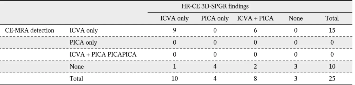
Table 1. Detection of Arterial Lesions in Lateral Medullary Infarction Patients on HR-CE 3D-SPGR and CE-MRA (n = 25)
HR-CE 3D-SPGR findings
ICVA only PICA only ICVA + PICA None Total
CE-MRA detection ICVA only 9 0 6 0 15
PICA only 0 0 0 0 0
ICVA + PICA PICAPICA 0 0 0 0 0
None 1 4 2 3 10
Total 10 4 8 3 25
Note.─ Data are numbers of patients
HR-CE 3D-SPGR; high-resolution contrast-enhanced three dimensional imaging with spoiled gradient-recalled sequence CE-MRA; contrast-enhanced magnetic resonance angiography
ICVA; intracranial vertebral artery PICA; posterior inferior cerebellar artery
ICVA + PICA; Arterial lesions in both ICVA and PICA
cm; matrix of 256 × 192, contrast material of 1.0 mmol of Gadobutrol (Gadovist; Bayer Schering Pharma, Berlin, Germany) per kilogram of body weight] were taken in all patients. In sequence, HR-CE 3D-SPGR [repetition time/echo time (TR/TE) msec of 6.9/1.7; flip angle of 20�; section thickness (ST) of 1 mm; field of view (FOV) of 20 × 20 cm; matrix of 256 × 256] was also obtained. Each HR-CE 3D-SPGR was transferred to the workstation software (Aquarius iNtuition Edition version 4.4.6.85.2800; TeraRecon, Inc, San Mateo, Calif). Transverse images perpendicu- lar to vascular course of ICVA or PICA were obtained using this software for further evaluation.
Arterial lesions in ICVA and PICA of LMI patients were evaluated with HR-CE 3D-SPGR and CE-MRA.
Arterial lesions in ICVA on HR-CE 3D-SPGR were subcategorized as follows: 1. atheroma, 2. dissection, 3. thromboembolism, 4. no arterial lesion (Fig. 1).
Atheroma on HR-CE 3D-SPGR was defined as thickness of wall between lumen showing high signal intensity (SI) and enhanced outer wall with additional consideration of simultaneous atheroma in intracranial vessels. Dissection on HR-CE 3D-SPGR was defined as double lumen containing true lumen and false lumen (10) with relatively intact other intracranial vessels. Thromboembolism in ICVA defined as very
c
a b
Fig. 1. Subcategory of arterial lesions in intracranial vertebral artery (ICVA) on HR-CE 3D-SPGR.
a. Atheroma, a lesion with low signal intensity between high signal intensity of arterial lumen and enhanced outer wall in both ICVAs. b. Dissection, two irregular high signal intensities in the arterial lumen with a low signal linear structure in between at Rt.
ICVA. c. Thromboembolism, very low signal intensity of the arterial lumen with enhancement of the outer wall in Rt. ICVA.
low SI arterial lesions with outer vessel wall, which meet neither atheroma nor dissection criteria. Arterial lesions in ICVA on CE-MRA were divided into subgroups as follows: 1. Stenosis, 2. Occlusion, 3. No arterial lesion. The presence or absence of arterial lesions in PICA was obtained on both HR-CE 3D- SPGR and CE-MRA.
It was divided into two groups of arterial lesion in ICVA relative to PICA involvement. Each group was compared about its demographics, the area of LMI, and cerebellar involvement. The area of LMI and lateral medulla were measured by manual tracing method in workstation. The extent of LMI was obtained from calculation of the area of LMI (ALMI) and lateral medulla (ALM) at the largest infarction level as follows: the area of LMI (%) = (ALMI/ALM) × 100.
These images were reviewed and analyzed indepen- dently blinded to clinical information and other brain MR images by two radiologists (Y.Y. and S.J.A.) with 3 and 6 years’ experience of radiology. In a disagree- ment of opinion, two radiologists reached consensus with discussion.
Statistical Analysis
Mann-Whitney U test was calculated in continuous variables for comparison between two groups. Fischer exact test was used for comparison of categorical variables in each group. All statistical analyses were performed with SPSS (version 19.0, IBM, Chicago, Illinois, USA). P-value was defined as significant at less than 0.05.
Table 2. Arterial Lesions in Intracranial Vertebral Artery on HR-CE 3D-SPGR and CE-MRA (n = 25)
HR-CE 3D-SPGR findings
Atheroma Dissection Thromboembolism None Total
CE-MRA findings Stenosis 6 5 0 0 11
Occlusion 0 2 2 0 4
None 2 1 0 7 10
Total 8 8 2 7 25
Note.─ Data are numbers of patients
HR-CE 3D-SPGR; high-resolution contrast-enhanced three dimensional imaging with spoiled gradient-recalled sequence CE-MRA; contrast-enhanced magnetic resonance angiography
Table 3. The Comparison of Demographic Characteristics, the Area of LMI and Cerebellar Involvement Between the Groups with or without Posterior Inferior Cerebellar Artery Involvement in Lateral Medullary Infarction Patients with Intracranial Vertebral Arterial Lesion
Characteristics PICA+ (n = 8) PICA- (n = 10) P value
Age (year-old)a 60.25 ± 14.36 60.40 ± 18.88 1.000
Men 5 9 1.000
Hyperlipidemia 3 3 1.000
Hypertension 6 6 0.638
Diabetes mellitus 5 2 0.145
Smoking 3 3 1.000
The area of LMI (%)a 12.98 ± 5.18 12.91 ± 7.18 1.000
Cerebellar involvement 3 0 0.069
Note.─aMean ± standard deviation PICA; posterior inferior cerebellar artery PICA+ ; arterial lesions in both ICVA and PICA
PICA- ; arterial lesions in ICVA without PICA involvement
Two readers detected symptomatic side arterial lesions in ICVA or PICA in 22 patients with LMI on HR-CE 3D-SPGR. But CE-MRA showed arterial lesions in only 15 patients. Three cases were commonly not identified as arterial lesions in ICVA or PICA on two images by both two radiologists. Arterial lesions in only ICVA were detected in 10 LMI patients on HR-CE 3D-SPGR. CE-MRA also detected nine of ten only ICVA arterial lesions. One arterial lesion was missed on CE-MRA due to minimal atheroma in ICVA (Table 1). But PICA arterial lesions on CE-MRA were
not visualized in 12 cases which were shown on HR- CE 3D-SPGR image (Fig. 2).
Among arterial lesions in ICVA on HR-CE 3D-SPGR image, there were 8 cases atheroma, 8 cases dissection and 2 cases thromboembolism (Table 2). Seven cases showed hypoplastic vertebral artery which was defined as less than 2 mm diameter in several litera- tures.
Group of ICVA arterial lesions with PICA involve- ment tended to simultaneously have cerebellar infarc- tion with LMI (p = 0.069). The area of LMI between groups of ICVA arterial lesions with or without PICA involvement statistically showed no difference. Other factors were not different statistically (Table 3).
RESULTS
c
a b
Fig. 2. A 84-year-old lateral medullary infarction patient with left posterior inferior cerebellar arterial lesion.
a. HR-CE 3D-SPGR showed focal aneurysmal dilatation in left distal posterior inferior cerebellar artery. b. Diffusion weighted image showed left lateral medullary infarction. c. Contrast- enhanced magnetic resonance angiography showed normal left vertebral artery and no visualization of posterior inferior cerebellar artery.
In LMI patients, we found that all arterial lesions in PICA which were shown on HR-CE 3D-SPGR image were not detected on CE-MRA. Concurrent cerebellar involvement was more shown in LMI patients with both ICVA and PICA arterial lesions than those with only ICVA arterial lesions.
There were several reports about causes of LMI with arterial lesions in ICVA or PICA (2, 11, 12). Fischer et al reported that pathologic finding of vessel involve- ments were 14.3% PICA disease, 38.1% VA disease and 26.2% both arteries diseases (13). Our results of 10 arterial lesions (40%) in only ICVA, 8 arterial lesions (32%) in both ICVA and PICA and 4 arterial lesions (16%) in only PICA were similar with that report. Our results showed 8 cases atheroma, 8 cases dissection, 2 cases thromboembolism on HR-CE 3D- SPGR, and 4 cases PICA lesions.
There were reports about non-invasive vessel imaging using CE-MRA which is able to depict lumen of intracranial artery such as ICVA (14, 15). However CE-MRA has technical limitation of the possibility in distinguishing of pathology in ICVA. In some cases, diffuse atherosclerosis or dissection in ICVA showed just luminal stenosis on CE-MRA. In one case, CE- MRA showed normal ICVA despite of presence of mild atherosclerosis. There was probably arterial remodeling that luminal narrowing did not occur until more than 50% atherosclerotic lesion in vessel wall existed (16, 17). There was a report that 3D-SPGR with T1W image was good to evaluate vertebral artery lesion such as dissection (10). To supplement this limitation, HR-CE 3D-SPGR was used for evaluation of arterial lesions in ICVA. In addition, our findings showed that HR-CE 3D-SPGR detected arterial lesions in PICA in 12 cases which were not shown on CE- MRA. HR-CE 3D-SPGR also helped to find arterial lesions in PICA which was small diameter using better spatial resolution than CE-MRA (18).
In our results cerebellar infarction was frequently shown in LMI patients with arterial lesion in ICVA with PICA involvement. Lateral medulla is largely supplied by distal vertebral artery and PICA (19). The PICA also supplies blood to the inferior cerebellum and pons (20). Some literatures which reported that
PICA territory infarction was related with significant vertebral artery stenosis or occlusion supported our results (21, 22).
There were several limitations for this study. First, study with small population had limitation to make generalizations with our results. Although LMI was a rare disease, we believe that it was not small popula- tion to study LMI in light of prevalence (23). Second, absence of pathological confirmation for arterial lesion in ICVA or PICA caused incorrect results. But recent other studies showed possibility of evaluating intracra- nial small arteries such as basilar and middle cerebral artery using HR-MRI. These reports supported our study of evaluating arterial lesion in ICVA or PICA using HR-CE 3D-SPGR to enable detect small arterial lesion (4-6, 24). Third, it is difficult to distinguish hypoplastic vertebral artery from the pathology. There were some reports that these factors play an additional role in ischemic stroke (25, 26). Without stenosis, these factors were considered as risk factor for ischemia (12). In conclusion, HR-CE 3D-SPGR can help evaluate arterial lesions in ICVA and PICA for LMI patients.
References
1. Kim JS. Pure lateral medullary infarction: clinical-radiological correlation of 130 acute, consecutive patients. Brain 2003;126:
1864-1872
2. Caplan LR. The intracranial vertebral artery: a neglected species. The Johann Jacob Wepfer Award 2012. Cerebrovasc Dis 2012;34:20-30
3. Wismer GL, Rosen BR, Buxton R, Stark DD, Brady TJ.
Chemical shift imaging of bone marrow: preliminary experi- ence. AJR Am J Roentgenol 1985;145:1031-1037
4. Klein IF, Lavallee PC, Schouman-Claeys E, Amarenco P. High- resolution MRI identifies basilar artery plaques in paramedian pontine infarct. Neurology 2005;64:551-552
5. Klein IF, Lavallee PC, Touboul PJ, Schouman-Claeys E, Amarenco P. In vivo middle cerebral artery plaque imaging by high-resolution MRI. Neurology 2006;67:327-329
6. Lou X, Ma N, Ma L, Jiang WJ. Contrast-enhanced 3T high- resolution MR imaging in symptomatic atherosclerotic basilar artery stenosis. AJNR Am J Neuroradiol 2013;34:513-517 7. Klein IF, Lavallee PC, Mazighi M, Schouman-Claeys E,
Labreuche J, Amarenco P. Basilar artery atherosclerotic plaques in paramedian and lacunar pontine infarctions: a high-resolution MRI study. Stroke 2010;41:1405-1409
8. Swartz RH, Bhuta SS, Farb RI, et al. Intracranial arterial wall imaging using high-resolution 3-tesla contrast-enhanced MRI.
Neurology 2009;72:627-634
9. Mirowitz SA. Intracranial lesion enhancement with gadolinium:
T1-weighted spin-echo versus three-dimensional Fourier
DISCUSSION
transform gradient-echo MR imaging. Radiology 1992;185:529- 534
10. Hosoya T, Adachi M, Yamaguchi K, Haku T, Kayama T, Kato T.
Clinical and neuroradiological features of intracranial vertebrobasilar artery dissection. Stroke 1999;30:1083-1090 11. Kim JS, Lee JH, Choi CG. Patterns of lateral medullary infarc-
tion: vascular lesion-magnetic resonance imaging correlation of 34 cases. Stroke 1998;29:645-652
12. Cloud GC, Markus HS. Diagnosis and management of vertebral artery stenosis. QJM 2003;96:27-54
13. Rahmouni A, Divine M, Mathieu D, et al. Detection of multiple myeloma involving the spine: efficacy of fat-suppression and contrast-enhanced MR imaging. AJR Am J Roentgenol 1993;
160:1049-1052
14. Yang CW, Carr JC, Futterer SF, et al. Contrast-enhanced MR angiography of the carotid and vertebrobasilar circulations.
AJNR Am J Neuroradiol 2005;26:2095-2101
15. Ersoy H, Watts R, Sanelli P, et al. Atherosclerotic disease distri- bution in carotid and vertebrobasilar arteries: clinical experience in 100 patients undergoing fluoro-triggered 3D Gd-MRA. J Magn Reson Imaging 2003;17:545-558
16. Dong L, Underhill HR, Yu W, et al. Geometric and composi- tional appearance of atheroma in an angiographically normal carotid artery in patients with atherosclerosis. AJNR Am J Neuroradiol 2010;31:311-316
17. Glagov S, Weisenberg E, Zarins CK, Stankunavicius R, Kolettis GJ. Compensatory enlargement of human atherosclerotic coronary arteries. N Engl J Med 1987;316:1371-1375
18. Carroll KW, Feller JF, Tirman PF. Useful internal standards for distinguishing infiltrative marrow pathology from hematopoietic marrow at MRI. J Magn Reson Imaging 1997;7:394-398 19. Chen WT, Shih TT, Chen RC, et al. Blood perfusion of vertebral
lesions evaluated with gadolinium-enhanced dynamic MRI: in comparison with compression fracture and metastasis. J Magn Reson Imaging 2002;15:308-314
20. Jung HS, Jee WH, McCauley TR, Ha KY, Choi KH.
Discrimination of metastatic from acute osteoporotic compres- sion spinal fractures with MR imaging. Radiographics 2003;23:
179-187
21. Adams RD, Victor M, Ropper AH. Principles of neurology.
Companion handbook. 6th ed. New York: McGraw-Hill, Health Professions Division, 1998
22. Lechtenberg R. Handbook of cerebellar diseases. New York: M.
Dekker, 1993
23. Norrving B, Cronqvist S. Lateral medullary infarction: prognosis in an unselected series. Neurology 1991;41:244-248
24. Ryu CW, Jahng GH, Kim EJ, Choi WS, Yang DM. High resolu- tion wall and lumen MRI of the middle cerebral arteries at 3 tesla. Cerebrovasc Dis 2009;27:433-442
25. Chaturvedi S, Lukovits TG, Chen W, Gorelick PB. Ischemia in the territory of a hypoplastic vertebrobasilar system. Neurology 1999;52:980-983
26. Giannopoulos S, Markoula S, Kosmidou M, Pelidou SH, Kyritsis AP. Lateral medullary ischaemic events in young adults with hypoplastic vertebral artery. J Neurol Neurosurg Psychiatry 2007;78:987-989
통신저자 : 정태섭, (135-720) 서울시 강남구 언주로 211, 연세대학교 의과대학 강남세브란스병원 영상의학과 Tel. (02) 2019-3510 Fax. (02) 3462-5472 E-mail: [email protected]
고해상도 조영증강 삼차원 회손기울기 회상 영상을 이용한 측면연수경색 환자의 두개내 척추동맥 및 뒤아래소뇌동맥 평가
연세대학교 의과대학 강남세브란스병원 영상의학과 윤영노∙안성준∙서상현∙박아영∙정태섭
목적: 측면연수경색 환자의 두개내 척추동맥 및 뒤아래소뇌동맥 평가에 있어 고해상도 조영증강 삼차원 회손기울기에 코 회상영상의 역할 규명하고자 한다.
대상과 방법: 임상시험심사위원회에서 승인한 측면연수경색으로 확진된 25명의 환자를 대상으로 하였다. 모든 환자 는 3T 자기공명 영상을 이용한 고해상도 조영증강 삼차원 회손기울기에코 회상영상과 조영증강 자기공명 혈관조영술 을 받았다. 두 명의 영상의학과 의사는 환자의 임상정보와 확산강조영상 없이 두개내 척추동맥 및 뒤아래소뇌동맥에 있는 동맥병변을 평가하였다. 두개내 척추동맥과 뒤아래소뇌동맥의 병변 동반유무에 따라 임상정보와 측면연수경색 의 넓이, 소뇌경색 동반여부를 비교하였다.
결과: 총 25명의 환자 중 22명의 환자가 고해상도 조영증강 삼차원 회손기울기에코 회상영상으로 두개내 척추동맥 및 뒤아래소뇌동맥에서 박리, 죽종, 혈전색전증을 보였다. 그러나 그 중 12개의 뒤아래소뇌동맥의 병변은 조영증강 자기공명 혈관조영술에서 보이지 않았다. 추가적인 소뇌경색은 두개내 척추동맥과 뒤아래소뇌동맥에 병변이 있을 경 우 두개내 척추동맥에만 병변이 있는 경우보다 빈번하게 나타났다.
결론: 고해상도 조영증강 삼차원 회손기울기에코 회상영상은 측면연수경색 환자의 두개내 척추동맥 및 뒤아래소뇌동 맥의 병변평가에 도움을 줄 수 있다.
대한자기공명의과학회지 18:17-24(2014)