Comparison of the fit accuracy of zirconia-
based prostheses generated by two CAD/CAM systems
Seok-Joon Ha, Jin-Hyun Cho*
Department of Prosthodontics, School of Dentistry, Kyungpook National University, Daegu, Republic of Korea
PURPOSE. The purposes of this study are to evaluate the internal and marginal adaptation of two widely used CAD/CAM systems and to study the effect of porcelain press veneering process on the prosthesis adaptation.
MATERIALS AND METHODS. Molar of a lower jaw typodont resin model was prepared by adjusting a 1.0 mm circumferential chamfer, an occlusal reduction of 2.0 mm, and a 5° convergence angle and was duplicated as an abrasion-resistant master die. The monolithic crowns and copings were fabricated with two different CAD/CAM system-Ceramil and Zirkonzahn systems. Two kinds of non-destructive analysis methods are used in this study.
First, weight technique was used to determine the overall fitting accuracy. And, to evaluate internal and marginal fit of specific part, replica technique procedures were performed. RESULTS. The silicone weight for the cement space of monolithic crowns and copings manufactured with Ceramil system was significantly higher than that from Zirkonzahn system. This gap might cause the differences in the silicone weight because the prostheses were manufactured according to the recommendation of each system. Marginal discrepancies of copings made with Ceramil system were between 106 and 117 μm and those from Zirkonzahn system were between 111 and 115 μm. Marginal discrepancies of copings made with Ceramil system were between 101 and 131 μm and those from Zirkonzahn system were between 116 and 131 μm. CONCLUSION. Marginal discrepancy was relatively lower in Ceramil system and internal gap was smaller in Zirkonzahn system. There were significant differences in the internal gap of monolithic crown and coping among the 2 CAD/CAM systems. Marginal discrepancy produced from the 2 CAD/CAM systems were within a reported clinically acceptable range of marginal discrepancy. [J Adv Prosthodont 2016;8:439-48]
KEYWORDS. Fit accuracy; Weight technique; Replica technique; Press-over technique
INTRODUCTION
Dental prostheses require physiologic stability, biocompatibil- ity, strength sufficient for the occlusal load, an esthetic similar to the shape and the color of real tooth, and, lastly but most
importantly, accurate fit to the abutment.1,2 Advances in com- puter-aided design and manufacturing (CAD/CAM) tech- niques in the last three decades have dramatically improved prosthetic devices machined directly in the dental clinic or laboratory.3,4 The excellent esthetic and biocompatible prop- erties of zirconia restoration make it an attractive metal-free alternative for durable prosthetic reconstructions in high demand by patients.5 The introduction of zirconia-based ceramic materials is one of the most significant advances in the field of restorative dentistry. Once only used in engi- neering, this zirconia material combines several properties critical for dental applications, including high esthetics, excellent biocompatibility, low plaque accumulation, low thermal conductivity, and high strength.5
Current zirconia-based ceramics contain yttria-stabilized tetragonal zirconia polycrystals (Y-TZP). This material can efficiently arrest crack propagation.6,7 Two types of CAD/
Corresponding author:
Jin-Hyun Cho
Department of Prosthodontics, College of Dentistry, Kyungpook National University, #2175 Dalgubeoldae-ro, Jung-gu, Daegu 41940, Republic of Korea
Tel. +82536007675: e-mail, [email protected]
Received March 27, 2016 / Last Revision October 1, 2016 / Accepted October 11, 2016
© 2016 The Korean Academy of Prosthodontics
This is an Open Access article distributed under the terms of the Creative Commons Attribution Non-Commercial License (http://creativecommons.
org/licenses/by-nc/3.0) which permits unrestricted non-commercial use, distribution, and reproduction in any medium, provided the original work is properly cited.
CAM systems are commonly used today, which employ either subtractive milling or an additive technique based on three-dimensional (3D) printing. However, the subtractive technique is more widely used because it is more economi- cal than the 3D printing technique. Both layered zirconia with coping and monolithic zirconia are popular due to developments in the material properties of zirconia.
Precise fit of fixed prostheses reduces the prevalence of diseases associated with abutment teeth and increases long- term survival of prostheses.8,9 The first requirement of CAD/CAM systems should be their ability to produce pros- thetic components with similar fit accuracy to that of con- ventional manufacturing processes using heat-pressed or casting techniques.8,10
Previous studies on the fit of conventional prostheses have reported marginal openings less than 120 µm to be clinically acceptable.11,12 However, a minimal space between the prosthesis and its abutment is necessary to ensure accu- rate insertion of the prosthetic component and to allow interposition of an even layer of luting cement with mean values from 25 to 50 µm.13
Holmes et al. first defined the perpendicular measure- ment between the internal surface of the casting and the axial wall of the preparation as the internal gap; the same measurement at the margin of the casting is defined as the marginal gap.14 Another important measurement, the abso- lute marginal discrepancy, is the angular combination of the marginal gap and extension error (over- or under-exten- sion); in other words, the combination of the vertical and horizontal marginal discrepancies.15
Various methods for measuring and evaluating the mar- ginal gap are described in the literature, all with individual pros and cons. Sorensen classified the available methods into four basic categories: direct view, cross-sectional view, impression technique, and use of explorer with visual exam- ination (x-rays).16
The silicone weight technique is one popular method currently used to evaluate marginal and internal misfit. With this technique, marginal and internal misfits are easily identi- fied using low-viscosity impression material. This technique is nondestructive, and measurements can be repeated for comparison of results.17 Another recently favored method for measuring the marginal and internal gap is the replica technique. A number of studies have evaluated the accuracy of crowns and fixed dental prosthesis in vivo as well as in vitro. This technique offers the advantage that neither resto- ration nor abutment are destroyed during the assess- ment.18,19
While many CAD/CAM systems are currently available, the present study evaluated the fit accuracy of prostheses generated by two widely used systems. This study assessed the Ceramill (Amann Girrbach AG, Koblach, Austria) and Zirkonzahn (Zirkonzahn GmbH, Bruneck, Italy) systems.
Partially sintered zirconia blocks were used in each system, and the press-over technique (IPS e.max ZirPress; Ivoclar Vivadent, Amherst, NY, USA) was used for porcelain veneering. Few studies have assessed the fit of monolithic
crowns, coping, and layered zirconia crowns, and even fewer have investigated press-over techniques for porcelain veneer- ing.20,21
The aims of this study are to evaluate the internal and marginal adaptation of two widely used CAD/CAM sys- tems and to evaluate the effect of porcelain pressing on prosthesis adaptation. Two non-destructive analysis meth- ods were used in this study. The first, the silicone weight technique, uses low-viscosity materials. The second is the replica technique. The null hypothesis was that there is no significant difference in the internal and marginal fit among restorations fabricated by the two different CAD/CAM sys- tems. Another null hypothesis was that there is no signifi- cant difference in the internal and the marginal fit between zirconia coping with or without veneering porcelain.
MATERIALS AND METHODS
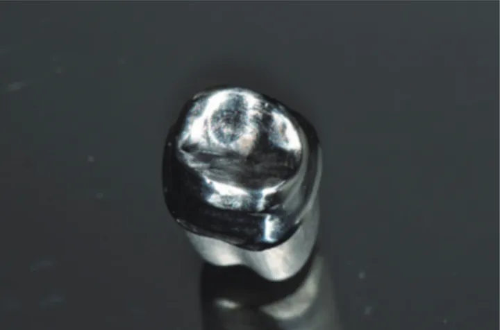
In master die preparation, the first molar of a lower jaw typodont resin model (KaVo Basic study model; KaVo Dental GmbH, Biberach, Germany) was prepared to accommodate an all-ceramic restoration. The model was prepared by adjusting for a 1.0 mm circumferential chamfer, an occlusal reduction of 2.0 mm, and a 5° convergence angle. The area of the resin model including the prepared tooth was then duplicated as an abrasion-resistant master model made of Ceramillsintron (Amann Girrbach AG, Koblach, Austria) using Ceramill system (Fig. 1). Based on 40 individual impressions of this master die (Aquasil Ultra XLV and Monophase, Dentsply, Milford, DE, USA), 40 stone dies in class IV stone (Fuji Rock, GC, Leuven, Belgium) were made and subsequently used to fabricate the Y-TZP restorations.
The 40 stone dies were randomly assigned to four groups (n
= 10). Each twenty stone die was used to fabricate copings and to manufacture monolithic crowns.
Fig. 1. Master die.
In the fabrication of monolithic crown and coping, cop- ings and crowns were fabricated using Ceramill and Zirkonzahn CAD/CAM systems. As this study is designed to compare two different CAD/CAM systems, burs and zir- conia blocks recommended by each system were used for the experiment. Designated cement space follows the guide- line of each system. The Ceramill system was used to manu- facture 10 copings and 10 monolithic crowns by CAD/
CAM. After each stone die was scanned by the Ceramill map400 (Amann Girrbach AG, Koblach, Austria), a mono- lithic crown and coping were designed using the Ceramill mind design program (Amann Girrbach AG, Koblach, Austria). The design of the final prosthesis after porcelain veneering was also drawn while designing the coping.
CeramillZolid FX (Amann Girrbach AG, Koblach, Austria), a partially sintered zirconia block, was processed using a CeramillMotion 2 (Amann Girrbach AG, Koblach, Austria), a five-axis milling machine, to manufacture the prostheses.
Three kinds of burs with diameters of 2.5, 1.0 and 0.6 mm were used in the milling machine in regular sequence. Finally, they were sintered at 1450°C for 11 hours in a Ceramill Therm furnace (Amann Girrbach AG, Koblach).
Another 10 copings and 10 monolithic crowns were manufactured using a Zirkonzahn system. In this system, a SCANNER S600 ARTI (Zirkonzahn GmbH, Bruneck, Italy) was used to scan the stone dies and a Zirkonzahn Modellier (Zirkonzahn GmbH, Bruneck, Italy) was used to design monolithic crowns and copings. As with the Ceramill
system, the design of the final prosthesis after porcelain veneering was also drawn while designing the coping.
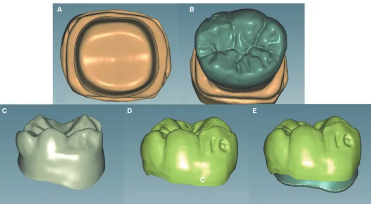
Milling burs with diameters of 2.0, 1.0, and 0.5 mm were used in a MILLING UNIT M5 (Zirkonzahn GmbH, Bruneck, Italy), a five-axis milling machine. Partially sintered Prettauzirconia (Zirkonzahn GmbH, Bruneck, Italy) blocks were used and the prostheses were finally sintered at 1600°C for 10 hours in a ZIRKONOFEN 600 furnace (Zirkonzahn GmbH, Bruneck, Italy) (Fig. 2).
In the analysis of fit accuracy, the copings and mono- lithic crown manufactured by both CAD/CAM systems fit- ted well to the master die without any adjustment by dental technicians. First, the weight technique was used to deter- mine the overall fit accuracy: the inner side of the mono- lithic crowns and copings were filled with light-body sili- cone (Aquasil Ultra XLV; Dentsply, Milford, DE, USA), simulating the clinical application of a luting agent. Then, the full monolithic crowns and copings were placed onto the master die in order. The restorations were then seated on the master die using finger pressure. Following the removal of excess unpolymerized material at the margins, finger pressure was applied again for four minutes. After polymerization of the impression material, the crowns were carefully removed from the master die, and the weight of the additional silicone was measured using an analytical bal- ance (Mettler AJ, San Francisco, CA, USA). A single opera- tor performed all measurements. The same procedure was performed three times for each specimen.
Fig. 2. (A) Scan of stone die, (B) Monolithic crown design, (C) Coping design, (D) Porcelain veneering form design, (E) Superposition of coping and porcelain veneering forms.
A B
C D E
C
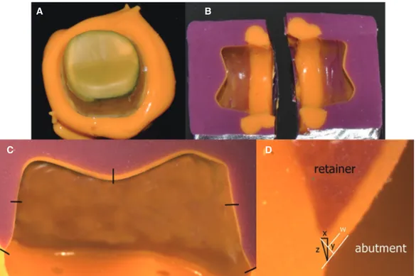
Replica technique procedures were used to evaluate the internal and marginal fit of each specific part. The first steps of the replica technique were similar to those of the silicone weight technique. The master die was lubricated with separating fluid (Microfilm; Kerr Italia Srl, Salerno, Italy) before each prosthesis was adapted. First, monolithic crowns and copings were filled with light-body silicone (Aquasil Ultra XLV; Dentsply, Milford, DE, USA) and seat- ed on the master die with finger pressure. After the light- body silicone had set, the restorations were removed from the master model while the thin silicone films representing the space between abutment teeth and retainers remained on the abutment teeth (Fig. 3A). The silicone films were then stabilized by contrasting heavy-body silicone (Aquasil Ultra Monophase; Dentsply, Milford, DE, USA) using a customized impression tray. The trays were designed to have generally equal space over the master die so that the light- body silicone is not deformed by the heavy-body silicone while making the replica. The replicas were then cut in the bucco-lingual direction in the center of the prosthesis using a No. 10 surgical blade to measure and photograph the internal and marginal gap with a stereoscopic microscope with surface illumination (MZ-16FA; Leica Microsystems, Wetzlar, Germany) using Leica microscope software (Fig.
3B).The silicone film thickness was recorded using a stereo- scopic microscope to measure five points for each replica.
Marginal discrepancies, internal gap between the two points, and occlusal gap at one point of occlusal surface were mea-
sured (Fig. 3C), as well as three different points at the mar- ginal area. The horizontal (x), vertical (y), and absolute mar- ginal discrepancies (z) were evaluated. Horizontal marginal discrepancy is the horizontal distance between abutment margin and prosthetic margin, while vertical marginal dis- crepancy is the vertical distance between abutment margin and prosthetic margin. Absolute marginal discrepancy is the summation of the two previously-explained discrepancies (Fig. 3D).
For evaluating of porcelain veneering effect, copings were subsequently veneered with their respective ceramic material to produce the final restoration. In this study, the press-over technique was used in order to reduce dental technician errors and to produce uniform porcelain veneer thickness. The veneer form designed with the coping was duplicated using the castable milling wax produced by each company (CeramillWax; Amann Girrbach AG, Koblach, Austria and ZirkonzahnWax; Zirkonzahn GmbH, Bruneck, Italy). The duplicated wax replicas and sprues were attached to each coping, which were then embeded. The wax was burnt out and the final prosthesis acquired by filling the space with IPS e.max ZirPress (Ivoclar Vivadent, Amherst, NY, USA), a pressable glass-ceramic ingot. Firing was per- formed in a proper ceramic furnace (Programat EP 5000, Ivoclar Vivadent, Amherst, NY, USA) at 910ºC. After recov- ery of the restorations, they were finished according to the manufacturer’s instructions and glazed at 750ºC. After the veneering process, the previously described measurements were performed on every veneered restoration.
Fig. 3. (A) Silicone films representing the space between the abutment teeth and prosthesis, (B) Stabilized replica after segmentation in the bucco-lingual direction, (C) Measuring points of the cut silicone replica, (D) Microscopic cross- sectional photograph of a replica showing the distances used to assess marginal accuracy.
A B
C D
Statistical analysis was performed using software (SPSS Statistics for Windows, Version 20.0; IBM Corp., Armonk, New York, USA). The Shapiro-Wilk test was used to con- firm the normal distributed of the silicone weight, internal gaps and marginal discrepancies. The mean values and stan- dard deviations per group were calculated. One-way analysis of variance (ANOVA) was used to assess the influence of the CAD/CAM systems and the porcelain veneering pro- cess on the silicone weight, internal gaps and marginal dis- crepancies. Levene’s tests were also performed to evaluate the equality of the variances. The level of significance was set at 0.05.
Results
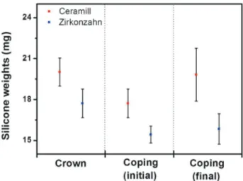
Table 1 shows the silicone weights for the cement space of the Ceramill (n = 10) and Zirkonzahn monolithic (n = 10) crowns measured using the weight technique. The average weights of silicone in the Ceramill and Zirkonzahn mono- lithic crown specimens were 20.02 ± 1.02 and 17.72 ± 1.05 mg, respectively (Fig. 4). This difference was statistically sig- nificant (Table 3). The initial silicone weight of the Ceramill
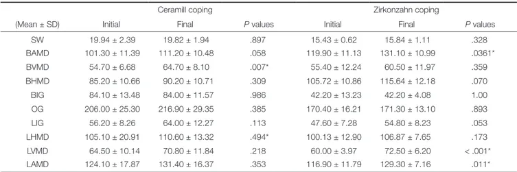
coping specimen was 19.94 ± 2.39 mg; the silicone weight after porcelain veneering 19.82 ± 1.94 mg. In comparison, the initial silicone weight for the Zirkonzahn coping speci- men was 15.43 ± 0.62 mg, which increased to 15.84 ± 1.11 mg after porcelain veneering (Table 1) (Fig. 4). The initial silicone weight and the silicone weight after porcelain veneer differed significantly between the Ceramill and Zirkonzahn systems. However, the silicone weight before and after por- celain veneering was not significantly different in both man- ufacturing system (Table 4, Table 5).
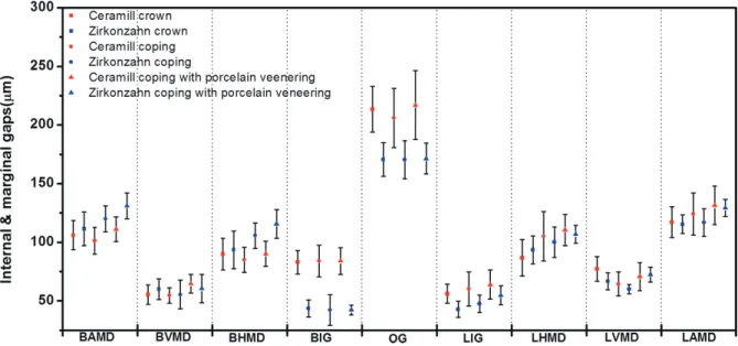
Analysis using the replica technique revealed internal gaps of the Ceramill monolithic crowns of 83.20 ± 10.0 µm at the bucco-axial surface, 213.40 ± 19.5 µm on occlusal surface, and 56.20 ± 8.2 µm at linguo-axial surface. The measured internal gaps of the Zirkonzahn crown were 43.70 ± 7.3 µm at the bucco-axial surface, 170.60 ± 14.2 µm on occlusal surface, and 42.90 ± 6.8 µm at linguo-axial sur- face (Table 2) (Fig. 5). The internal gaps measured at each position of the monolithic crowns differed significantly between the two manufacturing systems (Table 3).
The initial internal gaps of the Ceramill copings were 84.10 ± 13.48 µm at the bucco-axial surface, 206.00 ± 25.29 µm on the occlusal surface, and 60.20 ± 14.63 µm at the lin- guo-axial surface. After porcelain veneering, these gaps were 84.00 ± 11.57, 216.90 ± 29.35, and 64.00 ± 12.27 µm, respectively. In comparison, the initial internal gaps of the Zirkonzahn copings were 42.20 ± 13.23, 170.40 ± 16.21, and 47.60 ± 7.28 µm, respectively. After veneering, the gaps were 42.20 ± 4.08, 171.30 ± 13.10, and 54.80 ± 8.23 µm, respectively (Table 2) (Fig. 5). The internal gaps differed sig- nificantly before and after porcelain veneering; the coping also differed significantly between manufacturing systems (Table 4, Table 5).
The horizontal, vertical, and absolute marginal discrep- ancies were also measured. The discrepancies at the buccal margins of the Ceramill crowns were 89.91 ± 13.30, 55.40
± 8.33, and 106.00 ± 12.48 µm, respectively, and 86.92 ± 15.52, 77.30 ± 10.44, and 117.00 ± 13.16 µm at the lingual margins. The horizontal, vertical, and absolute marginal dis- crepancies at the buccal margins of the Zirkonzahn crowns were 93.47 ± 16.06, 60.0 ± 8.82, and 111.60 ± 14.30 µm, and 93.48 ± 11.84, 66.80 ± 7.18, and 115.40 ± 7.95 µm,
table 1. Silicone weights (mg)
Mean SD Min Max
Ceramill crown 20.02 1.02 19.03 22.03
Zirkonzahn crown 17.72 1.05 16.50 19.43
Ceramill coping Initial 19.94 2.39 16.50 23.07
Final 19.82 1.94 17.77 22.13
Zirkonzahn coping Initial 15.43 0.62 14.33 16.5
Final 15.84 1.11 14.83 18.27
SD: Standard deviation, Min: Minimum, Max: Maximum, Initial: Before porcelain veneering, Final: After porcelain veneering
Fig. 4. Silicone weights (mg). Initial: Before porcelain veneering, Final: After porcelain veneering.
respectively, at the lingual margins (Table 2) (Fig. 5).
Comparisons of the monolithic crowns revealed that only the linguo-vertical marginal discrepancies differed signifi- cantly between the two manufacturing systems (Table 4).
The horizontal, vertical, and absolute marginal discrep- ancies at the buccal margins of the Ceramill copings before porcelain veneering were 85.12 ± 10.62, 54.70 ± 6.68, and 101.30 ± 11.39 µm, respectively. The discrepancies at the lingual margins were 105.06 ± 21.03, 64.50 ± 10.15, and 124.10 ± 17.87, respectively. After veneering, the horizontal, vertical, and absolute marginal discrepancies at the buccal margins of the Ceramill copings were 90.09 ± 10.74, 64.70
± 8.10, and 111.20 ± 10.49 µm, respectively, and 110.50 ± 13.25, 70.80 ± 11.85, and 131.40 ± 16.37 µm at the lingual margins. The horizontal, vertical, and absolute marginal dis- crepancies at the buccal margins of the Zirkonzahn copings
before porcelain veneering were 105.72 ± 10.86, 55.40 ± 12.24, and 119.90 ± 11.13 µm, respectively, and 100.13 ± 12.90, 60.00 ± 3.97, and 116.90 ± 11.79 µm at the lingual margins. The horizontal, vertical, and absolute marginal dis- crepancies at the buccal margins of the Zirkonzahn copings after porcelain veneering were 115.64 ± 12.18, 60.50 ± 11.97, and 131.10 ± 10.99 µm, respectively. They were 106.87 ± 7.65, 72.50 ± 6.20, and 129.30 ± 7.17 µm, respec- tively, at the lingual margins (Table 2) (Fig. 5). There were significant differences in the marginal discrepancies of the copings between the two manufacturing systems. The mar- ginal discrepancies before and after porcelain veneering also differed significantly in the bucco-vertical margins of the Ceramill system and the bucco-absolute, linguo-vertical, and linguo-absolute margins in the Zirkonzahn system (Table 4, Table 5).
Fig. 5. Internal and marginal gaps (μm) of monolithic crowns, simple copings and copings with porcelain veneering.
Table 2. Internal and marginal gaps (μm)
Ceramill crown Zirkonzahn crown Ceramill coping Zirkonzahn coping
(Mean ± SD) Initial Final Initial Final
BAMD 106.00 ± 12.48 111.60 ± 14.30 101.30 ± 11.39 111.20 ± 10.49 119.90 ± 11.13 131.10 ± 10.99 BVMD 55.40 ± 8.33 60.00 ± 8.82 54.70 ± 6.68 64.70 ± 8.10 55.40 ± 12.24 60.50 ± 11.97 BHMD 89.91 ± 13.30 93.47 ± 16.06 85.12 ± 10.62 90.09 ± 10.74 105.72 ± 10.86 115.64 ± 12.18 BIG 83.20 ± 10.02 43.70 ± 7.36 84.10 ± 13.48 84.00 ± 11.57 42.20 ± 13.23 42.20 ± 4.08 OG 213.40 ± 19.57 170.60 ± 14.29 206.00 ± 25.29 216.90 ± 29.35 170.40 ± 16.21 171.30 ± 13.10 LIG 56.20 ± 8.26 42.90 ± 6.84 60.20 ± 14.63 64.00 ± 12.27 47.60 ± 7.28 54.80 ± 8.23 LHMD 86.92 ± 15.52 93.48 ± 11.84 105.06 ± 21.03 110.50 ± 13.25 100.13 ± 12.90 106.87 ± 7.65 LVMD 77.30 ± 10.44 66.80 ± 7.18 64.50 ± 10.15 70.80 ± 11.85 60.00 ± 3.97 72.50 ± 6.20 LAMD 117.00 ± 13.16 115.40 ± 7.95 124.10 ± 17.87 131.40 ± 16.37 116.90 ± 11.79 129.30 ± 7.17 BAMD: bucco-absolute margin discrepancy, BVMD: bucco-vertical margin discrepancy, BHMD: bucco-horizontal margin discrepancy, BIG: bucco-internal gap, OG:
occlusal gap, LIG: linguo-internal gap, LHMD: linguo-horizontal margin discrepancy, LVMD: linguo-vertical margin discrepancy, LAMD: linguo-absolute margin discrepancy, Initial: Before porcelain veneering, Final: After porcelain veneering
Table 3. Statistical comparison of Ceramill and Zirkonzahn monolithic crowns
(Mean ± SD) Ceramill crown Zirkonzahn crown P values
SW 20.02 ± 1.02 17.72 ± 1.05 < .001*
BAMD 106.00 ± 12.48 111.60 ± 14.30 .363
BVMD 55.40 ± 8.33 60.00 ± 8.82 .246
BHMD 89.91 ± 13.30 93.47 ± 16.06 .597
BIG 83.20 ± 10.02 43.70 ± 7.36 < .001*
OG 213.40 ± 19.57 170.60 ± 14.29 < .001*
LIG 56.20 ± 8.26 42.90 ± 6.84 < .001*
LHMD 86.92 ± 15.52 93.48 ± 11.84 .302
LVMD 77.30 ± 10.44 66.80 ± 7.18 .017*
LAMD 117.00 ± 13.16 115.40 ± 7.95 .747
SW: Silicone weight, BAMD: bucco-absolute margin discrepancy, BVMD: bucco-vertical margin discrepancy, BHMD: bucco-horizontal margin discrepancy, BIG:
bucco-internal gap, OG: occlusal gap, LIG: linguo-internal gap, LHMD: linguo-horizontal margin discrepancy, LVMD: linguo-vertical margin discrepancy, LAMD: linguo- absolute margin discrepancy, * Statistically significant P < .05
Table 4. Statistical comparison of Ceramill and Zirkonzahn copings
Initial Final
(Mean ± SD) Ceramill coping Zirkonzahn coping P values Ceramill coping Zirkonzahn coping P values SW 19.94 ± 2.39 15.43 ± 0.62 < .001* 19.82 ± 1.94 15.84 ± 1.11 < .001*
BAMD 101.30 ± 11.39 119.90 ± 11.13 .001* 111.20 ± 10.49 131.10 ± 10.99 < .001*
BVMD 54.70 ± 6.68 55.40 ± 12.24 .876 64.70 ± 8.10 60.50 ± 11.97 .370
BHMD 85.12 ± 10.62 105.72 ± 10.86 < .001* 90.09 ± 10.74 115.64 ± 12.18 < .001*
BIG 84.10 ± 13.48 42.20 ± 13.23 < .001* 84.00 ± 11.57 42.20 ± 4.08 < .001*
OG 206.00 ± 25.29 170.40 ± 16.21 .014* 216.90 ± 29.35 171.30 ± 13.10 < .001*
LIG 60.20 ± 14.63 47.60 ± 7.28 .029* 64.00 ± 12.27 54.80 ± 8.23 .645
LHMD 105.06 ± 21.03 100.13 ± 12.90 .536* 110.50 ± 13.25 106.87 ± 7.65 .465
LVMD 64.50 ± 10.15 60.00 ± 3.97 .208 70.80 ± 11.85 72.50 ± 6.20 .694
LAMD 124.10 ± 17.87 116.90 ± 11.79 .302 131.40 ± 16.37 129.30 ± 7.17 .716
SW: Silicone weight, BAMD: bucco-absolute margin discrepancy, BVMD: bucco-vertical margin discrepancy, BHMD: bucco-horizontal margin discrepancy, BIG:
bucco-internal gap, OG: occlusal gap, LIG: linguo-internal gap, LHMD: linguo-horizontal margin discrepancy, LVMD: linguo-vertical margin discrepancy, LAMD: linguo- absolute margin discrepancy, Initial: Before porcelain veneering, Final: After porcelain veneering, * Statistically significant P < .05.
Table 5. Statistical comparison of simple copings and copings with porcelain veneering
Ceramill coping Zirkonzahn coping
(Mean ± SD) Initial Final P values Initial Final P values
SW 19.94 ± 2.39 19.82 ± 1.94 .897 15.43 ± 0.62 15.84 ± 1.11 .328
BAMD 101.30 ± 11.39 111.20 ± 10.48 .058 119.90 ± 11.13 131.10 ± 10.99 .0361*
BVMD 54.70 ± 6.68 64.70 ± 8.10 .007* 55.40 ± 12.24 60.50 ± 11.97 .359
BHMD 85.20 ± 10.66 90.20 ± 10.71 .309 105.72 ± 10.86 115.64 ± 12.18 .070
BIG 84.10 ± 13.48 84.00 ± 11.57 .986 42.20 ± 13.23 42.20 ± 4.08 1.00
OG 206.00 ± 25.30 216.90 ± 29.35 .385 170.40 ± 16.21 171.30 ± 13.10 .893
LIG 56.20 ± 8.26 64.00 ± 12.27 .113 47.60 ± 7.28 54.80 ± 8.23 .053
LHMD 105.10 ± 20.91 110.60 ± 13.32 .494* 100.13 ± 12.90 106.87 ± 7.65 .173
LVMD 64.50 ± 10.14 70.80 ± 11.84 .218 60.00 ± 3.97 72.50 ± 6.20 < .001*
LAMD 124.10 ± 17.87 131.40 ± 16.37 .353 116.90 ± 11.79 129.30 ± 7.16 .011*
SW: Silicone weight, BAMD: bucco-absolute margin discrepancy, BVMD: bucco-vertical margin discrepancy, BHMD: bucco-horizontal margin discrepancy, BIG:
bucco-internal gap, OG: occlusal gap, LIG: linguo-internal gap, LHMD: linguo-horizontal margin discrepancy, LVMD: linguo-vertical margin discrepancy, LAMD: linguo- absolute margin discrepancy, Initial: Before porcelain veneering, Final: After porcelain veneering, * Statistically significant P < .05.
DISCUSSION
Despite careful preparation of full-coverage restorations and precise cementation, small gaps will remain between the margins of the restorations and the finish lines of the pre- pared teeth, predisposing the teeth to caries and periodontal disease. The closer the margin of restoration to the finish line of the preparation, the smaller the marginal gap and thickness of the exposed cement layer at the margin.22 The clinically allowable marginal gaps of dental prostheses have been described in numerous studies. Some authors have reported marginal gaps under 120 µm to be clinically allow- able for traditional fixed prostheses.13 Others have reported marginal gaps of 160 - 172 µm to be clinically acceptable for conventional crowns, with an acceptable marginal gap range of under 200 µm and occlusal gap range of under 250 - 300 µm.23-26 In this study, finger pressure was used to seat the crowns and copings to the master die to reproduce the clinical situations. Some previous literatures also used this method.27
While planning this research, micro-CT was also consid- ered for measurement of the fit accuracy. However, it was excluded due to some limitations. First of all, it is not easy to find specific measuring point for internal gap and mar- ginal discrepancy because the exact interface between mate- rials with different radiation absorption coefficient is hardly defined in micro-CT. Furthermore, radiographic images could have artificial defects due to the reflection of radioac- tive rays.28
Kokubo et al.29 used a light-body silicone in place of lut- ing cement to determine the relative marginal gaps in ceramic crowns. McLean and von Fraunhofe also used a light-body silicone to measure the thickness of the cement film, concluding that it is a convenient method to evaluate three-dimensional volume of the luting cement space.30 Nakamura et al.31 and May et al.32 both used a test-fit silicone paste to measure internal gaps.
In this study, the silicone weights of the cement spaces of crowns manufactured with the Ceramill system were sig- nificantly higher than those of the Zirkonzahn system. The Ceramill system recommends 50 μm of cement space, while the Zirkonzahn system recommends 35 μm. This gap might have contributed to the observed differences in silicone weights, as the prostheses were manufactured according to the recommendations for each system. However, higher sili- cone weight does not mean lower prosthesis fit accuracy.
Excessively small cement space can disturb precise setting of the prosthesis. Therefore, the fit accuracy of prostheses cannot be determined only by the cement space. In this study, both replica and silicone weight techniques were used to more accurately measure the prosthesis fit accuracies.
Impression techniques with low-viscosity impression material (replica technique) are popular methods for evalu- ating marginal discrepancies between crowns and teeth. The replica technique is a methodology applicable to both in vitro and in vivo measurements of precision. The advantages of replica technique include the small probability of damag-
ing the sample and abutment in the process, which makes it a non-destructive methodology. The majority of authors agree that, compared with other techniques, the replica technique offers more potential for verifiable and accurate results.33,34 The replica technique can be used to measure the fit accuracy of prosthesis in each position and provide information about specific margins with critical impact on the fitness and prognosis of prostheses.
In this study, the marginal discrepancies of the mono- lithic crowns manufactured by both systems were less than 120 μm, well within the clinically acceptable range. The marginal discrepancies were relatively lower in the Ceramill system, and the internal gaps were smaller in the Zirkonzahn system.
The marginal discrepancies of copings made with the Ceramill and Zirkonzahn system were between 101 and 131 μm and 116 and 131 μm, respectively. Both were within the clinically allowable range, based on less strict criteria.35,36 Similarly, analysis of the monolithic crowns showed that the Ceramill system had lower marginal discrepancies, while the Zirkonzahn system had smaller internal gaps. It can be assumed one of the factors contributed to the lower mar- ginal discrepancies in the Ceramill system is that the pros- theses are set to the correct position without any friction against the abutments because the Ceramill system requires 15 µm more cement space. Additional studies are necessary to investigate the optimal cement space required for lower marginal discrepancies with internal gaps within clinically acceptable ranges.
In this study, the press-over technique was used for por- celain veneering process. This technique resulted in porce- lain veneers with equal shape and thickness, and also reduced the potential for dental technician errors.
Several studies have reported conflicting results on the effects of porcelain veneering on marginal fit. Balkata et al.37 compared the marginal fit of three all-ceramic systems. The results showed that porcelain firing significantly altered the marginal fit of the crowns. They concluded that the copings were not completely stable during the porcelain firing cycle and that the distortion might have been due to a nonuni- form porcelain mass. Gemalmaz and Alkumru reported a small increase in the metal-ceramic crown’s gap size after firing the body porcelain; this distortion was most evident during the first firing cycle.38
In a study comparing all-ceramic and porcelain-fused-to- metal (PFM) crowns, Castellani et al.39 reported a firing cycle to significantly influence the vertical discrepancy in both types of prosthesis. The results of the study showed the influence to be greater in all-ceramic crowns. Another study also reported that thermocycling changed the marginal dis- crepancies of all three crown types they tested.40
However, other studies argue that there are no changes in marginal discrepancies after veneering because the volu- metric stabilities of the copings are maintained during the porcelain veneering procedure.41,42
Torabi et al.43 measured changes in marginal fit of zirco- nia copings using three porcelain-veneering techniques,
including layering, press-over, and CAD-on techniques. The results showed significant alterations in all three techniques;
however, these alterations were in the range of clinically allowable marginal fit. The changes were smallest using the press-over technique, and significant compared with the conventional layering technique.
This difference may be explained by the fact that porce- lain particles melt and gather to fill voids during the porce- lain veneering procedure, and the resulting contraction of the porcelain mass causes a compressive force on the cop- ing. The deformation of the coping under the stress of contracting porcelain is spread around the entire marginal circumference.Generally, the marginal openings of these crowns after porcelain veneering were within clinically acceptable standards; thus, the amount of distortion does not exclude their use in clinical applications.44
The results of this study revealed that the overall vol- ume change before and after porcelain veneering was not statistically significant. Differences in marginal discrepancies before and after porcelain veneering were statistically signif- icant only at some positions, and the effect of porcelain veneering was greater in the Zirkonzahn system than in the Ceramill system. Nevertheless, these differences do not exceed the acceptable limits in clinical settings.
The results of this study showed that full zirconia crowns and porcelain-fused zirconia coping crowns manu- factured with Ceramill and Zirkonzahn systems are suffi- cient for use in clinical settings. The fit accuracy of the prostheses made by both systems compared favorably with that of conventional prostheses. Differences between the results of the present study and those of other studies may be related to differences in measurement methodologies, microscopes, microscope magnification, measurement loca- tion and number, or luting agents.
However, this experiment only compared marginal dis- crepancy values at two points of zirconia-based prostheses, it is hard to say that these values represent fit accuracy of the entire prostheses. More accurate and meaningful conclu- sions will be drawn if marginal discrepancy values are com- pared in more points.
CONCLUSION
The results of this study reject the null hypotheses that the internal and marginal fits of dental prostheses are not affected by different CAD/CAM systems or porcelain veneering process. There were significant differences in the internal crown and coping gaps between the two CAD/
CAM systems. Porcelain press veneering did not significant- ly influence the internal gap, but did significantly affect the marginal fit at some position. And, the marginal discrepan- cies produced by two CAD/CAM systems were within the reported clinically acceptable ranges.
ORCID
Seok-Joon Ha http://orcid.org/0000-0001-7532-5000
REFERENCES
1. Hannink RHJ, Kelly PM, Muddle BC. Transformation tough- ening in zirconia-containing ceramics. J Am Ceram Soc 2000;
83:461-87.
2. Conrad HJ, Seong WJ, Pesun IJ. Current ceramic materials and systems with clinical recommendations: a systematic re- view. J Prosthet Dent 2007;98:389-404.
3. Baig MR, Tan KB, Nicholls JI. Evaluation of the marginal fit of a zirconia ceramic computer-aided machined (CAM) crown system. J Prosthet Dent 2010;104:216-27.
4. Beuer F, Schweiger J, Edelhoff D. Digital dentistry: an over- view of recent developments for CAD/CAM generated res- torations. Br Dent J 2008;204:505-11.
5. Luthardt RG, Holzhüter M, Sandkuhl O, Herold V, Schnapp JD, Kuhlisch E, Walter M. Reliability and properties of ground Y-TZP-zirconia ceramics. J Dent Res 2002;81:487-91.
6. Martínez-Rus F, Suárez MJ, Rivera B, Pradíes G. Evaluation of the absolute marginal discrepancy of zirconia-based ce- ramic copings. J Prosthet Dent 2011;105:108-14.
7. Kohorst P, Junghanns J, Dittmer MP, Borchers L, Stiesch M.
Different CAD/CAM-processing routes for zirconia restora- tions: influence on fitting accuracy. Clin Oral Investig 2011;
15:527-36.
8. Tan K, Pjetursson BE, Lang NP, Chan ES. A systematic re- view of the survival and complication rates of fixed partial dentures (FPDs) after an observation period of at least 5 years. Clin Oral Implants Res 2004;15:654-66.
9. Felton DA, Kanoy BE, Bayne SC, Wirthman GP. Effect of in vivo crown margin discrepancies on periodontal health. J Prosthet Dent 1991;65:357-64.
10. Tinschert J, Natt G, Hassenpflug S, Spiekermann H. Status of current CAD/CAM technology in dental medicine. Int J Comput Dent 2004;7:25-45.
11. Molin MK, Karlsson SL, Kristiansen MS. Influence of film thickness on joint bend strength of a ceramic/resin compos- ite joint. Dent Mater 1996;12:245-9.
12. McLean JW, von Fraunhofer JA. The estimation of cement film thickness by an in vivo technique. Br Dent J 1971;131:
107-11.
13. Boitelle P, Mawussi B, Tapie L, Fromentin O. A systematic re- view of CAD/CAM fit restoration evaluations. J Oral Rehabil 2014;41:853-74.
14. Holmes JR, Bayne SC, Holland GA, Sulik WD. Considerations in measurement of marginal fit. J Prosthet Dent 1989;62:405- 8.
15. Holmes JR, Sulik WD, Holland GA, Bayne SC. Marginal fit of castable ceramic crowns. J Prosthet Dent 1992;67:594-9.
16. Sorensen JA. A standardized method for determination of crown margin fidelity. J Prosthet Dent 1990;64:18-24.
17. Moldovan O, Luthardt RG, Corcodel N, Rudolph H. Three- dimensional fit of CAD/CAM-made zirconia copings. Dent Mater 2011;27:1273-8.
18. Falk A, Vult von Steyern P, Fransson H, Thorén MM. Reliability of the impression replica technique. Int J Prosthodont 2015;
28:179-80.
19. Trifković B, Budak I, Todorović A. Application of replica
technique and SEM in accuracy measurement of ceramic crowns. Meas Sci Rev 2012;12:90-7.
20. Grenade C, Mainjot A, Vanheusden A. Fit of single tooth zir- conia copings: comparison between various manufacturing processes. J Prosthet Dent 2011;105:249-55.
21. Subasi G, Ozturk N, Inan O, Bozogullari N. Evaluation of marginal fit of two all-ceramic copings with two finish lines.
Eur J Dent 2012;6:163-8.
22. Tinschert J, Natt G, Mautsch W, Spiekermann H, Anusavice KJ. Marginal fit of alumina-and zirconia-based fixed partial dentures produced by a CAD/CAM system. Oper Dent 2001;
26:367-74.
23. Felton DA, Kanoy BE, Bayne SC, Wirthman GP. Effect of in vivo crown margin discrepancies on periodontal health. J Prosthet Dent 1991;65:357-64.
24. Reich S, Wichmann M, Nkenke E, Proeschel P. Clinical fit of all-ceramic three-unit fixed partial dentures, generated with three different CAD/CAM systems. Eur J Oral Sci 2005;113:
174-9.
25. Souza RO, Özcan M, Pavanelli CA, Buso L, Lombardo GH, Michida SM, Mesquita AM, Bottino MA. Marginal and inter- nal discrepancies related to margin design of ceramic crowns fabricated by a CAD/CAM system. J Prosthodont 2012;21:
94-100.
26. Borba M, Cesar PF, Griggs JA, Della Bona Á. Adaptation of all-ceramic fixed partial dentures. Dent Mater 2011;27:1119- 26.
27. Ucar Y, Akova T, Akyil MS, Brantley WA. Internal fit evalua- tion of crowns prepared using a new dental crown fabrication technique: laser-sintered Co-Cr crowns. J Prosthet Dent 2009;
102:253-9.
28. Pelekanos S, Koumanou M, Koutayas SO, Zinelis S, Eliades G. Micro-CT evaluation of the marginal fit of different In- Ceram alumina copings. Eur J Esthet Dent 2009;4:278-92.
29. Kokubo Y, Nagayama Y, Tsumita M, Ohkubo C, Fukushima S, Vult von Steyern P. Clinical marginal and internal gaps of In-Ceram crowns fabricated using the GN-I system. J Oral Rehabil 2005;32:753-8.
30. McLean JW, von Fraunhofer JA. The estimation of cement film thickness by an in vivo technique. Br Dent J 1971;131:
107-11.
31. Nakamura T, Dei N, Kojima T, Wakabayashi K. Marginal and internal fit of Cerec 3 CAD/CAM all-ceramic crowns. Int J Prosthodont 2003;16:244-8.
32. May KB, Russell MM, Razzoog ME, Lang BR. Precision of fit: the Procera AllCeram crown. J Prosthet Dent 1998;80:
394-404.
33. Coli P, Karlsson S. Fit of a new pressure-sintered zirconium dioxide coping. Int J Prosthodont 2004;17:59-64.
34. Laurent M, Scheer P, Dejou J, Laborde G. Clinical evaluation of the marginal fit of cast crowns-validation of the silicone replica method. J Oral Rehabil 2008;35:116-22.
35. Fransson B, Oilo G, Gjeitanger R. The fit of metal-ceramic crowns, a clinical study. Dent Mater 1985;1:197-9.
36. Boening KW, Walter MH, Reppel PD. Non-cast titanium res- torations in fixed prosthodontics. J Oral Rehabil 1992;19:281- 7.
37. Balkaya MC, Cinar A, Pamuk S. Influence of firing cycles on the margin distortion of 3 all-ceramic crown systems. J Prosthet Dent 2005;93:346-55.
38. Gemalmaz D, Alkumru HN. Marginal fit changes during por- celain firing cycles. J Prosthet Dent 1995;73:49-54.
39. Castellani D, Baccetti T, Clauser C, Bernardini UD. Thermal distortion of different materials in crown construction. J Prosthet Dent 1994;72:360-6.
40. Hung SH, Hung KS, Eick JD, Chappell RP. Marginal fit of porcelain-fused-to-metal and two types of ceramic crown. J Prosthet Dent 1990;63:26-31.
41. Shearer B, Gough MB, Setchell DJ. Influence of marginal configuration and porcelain addition on the fit of In-Ceram crowns. Biomaterials 1996;17:1891-5.
42. Song TJ. Marginal fit of the auro galvano crown system made using the electroforming technique. Master thesis, Seoul, Korea; Seoul National University, 2004.
43. Torabi K, Vojdani M, Giti R, Taghva M, Pardis S. The effect of various veneering techniques on the marginal fit of zirco- nia copings. J Adv Prosthodont 2015;7:233-9.
44. Weaver JD, Johnson GH, Bales DJ. Marginal adaptation of castable ceramic crowns. J Prosthet Dent 1991;66:747-53.