Multilock 대퇴스템을 이용한 고관절 전치환술
조선대학교 의과대학 정형외과학교실, 방사선학교실*
이상홍・김성진・변주남*
- Abstract -
Cementless Total Hip Arthroplasty using the Multilock Femoral Stem
Sang-Hong Lee, M.D., Seong-Jin Kim, M.D. and Joo-Nam Byun, M.D.*
Department of Orthopaedic Surgery, Department of Diagnostic Radiology* Medical School, Chosun University, Kwangju, Korea
We reviewed the clinical records, preoperative and postoperative radiographs of 37 patients(39 hips) who were treated with total hip arthroplasty using the Multilock femoral stem from Jan, 1994 to Jan, 1997 in order to evaluate the clinical and radiographic results. The follow up period ranged from 2 years to 5.5 years(average, 34 months). The age of the patients ranged from 32 to 67 years(average, 49 years). Preoperative diagnoses were avascular necrosis in 30 cases(77%), post-traumatic arthritis in 6 cases(15%) and others in 3 cases(8%). The average Harris hip score improved from 58 point preopera- tively to 93 point postoperatively. The last follow up radiographic findings showed that femoral stabili- ty was considered stable with bony ingrowth in 36 cases(92%) and stable with fibrous ingrowth in 3 cases(8%). The radiolucent line around the femoral stem less than 2mm in width along the radiopaque line was found in 14 cases, and focal osteolysis was found in one. The stress shielding was not found during the follow up period. Postoperative complications were intraoperative fracture in two, polyeth- ylene liner dissociation in two, dislocation in one and heterotopic ossification in one case.
This study showed good clinical and radiographic results of total hip arthroplasty using the Multilock hip prosthesis after the average follow up of 34 months, but a long term follow up is need- ed for clinical and radiographic evaluation.
Key Words : Total hip arthroplasty, Multilock femoral stem.
※통신저자: 이 상 홍
광주광역시동구서석동5 8 8 조선대학교의과대학정형외과학교실 Tel : 062) 220-3140, Fax : 062) 226-3379
서 론
무시멘트형 인공 관절 치환술에서 좋은 결과를 얻기 위해서는대퇴 스템의 압박고정을 통한 초기안정성과
인공삽입물표면의 이차적인 생물학적고정을얻는 것 이중요하다.
대퇴스템 근위부가환형으로 미세포말된 Mul-tilock 대 퇴스템은근위부의골성장을통한마모입자의골수강내 이동을막아골용해를억제하고, 원위부가플룻형으로대
퇴골 협부의 압박고정을 통해 torsional stability를증진시 키고, 티타늄재질을이용하여스템s t i f f n e s s를감소시키 게고안되어있어골흡수를감소시키고, 좋은고정력과 미세운동을감소시키게되어있다.
이에 저자들은 Multilock 대퇴스템을 이용하여 무시멘 트형 인공 고관절 전치환술을 시행한 환자 중 최소 2년 이상 추시가 가능했던 환자의 임상적, 방사선학적 추시 결과를보고하고자한다.
연구대상및방법
1 9 9 4년1월부터 1 9 9 7년1월까지대퇴골협부의압박 고정을 얻도록 고안된 Multilock 대퇴 스템과 H a r r i s - Galante(Zimmer Warsaw, Indiana, USA)형 비구컵을 가지 고인공고관절전치환술을시행한 3 7명( 3 9례)의환자를 대상으로하였다.
연령은최소3 2세에서6 7세까지로평균4 9세였고, 성별 은남자가 3 3명( 3 4례), 여자가4명( 5례)였으며추시기간은 2년에서최장5 . 5년으로평균3 4개월이었다.
수술전진단명은대퇴골두무혈성괴사3 0례(77%), 후 외상성관절염6례(15%), 결핵성관절염과 화농성 고관 절염의 후유증이 각각 1례, Legg-Calve Perthes병이 1례 였다.
대퇴스템의크기는 1 0㎜에서1 6㎜로평균 1 3㎜였고, 비구컵은5 0㎜에서6 8㎜로평균 5 6㎜였다.
수술방법은 후측방 도달법을 이용하였으며대퇴스템 은 압박고정을 위해 골수강을 0 . 5㎜ 적게 reaming 후 Multilock 대퇴스템을삽입하였으며, 젊은환자와큰대퇴 스템을삽입하는경우는reaming 크기와같은크기의대퇴 스템을삽입하였다. 비구컵은reaming 크기와동일한크기 의컵을사용하였으며비구컵과비구변연부사이의간격 이없이접촉을시도하였고2개의나사못고정을시행하 였다.
임상적기능평가는술전, 술후Harris hip score를측정하 였으며대퇴부동통유무를확인하였다. 방사선학적평가 는수술전후, 술후3개월, 술후6개월, 술후매1년마다고 관절전후면, 측면, frog leg 사진등을촬영하였다. 수술직 후 고정도는 C a l l a g h a n등7 )의방법에 의한전후면 및측면 사진상피질골과간격없이접촉된경우를우수로, 2㎜이 내의간격을보이는경우를양호로, 3㎜이상의간격을보 이는경우를불량으로구분하였으며, 최종추시고정상태 는 E n g h등1 4 )의방법에따라대퇴스템과피질골사이의방
사선투과선이없는경우를골성고정, 대퇴스템과평행 한방사선투과선이존재하나진행하지않은경우를섬유 성고정으로분류하고, 진행하는방사선투과선이나대퇴 스템의이동이있는경우를불안정고정으로분류하였다.
골용해증은H u d d l e s t o n1 8 )의분류에따라국소형, 다발성국 소형, 미만형으로분류하여조사하였으며골용증의최대 직경에따라4등급으로 구분하였다. Gruen등1 7 )의분류에 따른스템주위의각 Z o n e에서의방사선투과선및말단부 의피질골경화, 골용해증, 대퇴삽입물의해리여부등을 방사선과전문의1인과함께조사하였다.
비구컵의 방사선분석은 D e L e e와 C h a r n l e y1 0 )의 방법을 이용하여각각의 z o n e의 방사선투과선, 골용해증을검 사하고 비구컵의 수직 및 수평이동, 골내막 골형성등을 분석하였다.
인공삽입물의해리는대퇴부는5㎜이상의수직침강, 2
㎜이상의방사선투과선, 계속진행하는내반변형이있는 경우로정의하였고, 비구부는M a r t e l l등2 0 )의평가방법에의 하여수직전이가2㎜이상이거나경사각의변화가5도이상 일때또는2㎜이상의방사선투과선이1구역또는그이상 에서존재할때로정의하였다. 그리고술중과술후합병증에 대해서도분석하였다.
결 과 1. 임상적결과
Harris hip score는수술전 3 4 ~ 7 1점으로평균 5 8점이었 고최종추시시평균9 3점( 7 8 ~ 9 8점)으로향상되었으며우 수3 6례(92%), 양호2례(5%), 보통1례( 3 % )였다.
대퇴부 동통은 추시기간중 2례에서 관찰 되었으며 1 례는 1 5㎜ 스템을 삽입하였던 경우로 섬유성 안정고정 소견을보였으며술후1년에대퇴부동통이소실되었으 며, 다른1례는술전약5㎝이상하지단축에대한하지연 장교정결과로사료되며술후6개월경에동통의소실을 관찰하였다.
2. 방사선학적결과
1) 대퇴스템의방사선학적소견
대퇴 스템의 초기고정도는수술 직후 방사선 사진상 우수2 7례(69%), 양호 1 2례( 3 1 % )였으며, 최종추시상골 성 고정이 3 6례(92%), 섬유성 안정고정이 3례( 8 % )였으 며불안정성고정은1례도없었다.
대퇴스템의수직침강은전례에서없거나2㎜이하로
5㎜이상임상적으로의의를가진경우는없었으며최종 추시시평균수직침강은약 0 . 3㎜이었다.
방사선투과선은G r u e n의zone Ⅳ에서1 1례( 2 8 % )로가장 많았고미세포말형패드가부착된Zone Ⅰ과Ⅶ에서는전 후면상에서Zone Ⅰ1례, Zone Ⅶ이2례였고, 측면상에서 Zone Ⅶ에서1례가관찰되었다(Fig. 1-A). 골용해증은1례 ( 3 % )에서국소형골용해증이발견되었으며(Fig. 2-A,B) 대 퇴스템의무균성해리는1례도관찰되지않았다.
추시기간중 스트레스 방패에 의한 골흡수 소견은 관 찰할수없었다.
2) 비구컵의방사선학적소견
비구컵 주위의 2㎜이하의 방사선 투과선은 총 4례 ( 1 0 % )에서관찰되었고, DeLee와C h a r n l e y의Zone Ⅲ에서4 례로가장많았고Zone Ⅱ3례, Zone Ⅰ에서1례가관찰되 었다(Fig. 1-B).
2㎜이상 비구컵 수직전이 및 5도이상 비구컵 경사각 변화는관찰되지않았으며, 2㎜이하의비구컵의수직전 이는5례( 1 3 % )에서관찰되었고경미한비구컵의경사각 변화는1례( 3 % )에서관찰되었다.
비구컵주위의 골용해증은 관찰되지 않았으며 2례에 서비구부의폴리에틸렌컵분리가발생하여폴리에티렌 컵만재치환술을실시하였다.
3. 합병증
수술중합병증으로는대퇴경부주위의선상골절이2 례에서발생하여환형강선고정술을시행하였으며추시 관찰시골유합소견보였다(Fig. 3-A,B). 술후합병증으로는 폴리에틸렌라이너분리2례가발생하였는데2례모두과 거력상농부였으며쭈구려앉아일하던도중“뚝”하는느 낌을경험하였으며, 다시한차례의이상경험을한후외래 에방문한경우로방사선사진상비구컵내연상외측에대 퇴골두가위치한 소견을관찰할수있었으며, 술중폴리 에틸렌라이너의전방변연부의심한마모와함께라이너 분리소견이관찰되어라이너만교환하였다. 그리고고관 절탈구1례, Brooker등6 )의제Ⅱ형이소성골이형성이1례 에서발생하였다(Fig. 4-A,B).
고 찰
Fig. 1-A. The location and frequency(%) of radiolucent lines less than 2㎜in width along the radiopaque line in Gruen’s seven zones on the anteroposterior and lateral radiograph.
Fig. 1-B. The location and frequency(%) of radiolucent lines less than 2㎜in width in each three zones of DeLee and Charnley.
A B
현재까지많은종류의무시멘트형인공고관절이개발 되어왔으며특히일차적고관절전치환술시압박고정을 얻고무시멘트형구조물의골내성장을향상시킬수있는 방법에대한많은연구가진행되어왔다.
무시멘트형 인공 고관절 전치환술후 근위 대퇴부의 스트레스로인한 이차적인골 흡수로 삽입물의지지구 조물의손실, 침강그리고삽입물또는주위골절이발생 한다고하였으며5 )이것은시멘트형인공고관절치환술 보다증가된스템의크기및 s t i f f n e s s가원인이다고하였
다. 최근들어 삽입물 주위의 대퇴골 흡수를 감소시키기 위해 정상적인 대퇴골의 스트레스에 대해 충분한 유연 성을 갖으면서 강도를 제공하는데 많은 연구가 진행되 어 왔으며, 대퇴 스템 근위부가 환형 미세포말 처리된 Multilock 대퇴스템과AML 대퇴스템에대한고정물주위 의골흡수를비교한결과Multilock 대퇴스템에서골흡수 가 적은 것으로 나타났는데 이는 Multilock 대퇴 스템의 s t i f f n e s s감소로 인한근위부로 부하가 전달되어스트레 스방패를감소시킨결과라고하였다4 , 2 1 ).
Fig. 3-A. Intraoperative fracture of proximal third of femur(A.A.O.S type I) was treated by circlage wiring.
Fig. 3-B. At 3-year follow up, the radiograph shows fracture union and no evidence of loosening or subsidence of femoral stem.
A B
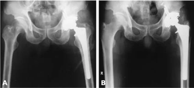
Fig. 2-A. Preoperative AP radiograph of 53-year-old male showed post-traumatic AVN due to previous femoral neck fracture.
Fig. 2-B. 2-year follow up radiograph shows linear osteolysis on Zone Ⅰ, Ⅶ.
A B
저자들은초기고정도를얻기위해가능한한큰크기 의 인공 삽입물을 삽입하여 피질골과 압박 고정하도록 노력하였는데수술직후고정도에서우수 2 7례(69%), 양 호 1 2례( 3 1 . % )로나타났으며, 압박고정과정중2례에서 근위 대퇴부에 선상골절이 발생하였으나 환형 강선 고 정을시행한후추시결과양호한임상결과및골유합소 견을 관찰 할 수 있었다. Fitzgerald등1 5 )은 골절의 안정성 을얻지 못하면실패율이높다고 하였으며 일차적고관 절 전치환술시 근위 대퇴부에 3.5%, 재치환술시 1 7 . 6 % 가발생하였다고보고 하면서모든 골절에 대해골절의 진행을막는예방적환형강선고정술을추천하였다.
Multilock 대퇴스템을이용한고관절전치환술후평균 Harris hip score는 박등1 )은 4 8개월 추시 결과에서 평균 9 4 . 7점의우수한결과를보고하였으며저자들의경우에 도평균 3 4개월추시결과 9 3점의우수한결과를보였다.
대퇴삽입물주위의방사선투과선은골과내고정물과 의탄력성차이에의하거나, 적어도한때삽입물이움직 임이있었다는증거이지만해리로간주해서는안된다고 하였으며, 백선경화선을동반한2㎜이하의방사선투과 선은골조직이아닌섬유성조직으로서스템과골조직사 이의미세운동에의해생기나더욱진행하지않으면기 능과는 관련이 없다고 하였다1 1 , 2 1 ). Martell등은 전후면상 Zone Ⅳ, 측면상에서는Zone Ⅳ, Ⅴ에서가장많이관찰되 었다고 하였으며 박등은 전후면상 Zone Ⅳ, 측면상에서
Ⅳ가가장많이관찰되었다고하였다. 저자들의경우전
후면상, 측면상모두Zone Ⅳ에서1 1례( 2 8 % )로가장많이 관찰되었고 미세 포말형 패드가 부착된 Zone Ⅰ과 Z o n e
Ⅶ에서각각1례씩관찰되었고비구부도Ⅰ, Ⅱ, Ⅲ구역 에서각각1, 3, 4례관찰되었다.
대퇴스템말단부의골내막경화상은국소체중부하에 의해발생하며1 9 ), Martell등은72%, 박등은4 0 %에서관찰 되었다고보고하였으며저자들의경우 3 8 %에서관찰되 었다.
술후 대퇴부의 동통은 원인 불명설과 대퇴 삽입물의 원위부와 골사이의 미세운동, 대퇴 삽입물의 근위부 고 정의 불안정으로 원위부로 체중 부하 전달시 발생한다 고보고되었는데2 , 8 )저자들의경우에는2례에서대퇴부 동통이발생하였는데1례는술전약5㎝이상하지단축 에대한하지연장교정결과로사료되며, 나머지1례에 서는무균성해리 소견은보이지않았지만 섬유성 고정 으로추정되는환자에서대퇴부동통을호소하였다.
골용해증은내고정물의움직임, 마멸입자혹은금속에 대한이물반응등으로인해유발되며내고정물근위부에 환형패드가있으면근위부골내성장으로용해를일으킬 수있는미세입자의원위부골내강으로의침투를방지한 다고보고되고있는데1 6 )골시멘트를사용하지않는고관 절전치환술후골용해증의발생빈도는 1 0 %에서3 0 %가 보고되고있으며1 , 3 , 2 2 , 2 3 ), 박등은9 . 2 %에서관찰되었다고보 고하였으며저자들의경우1례( 3 % )에서Gruen Zone Ⅰ, Ⅶ 에서관찰되었는데이는향후좀더추시관찰을요한다.
Fig. 4-A. Postoperative AP radiograph of 42-year-old male with Lt hip AVN showed good initial stability
Fig. 4-B. 12 months after surgery, Grade II heterotopic ossification is present, but functional range of motion was retained.
A B
최종추시시대퇴스템의안정도는E n g h등의평가방법 에 의해 골성 고정이 3 6례(92%), 섬유성 고정이가 3례 ( 8 % )였으며 불안정성 고정된 예는없었다. 방사선 투과 선은 1 4례( 3 6 % )에서 관찰되었으며 2㎜이상의 방사선 투과선은1례도관찰되지않았다.
비구컵주위의백선경화선을동반한방사선투과선은 비구컵이움직였다는증거이지만진행하지않으면의미 가없다고하였으며1 3 ), 박등은2 0 %에서관찰되었다고하 였고저자들의경우Zone Ⅲ가4례( 1 0 % )로가장많았고2
㎜이상의방사선투과선과골용해증은1례도없었다.
합병증으로는 수술중 발생한 대퇴골 골절이 2례에서 발생하였는데 A.A.O.S 분류9 )제1형으로 환형 강선 고정 술을 시행하여 골유합을 얻을 수 있었으며 폴리에틸렌 라이너분리2례는재치환술을시행하였고고관절탈구 1례는보존적요법으로치료하였고이소성골이형성1 례는Brooker Ⅱ형으로고관절기능에영향이없었다.
결 론
Multilock 대퇴스템은근위부의환형미세포말처리및 플룻형원위스템을통한협부고정을얻도록고안되었 으며 초기 우수한 고정력과 함께 stiffness 감소로 스템 주위의 골흡수를 감소시키며, 근위부 골내성장으로 골 용해 마모 입자의 원위부 골내강으로의 침투를 방지하 는장점이있다. 비교적단기추시결과이지만Multilock 대 퇴스템을 이용한 인공 고관절 치환술에서 임상적, 방사 선학적우수한결과를얻을수있었으며, 보다더장기적 인추시관찰이요하리라사료된다.
REFERENCES
01) 박상원,이광석,이순혁,백종륜: Multilock 대퇴스템 을 이용한 고관절 전치환 성형술. 대한고관절학회지, 9:92-98, 1997.
02) Barrack RL, Jasty M, Bragdon C, Haire T and Harris WH : Thigh pain despite bone ingrowth into uncemented femoral stems. J Bone Joint Surg, 74- B:507-510, 1992.
03) Beauchesne R, Engh CA, Knezevich S, Suthers K and Kukzta Y : Roentgenographic evaluation after AML porous coated acetabular components. A six year minimum follow up study. Orthop Trans, 16:834, 1 9 9 2 - 1 9 9 3 .
04) Bobyn JD, Galante JO, Jordan LR, Rosenberg AG, Rubash HE and White RE : A radiographic assess- ment of bone resorpotion and biologic fixation with the Multilock Hip Prosthesis A multi-Center study. A sci - entific Monograph Prepared for the 61st annual Meeting of the American Academy of Orthopaedic sur - g e o n s, New Oleans Feb, 24-28, 1994.
05) Bobyn JD, Mortimer Es, Glassman AH, Engh CA, Miller JE and Brooks CE : Producing and avoiding stress shielding. Laboratory and clinical observations of noncemented total hip arthroplasty.
Clin Orthop, 274: 79-96 , 1992.
06) Brooker AF, Bowerman JW and Robinson RA : Ectopic ossificaion following total hip arthroplasty.
J Bone Joint Surg, 55-A:1629-1632, 1973.
07) Callaghan JJ, Dysart SH and Savory CG : The uncemented porous-coated anatomic total hip pros- thesis. J Bone Joint Surg, 70-A:337-346, 1988.
08) Campell ACL, Rorabeck CH, Bourne RB, Chess D and Nott L : Thigh pain after cementless hip arthroplasty. J Bone Joint Surg, 74-B:63-66, 1992.
09) Committee on the hip : Classification and manage- ment of femoral defects in total hip replacement[e- xhibit]. Presented at the 57th Annual Meeting of the Amerian Academy of Orthopaedic Surgeons, New Orleans, Feb, 8-13, 1990.
10) DeLee JG and Charnley J : Radiological demar- cation of cemented sockets in total hip replacement.
Clin Orthop, 121:21-32, 1976.
11) Engh CA : Hip arthroplasty with a Moore prostheses with porous coating. Clin Orthop, 176:52-66, 1983.
12) Engh CA and Bobyn JD : The influence of stem size and extent of porous coating on femoral bone resorption after primary cementless hip arthroplasty.
Clin Orthop, 231:7-28, 1988
13) Engh CA and Griffin WL : Cementless acetabular components. J Bone Joint Surg, 72-B:53-59, 1990.
14) Engh CA, Massin P and Suthers KE : Roentgeno- graphic assessment of the biologic fixation of porous- surfaced femoral components. Clin Orthop, 257:107- 128, 1990.
15) Fitzgerald RH Jr, Brindley GW and Kavanagh BF : The uncemented total hip arthroplasty. Clin Orthop, 235:61-67, 1988.
16) Friedman RJ, Jonathan Black C, Galate JO Ja- cobs JJ and Skinner HB : Current concepts in ortho- paedic biomaterials and implant fixation. J Bone Joint S u r g, 75-A:1086-1109, 1993.
17) Gruen TA, McNeice GM and Amstutz HC : “Mode
of failure” of cemented stem-type femoral compo- nents. Clin Orthop, 141:17-27, 1979.
18) Huddleston HD : Femoral lysis after cemented hip arthroplasty. J Arthroplasty, 3:285-297, 1988.
19) Kaplan PA, Montesi SA, Jardon OM and Grego- ry PR : Bone ingrowth hip prosthesis in asympto- matic patients. Radiographic features, Radiology, 169:221-227, 1988.
20) Martell JM, Pierson RH, Jacobs JJ, Rosenberg AG, Maley M and Galante JO : Primary total hip reconstruction with a titanium fiber coated prosthe- sis inserted without cement. J Bone Joint Surg, 75- A:554-571, 1993.
21) Mittelmeier H : Ceramic prosthetic devices. T h e Hip, pp. 165-189, Mosby Co, 1984.
22) Tanzer M, Maloney WJ, Jasty M and Harris WH : The progression of femoral osteolysis in associa- tion with total hip arthroplasty without cement. J Bone Joint Surg, 74-A:404-410, 1992.
23) Zicat B, Engh CA and Gokcen J : Pattern osteoly- sis around total hip components inserted with and
without cement. J Bone Joint Surg, 77-A:432-439, 1995.